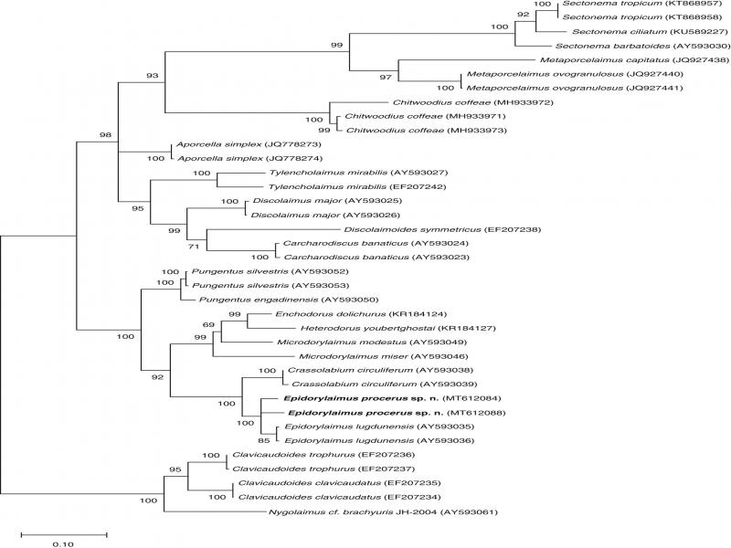
The genus Epidorylaimus Andrássy, 1986 is a widespread taxon in Dorylaimida. Originally proposed to accommodate 12 species previously included in Eudorylaimus Andrássy, 1959 its taxonomy was later updated by several authors (Andrássy, 1992; Jairajpuri and Ahmad, 1992; Vinciguerra, 2006; Andrássy, 2009). More recently, Ahmad et al. (2016) presented an emended diagnosis, a list of 15 valid species with a key to their identification, and a compendium of their more relevant morphometrics.
In spite of the nearly cosmopolitan distribution of the genus, none of its species was hitherto recorded in Vietnam. However, a nematological survey conducted to study nematode fauna associated with natural habitats in the country located an interesting Epidorylaimus species. Its study revealed that it belonged to an unknown species that is hereunder described.
Materials and methods
Nematode extraction and processing
Soil samples were collected in Cao Bang Province, a natural area of Northern Vietnam, and temporarily stored in plastic bags for transport to the laboratory. Nematode extraction was done following the methods of Baermann (1917) and Flegg (1967). Specimens were relaxed and killed with heat, fixed in 4% formaldehyde, processed to anhydrous glycerin according to Siddiqi’s (1964) technique, and mounted on permanent glass slides for handling and observation with light microscopy.
Light microscopy (LM)
Specimens were measured, drawn, and identified with a Nikon Eclipse 80i light microscope equipped with differential interference contrast (DIC) optics and a drawing tube (camera lucida). Morphometrics included Demanian ratios and other measurements. Microphotographs were taken with the same microscope provided with a Nikon Digital Sight DS-U1 camera. Raw photographs were edited using Adobe® Photoshop® CS.
DNA extraction, PCR, and sequencing
DNA was extracted from single individuals using the proteinase K and Worm Lysis Buffer protocol (William et al., 1992). Each nematode was transferred to a 0.5-ml Eppendorf tube containing 18 µl of Worm Lysis Buffer (WLB) (50 mM KCL, 10 mM Tris, pH 8.3, 2.5 mM MgCl2, 0.45% NP 40, and 0.45% Tween 20) and 2 µl of proteinase K (600 µg ml−1) (Thermo Scientific). The tubes were incubated at 65°C (1 hr) and then at 95°C (15 min). PCR was performed in a 30-µl final volume containing 24.9 µl of sterile water, 0.6 µl of each PCR primer, 0.6 µl of dNTP, 0.3 µl of Taq-polymerase, 3 µl of Buffer 10 x Thermo Scientific Green, and 1 µl of DNA-extracted solution. The PCR amplification profile consisted of 4 min at 94°C, 35 cycles of 1 min at 94°C, 1.5 min at 55°C, and 2 min at 72°C, followed by a final step of 10 min at 72°C. The primers used for amplification were D2A (5´-ACAAGTACCGTGAGGGAAAGTTA-3´) and D3B (5´-TCCTCGGAAGGAACCAGCTACTA-3´) for amplification of the D2-D3 region of 28S (Subbotin et al., 2006).
PCR products were purified with the GeneJET PCR Purification Kit (#K0701, Thermo Scientific, USA), following the manufacturer’s manual. The sequencing reaction was performed with 15 ng of purified template, 4 µL of BigDye Terminator v3.1 Ready Reaction Mix, 2 µL of 5X Sequencing Buffer, and 3.2 pmol of forward/reverse primers for a total of 10-µL volume. The mixture was heated for 10 sec at 96°C, then 5 sec at 55°C, repeated for 32 cycles followed by 4 min at 60°C. The sequencing was performed on 3500 x L Genetic Analyzers (Applied Biosystems, Foster City, California) at the National Key Laboratory of Gene Technology (IBT – VAST, Hanoi). The sequences obtained were submitted to the GenBank database under accession numbers MT612084 and MT612088.
Phylogenetic analysis
The obtained sequences were aligned with 35 other D2-D3 expansion segments of 28S rDNA gene sequences available in GenBank, using SeaView with the muscle algorithm followed by manual refining (V4.5.3, Gouy et al., 2010). Outgroup taxa were chosen according to previously published data (Holterman et al., 2008; Álvarez-Ortega et al., 2013). The sequence dataset was analyzed with Bayesian inference (BI) and maximum likelihood (ML) using MrBayes 3.1.2 (Huelsenbeck and Ronquist, 2001; Ronquist and Huelsenbeck, 2003). We chose the commonly used model GTR + I + G that then was used to calculate phylogenetic trees in PhyML 3.1 with 100 replicates and MrBayes with a burn-in of 25% and the final split frequencies of less than 0.01 (settings: mcmc ngen = 1,000,000; sample freq = 500; print freq = 500; diagn freq = 5,000; Ronquist and Huelsenbeck, 2003; Altekar et al., 2004). Both trees where then combined into one reconstructed phylogeny in Adobe Illustrator© (Creative Suite 6).
Results
Epidorylaimus procerus sp. n.
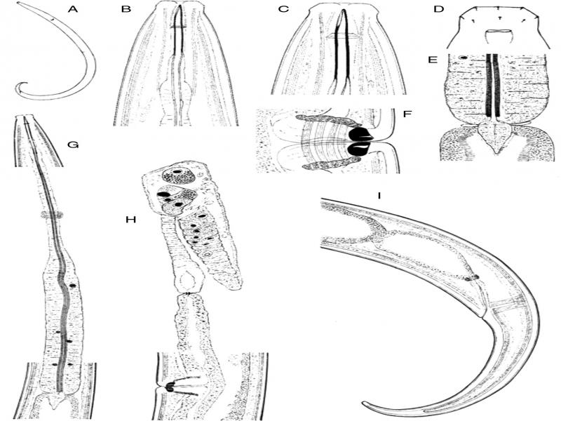
Figure 1:
Epidorylaimus procerus sp. n. (female). A: Entire. B, C: Anterior region in lateral median view. D: Lip region in lateral surface view. E: Pharyngo-intestinal junction. F: Vagina. G: Neck region. H: Anterior genital branch. I: Posterior body region [scale bars: A = 500 µm; B, C = 10 µm; D, F = 5 µm; E = 20 µm; G-I = 50 µm].
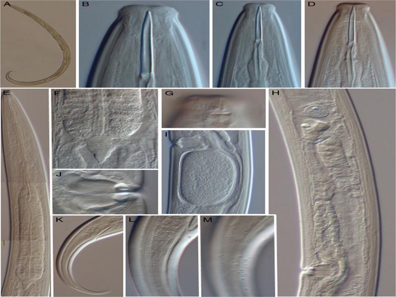
Figure 2:
Epidorylaimus procerus sp. n. (female, LM). A: Entire. B-D: Anterior region in lateral median view. E: Neck region. F: Pharyngo-intestinal junction. G: Lip region in lateral surface view. H: Anterior genital branch. I: Uterine egg. J: Vagina. K: Caudal region. L: Anterior portion of the caudal region in median view. M: Same in submedian view, showing the blister-like elements mainly subventrally and ventrally [scale bars: A = 500 µm; B-D = 10 µm; E, H, K = 50; F, I, L, M = 20 µm; G, J = 5 µm].
Material examined
In total, there were 15 females from one location in excellent state of preservation.
Morphometricssee Table 1.
Table 1.
Morphometrics of Epidorylaimus procerus sp. n. from Vietnam.
| Holotype | Paratypes | |
|---|---|---|
| ♀ | 15♀♀ | |
| Character | ||
| L | 2.37 | 2.28 ± 0.09 (2.16-2.46) |
| a | 30.8 | 31.4 ± 1.2 (28.8-32.9) |
| b | 5.5 | 5.2 ± 0.2 (4.9-5.6) |
| c | 13.0 | 13.3 ± 0.7 (12.1-14.4) |
| V | 41.5 | 41.2 ± 0.8 (39.8-42.6) |
| c′ | 5.4 | 4.9 ± 0.4 (4.4-5.5) |
| Lip-region diameter | 17 | 15.9 ± 0.6 (15-17) |
| Odontostyle length | 32 | 33.4 ± 1.2 (32-35) |
| Odontophore length | 32 | 30.2 ± 1.5 (28-33) |
| Neck length | 432 | 435 ± 18 (415-461) |
| Pharyngeal expansion length | 204 | 212 ± 13 (194-238) |
| Body diam. at neck base | 61 | 65.0 ± 4.0 (61-73) |
| Mid-body | 77 | 72.6 ± 3.4 (68-79) |
| Anus/cloaca | 36 | 35.2 ± 1.3 (32-36) |
| Distance vulva – anterior end | 982 | 938 ± 43 (885-1012) |
| Prerectum length | 91 | 91.8 ± 18.4 (70-123) |
| Rectum length | 42 | 42.5 ± 4.6 (35-49) |
| Tail length | 182 | 171 ± 9.2 (157-186) |