
The ascomycete fungus Hyalorbilia aff. multiguttulata DoUCR50 (basionym: Dactylella oviparasitica DoUCR50) is a hyperparasite of the sugarbeet cyst nematode, Heterodera schachtii. It is the causal agent of a long-term population suppression of this nematode in field 9E at the University of California Riverside Agricultural Operations (Borneman and Becker, 2007). Dactylella oviparasitica was re-named Brachyphoris oviparasitica (Chen et al., 2007) and subsequently assigned to the teleomorphic species Hyalorbilia oviparasitica (Baral et al., 2018). DoUCR50 was most recently re-named as Hyalorbilia aff. multiguttulata (H.-O. Baral, Tübingen, Germany, pers. comm.).
Microscopic studies on the mode of action of DoUCR50 parasitism of H. schachtii raised in Arabidopsis or cabbage root tissue culture revealed that sedentary fourth-stage juveniles exposed to the outside of the root are especially susceptible to parasitism (Smith Becker et al., 2013). In contrast, isolated eggs and eggs containing early juvenile stages were less susceptible. In cabbage root culture, DoUCR50 reduced the number of H. schachtii females with eggs by more than 95% and the number of eggs per female by almost 60% compared with females developing in culture without the fungus. Similarly, parasitism of Heterodera glycines by a sterile nematophagous fungus, ARF-L, was detected at early developmental stages after inoculation of soybean with nematode eggs and juveniles (Timper et al., 1999). Mycelial mats were observed on the surface of sedentary juvenile stages and females. The authors hypothesized that the fungus penetrates the cuticle of the developing nematode directly, similar to the mechanism of infection described for cyst parasitism (Kim et al., 1992). These observations strongly suggest that parasitism of developing late juvenile stages is an important mode of action in the population suppression of cyst nematodes by these fungi.
ARF-L isolate TN-14 (hereafter referred to as ARF) has now also been assigned to the genus Hyalorbilia (H.-O. Baral, Tübingen, Germany, pers. comm.). ARF was isolated from 9 out of 95 populations of H. glycines in Arkansas in the late 1980s (Kim and Riggs, 1991). Field soil naturally infested with ARF suppressed the number of cysts developing on soybean seedlings by more than 50% compared with seedlings grown in autoclaved soil. In tests with females of Heterodera species or egg masses of Meloidogyne incognita placed on cultures of the fungus, ARF parasitized eggs of M. incognita, Cactodera betulae, H. graminophila, H. lespedezae, H. leuceilyma, H. trifolii, and H. schachtii. An electron microscopic study of the ARF infection process on H. glycines females revealed the presence of holes in the cuticle at infection sites, suggesting the involvement of enzymatic degradation. This was the first report of direct penetration by a fungal parasite on a cyst nematode host (Kim et al., 1992). Despite culture on 28 different media and attempts to combine different mating types, no sporulation of ARF strains was observed.
In a subsequent study, 17 isolates of ARF-like fungi collected from H. glycines were grouped into two morphological phenotypes, ARF18-C and ARF18-L (Kim et al., 1998). The two phenotypes differed in the appearance of sclerotium-like structures (SLS) produced on CMA. ARF18-C isolates produced compact SLS while ARF18-L produced loosely formed SLS. ARF18-C and ARF18-L also differed in their pathogenicity on soybean cyst nematodes at different stages of development, depending on the type of assay used. In pathogenicity tests using CMA-grown fungi placed on water agar, ARF18-L parasitized approximately half as many eggs in cysts as ARF18-C. In contrast, ARF18-L was more effective at suppressing H. glycines nematode numbers and infected more eggs, juveniles, and young females than ARF18-C in assays conducted in the soybean rhizosphere (Timper and Riggs, 1998; Timper et al., 1999). These results highlight the importance of rhizosphere assays for the selection of potential biocontrol strains.
A fungus closely related to DoUCR50 and ARF was suggested to be the causal agent of M. incognita suppression in orchards of Lovell peach growing in the San Joaquin Valley of California in 1976. According to the authors, it was the first report of a fungal parasite of root-knot nematode eggs (Ferris et al., 1976; Stirling and Mankau, 1978a). Sporulation in culture was achieved after extensive experimentation with a variety of complex media. Culture on Yeast Peptone Soluble Starch agar enriched with egg yolk and incubation under fluorescent light resulted in good sporulation (Stirling and Mankau, 1978b). The authors identified the novel fungus as Dactylella based on conidia morphology, and provided the name Dactylella oviparasitica, hereafter referred to as StM. Several distinct strains of similar fungi were obtained which exhibited differences in virulence on M. incognita, growth rate, and sporulation. StM preferentially parasitized eggs in clumps or clusters upon removal from the gelatinous matrix of the M. incognita female and was less effective at parasitizing single eggs. StM parasitism was not unique to M. incognita. The fungus parasitized all Meloidogyne species tested, as well as eggs of Diplenteron sp., Acrobeloides sp., Tylenchulus semipenetrans, and H. schachtii. StM was also observed to invade white females and cysts of H. schachtii, but eggs of H. schachtii in older cysts containing J2 were less susceptible to parasitism. Microscopic observation of StM during parasitic interaction with M. incognita revealed the presence of appressoria formation on eggs, suggesting a direct penetration mechanism. The ability of the fungus to produce cleared zones on medium containing colloidal chitin raised the possibility that direct penetration of chitin containing eggs was facilitated by chitinase (Stirling and Mankau, 1979). Neither StM, ARF nor our strain DoUCR50 produced trapping structures on dilute Corn Meal Agar (CMA) or water agar, unlike the majority of previously described nematophagous Dactylella species (Pramer, 1964).
A phylogenetic analysis of DoUCR50, StM, and ARF revealed that they are members of a clade of similar nematophagous fungi (Yang et al., 2012). This rRNA ITS analysis showed that these three fungi belong to an assemblage of organisms, originally called the DO Clade, with global distribution. In addition to confirming the phylogenetic placement of ARF within the DO Clade, Sharma et al. (2017) created a draft genome assembly of the fungus. Gene prediction analysis was performed, and a total of 14,461 protein-encoding genes were identified. Among the genes identified through BLAST analyses were hydrolytic enzymes, such as serine proteases and chitinases, which have been previously associated with virulence of nematophagous fungi (Yang et al., 2007).
The objective of this study was to compare the parasitism of three genetically similar species from the genus Hyalorbilia on H. schachtii females and eggs. Previous studies on cyst parasitism have shown that susceptibility to fungal parasitism decreases as females and eggs mature. Our goal was to quantify this effect by testing the susceptibility of females and eggs of uniform age. The role of extracellular hydrolytic enzymes in parasitism was also investigated by testing for the activity of enzymes both in fungi grown on PDA and H. schachtii females.
Materials and methods
Fungal strains
Three Hyalorbilia strains were the focus of the investigations. Strain DoUCR50 originated from the H. schachtii-suppressive field 9E at Agricultural Operations, University of California, Riverside, CA (Olatinwo et al., 2006). Strain ARF was initially isolated from parasitized eggs of the soybean cyst nematode, H. glycines. It was one of several strains collected in the mid-south region of the United States from 1987 to 1994 (Timper and Riggs, 1998) and was provided by Patricia Timper, USDA-ARS, Tifton, GA. The third strain was first isolated from parasitized root-knot nematode eggs in a Meloidogyne incognita-suppressive peach orchard (Stirling and Mankau, 1978a). It was purchased as ATCC 38908 from the American Type Culture Collection (Manassas, VA) and named here StM. During this project, all strains were cultured on PDA or water agar in the dark at 23°C and subcultured monthly. Water agar cultures of the fungi were grown by transferring a 1 cm square block of agar from the growing edge of a PDA culture of the fungus to the center of a 1.5% water agar plate. To help ensure the preservation of parasitism, three-week-old females of H. schachtii raised in aseptic root culture were placed on fungal cultures grown on water agar every 6 months. After 10 days, parasitized females were placed individually at the center of PDA plates and were incubated in the dark at 23°C for 30 days prior to subculture.
Effect of temperature and nutrients on fungal growth
One cm square blocks of agar taken from the growing edge of one-month-old PDA-grown cultures of each fungus were placed in the center of water agar or PDA plates. Plates were placed in incubators at 18, 23, 28, or 33°C. After 10 days, the growth of each culture was measured from the edge of the agar block to the edge of the mycelial growth margin. Three plates of each fungus were used for each temperature tested, and the experiment was repeated twice.
Phylogenetic and pairwise nucleotide sequence analyses
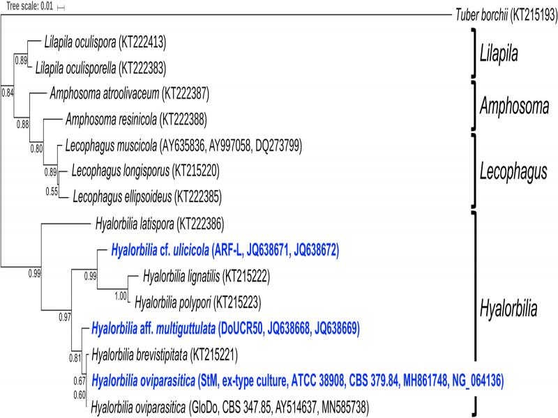
The sequences used were from Hyalorbilia spp. and related groups described in Baral et al. (2018) and from prior studies examining nematophagous fungi (Stirling and Mankau, 1978a; Timper and Riggs, 1998; Yin et al., 2003; Olatinwo et al., 2006). The analyses used the 5.8S rRNA gene and partial 28S rRNA gene sequences. The phylogenetic tree was constructed by performing the following analyses using GenomeNet (https://www.genome.jp/). Sequences were aligned using the CLUSTAL W algorithm (Thompson et al., 1994). The final alignment had a length of 660 nucleotides, and the sequences ranged from 648 to 654 nucleotides. Alignment and phylogenetic reconstructions were performed using the function ‘build’ of ETE3 v3.0.0b32 (Huerta-Cepas et al., 2016) as implemented on the GenomeNet (https://www.genome.jp/tools/ete/). Maximum likelihood trees were inferred using RAxML v8.1.20 that was run with model GTRGAMMA and the default parameters (Stamatakis, 2014). Branch supports were computed out of 100 bootstrapped trees. The tree figure was created using the Interactive Tree of Life (Letunic and Bork, 2007) (https://itol.embl.de/). Pairwise analyses using EMBOSS Water (Smith and Waterman, 1981; Madeira et al., 2019) were performed to determine the % nucleotide sequence identity of rRNA genes and the rRNA ITS region among the three strains. The accession numbers of the sequences used in this analysis are shown in the phylogenetic tree.
Surface sterilization of Heterodera schachtii females
Three-week-old cabbage seedlings grown in 200 cm3 sandy loam in cone-tainers (Stuewe & Sons Inc., Tangent, OR) were inoculated with 1,000 freshly hatched J2 of H. schachtii per cone and maintained in a greenhouse at 25 ± 2°C with ambient light. At 3, 4, or 5 weeks post-inoculation, roots from two cones were vigorously washed in a 24.5 cm diameter × 5 cm deep stainless-steel pan to dislodge females. The supernatant slurry was poured through a 250 µM mesh sieve (No. 60) to collect females. Females of uniform size were removed from debris using tweezers and placed into 30 ml sterile distilled water overlaid onto a water agar plate containing 100 µg/ml rifampicin and 100 µg/ml ampicillin. Females were stored overnight at 4°C on the antibiotic plates. Forceps were used to individually transfer females to a 70 µM mesh cell strainer (BD Falcon, Becton Dickinson, Franklin Lakes, NJ) placed in 30 ml sterile distilled water in a petri dish. Approximately 200 white females were collected for each experiment. The cell strainer was transferred aseptically to a 100 mm × 15 mm petri plate containing 30 ml of 0.4% sodium hypochlorite. Females were incubated in the dilute bleach solution for 2 min with gentle agitation then transferred in the mesh strainer to a second petri plate containing 30 ml sterile distilled water to rinse. The strainer containing surface-sterilized females was then placed on a water agar plate, and the mesh was cut from the base of the strainer using a flame-sterilized scalpel to facilitate the transfer of the females.
Effect of female age on J2 hatch after fungal parasitism
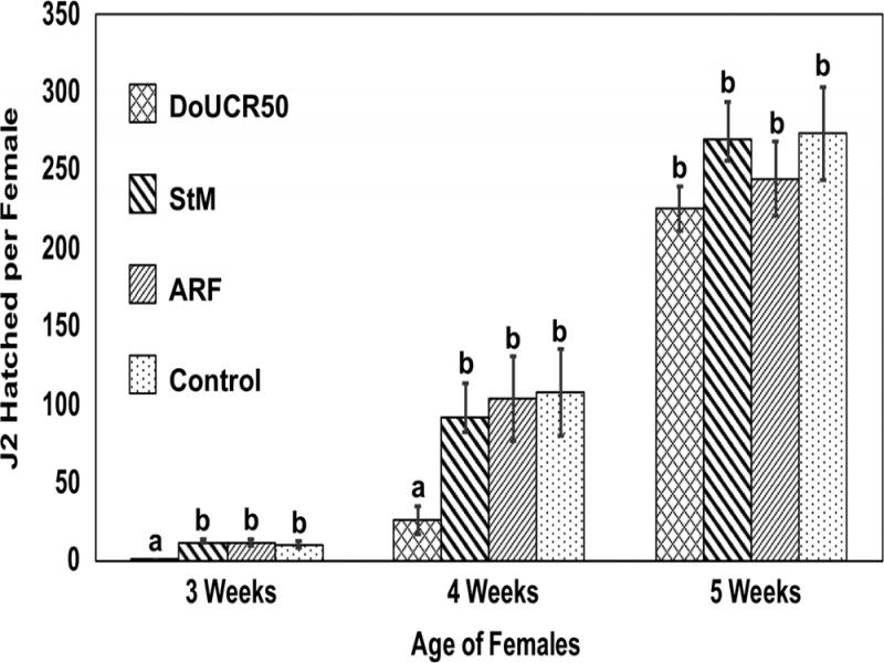
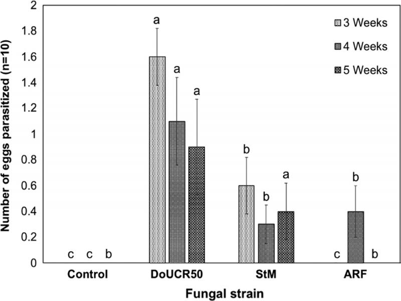
Fungal parasitism was evaluated for H. schachtii females collected 3, 4, and 5 weeks or approximately 357, 476, and 595 degree-days (DD, 8°C basal temperature (Curi and Zmoray, 1966)) respectively, post-inoculation by measuring J2 hatch inhibition. Surface-sterilized females were distributed to plates containing one-month-old water agar cultures of DoUCR50, ARF, StM, or no fungus as the control. Approximately 30 to 40 females were arranged evenly spaced around the center of the plate, 0.5 to 1 cm from the edge of the fungal block, and plates were incubated in the dark at 23°C. Healthy surface-sterilized females were placed on water agar plates as controls. After 3 days, 25 parasitized or control females from each plate were transferred to the center of water agar plates, 5 females per plate, and 5 plates per fungal strain or control. Plates were incubated in the dark at 23°C for 6 days, after which females were removed from plates and used for enzyme assays. Hatched J2 were rinsed from plates with 3 × 5 ml water and counted. The experiment was repeated twice.
Effect of female age on parasitism of isolated H. schachtii eggs
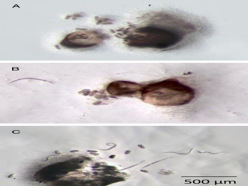
Approximately 100 surface-sterilized females collected 3, 4, or 5 weeks post-inoculation were placed into a sterile 70 µM mesh cell strainer (Falcon) on a water agar antibiotic plate (100 µg/ml rifampicin, 100 µg/ml ampicillin) overlaid with 20 ml sterile distilled water. Females were ruptured on the sieve using sharp flame-sterilized tweezers. The sieve was agitated to allow eggs to pass through the mesh onto the agar. Eggs were rinsed five times with sterile distilled water to remove debris. For each rinse, 30 ml sterile water was added to the plate and eggs were gently agitated. Water and debris were removed using a 23 cm sterile glass Pasteur pipette. After the final rinse, eggs were resuspended in 10 ml sterile water and transferred to a 50 ml sterile disposable centrifuge tube. The volume of water in the tube was adjusted to provide 1 egg/µl water. Each of the fungal strains (DoUCR50, ARF, and StM) was grown on water agar at 23°C for 20 days, after which the mycelia covered about half the plate or more. Ten µl of egg suspension was spotted onto five sites on each plate, approximately 2 cm from the center of the plate. Each of these sites served as replicates for the experiment. A fine pointed dental tool was used to move eggs such that each spot contained 10 eggs, and they were arranged such that they were not touching each other. Plates were incubated for 10 days at 23°C in the dark, and the eggs were monitored for parasitism and hatching of J2. The experiment was repeated twice.
Production of hydrolytic enzymes by fungi grown on PDA or H. schachtii females
The API ZYM test kit (bioMérieux, France) provides a semi-quantitative assay for the presence of 19 hydrolytic enzymes: alkaline phosphatase, esterase, esterase lipase, lipase, leucine arylamidase, valine arylamidase, cysteine arylamidase, trypsin, chymotrypsin, acid phosphatase, naphthol-AS-BI-phosphohydrolase, α-galactosidase, b-galactosidase, α-glucosidase, β-glucosidase, N-acetyl-β-glucosaminidase, α-mannosidase, and α-fucosidase. DoUCR50, ARF, and StM strains grown either on PDA or 3-week-old H. schachtii females were tested for extracellular hydrolytic enzyme production as follows. Sixty-five µl of 0.85% NaCl suspension medium was added to each well of the API ZYM test strips. For assay of enzymes produced by PDA-grown fungi, 5 mm cubes of PDA containing mycelia from 1-month-old cultures were placed into the test wells, one cube per well. Cubes of PDA were used as control. Enzyme production by fungi grown for 9 days on 3-week-old H. schachtii females was assayed by placing a single parasitized female or healthy control female from the hatch inhibition assay into each well of the test strips. Test strips were incubated at 37°C in the dark for 4 hr. The reaction was stopped by the addition of one drop of ZYM A reagent followed by one drop of ZYM B. Color development was allowed to proceed for 10 min at 23°C before recording. The activity of each enzyme was recorded on a colorimetric scale provided by the manufacturer ranging from 0 (no activity) to 5 (maximum), and the experiment was repeated three times.
Statistical analysis
Data sets of repeated experiments were analyzed for homogeneity of variance with Levene’s test (Real Statistics Using Excel: © 2012-2019, Charles Zaiontz, http://www.real-statistics.com). As there were no statistical differences between the variances of the repeated experiments (P ≤ 0.05), the homogeneity of variance assumption was met. Treatment effects were analyzed using a t-test or one-way analysis of variance (ANOVA) procedure. If appropriate, means were compared using Fisher’s protected least significant difference (LSD) test (P ≤ 0.05). Standard error bars with the same lowercase letter indicate no significant difference. Enzyme color rating means were analyzed by the nonparametric Kruskal–Wallis test, followed by Dunn’s post hoc test to determine the differences among the groups (P < 0.05).
Results
Effect of temperature and nutrients on fungal growth
All three fungal strains grew fastest at 28°C on both water agar and PDA, and growth was strongly attenuated at 33°C (Fig. 1). The three strains differed in their rate of growth on water agar and PDA. Both DoUCR50 and StM grew faster on water agar than on PDA at 28°C, while the growth of ARF on water agar at 28°C was approximately 50% that of DoUCR50 and StM. On PDA, the rate of growth of ARF at 23°C or 28°C was almost twice that of DoUCR50 or StM.
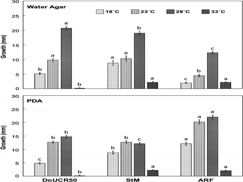
Figure 1:
Effect of temperature and nutrients on rate of growth of Hyalorbilia strains DoUCR50, StM, and ARF in agar culture. Mycelial growth from blocks of PDA cultures placed on PDA or water agar and grown at 18, 23, 28, or 33°C was measured after 10 days. The experiment was repeated twice, and data were pooled. Standard error bars are shown.
Phylogenetic and pairwise nucleotide sequence analyses
A phylogenetic analysis of rRNA genes and the rRNA ITS region showed that all three fungi belong to an assemblage of fungi in the genus Hyalorbilia, which is a member of the class Orbiliomycetes (Fig. 2). This group also includes a closely related fungus (H. oviparasitica, GloDo, CBS 347.85) isolated from Globodera rostochiensis in the Netherlands (Chen et al., 2007), suggesting that it might be nematophagous. This phylogenetic analysis and a pairwise sequence analysis (Table 1) both showed that DoUCR50 and StM are more closely related to each other than they are to ARF.
Table 1.
Percent nucleotide sequence identities of Hyalorbilia strains DoUCR50, StM, and ARF.
| DoUCR50 (%) | StM (%) | ARF (%) | |||
|---|---|---|---|---|---|
| DoUCR50 | 100 | 98.0 | 93.9 | ||
| StM | 75.5 | 100 | 93.4 | ||
| ARF | 72.6 | 69.5 | 100 |
| Treatmenta | Trypsin | Acid phosphatase | α-glucosidase | β-glucosidase | N-acetyl-β-glucosamidase |
|---|---|---|---|---|---|
| C PDA | 0.0 ± 0.00 c | 0.0 ± 0.00 b | 0.0 ± 0.00 c | 0.0 ± 0.00 c | 0.0 ± 0.00 c |
| C Fem | 0.0 ± 0.00 c | 1.3 ± 0.33 ab | 0.7 ± 0.67 bc | 0.3 ± 0.33 bc | 0.0 ± 0.00 c |
| DoUCR50 PDA | 2.3 ± 0.33 b | 3.7 ± 1.33 a | 3.7 ± 0.33 a | 1.7 ± 0.88 abc | 1.3 ± 0.33 b |
| DoUCR50 Fem | 3.7 ± 0.33 a | 3.0 ± 0.00 ab | 2.3 ± 0.67 ab | 3.3 ± 0.33 a | 3.3 ± 0.33 a |
| ARF PDA | 0.0 ± 0.00 c | 3.0 ± 0.00 ab | 3.3 ± 0.33 a | 1.3 ± 0.33 abc | 1.7 ± 0.33 b |
| ARF Fem | 0.0 ± 0.00 c | 3.4 ± 0.33 ab | 2.0 ± 0.58 abc | 3.0 ± 0.00 ab | 3.3 ± 0.33 a |
| StM PDA | 0.0 ± 0.00 c | 3.7 ± 1.33 a | 3.3 ± 0.67 a | 2.3 ± 0.33 ab | 2.0 ± 0.58 ab |
| StM Fem | 0.0 ± 0.00 c | 3.4 ± 0.33 ab | 1.0 ± 0.00 abc | 2.7 ± 0.88 ab | 3.3 ± 0.33 a |