
The barley root-knot nematode, Meloidogyne naasi Franklin, 1965 was originally described from field crops (cereals, grasses, and sugarbeet, Beta vulgaris L.) in England and Wales (Franklin, 1965). According to the EPPO Global Database, this nematode is present in temperate regions in all continents: Africa (Libya); Asia (Iran); Europe (Belgium, Czech Republic, Denmark, France, Germany, Hungary, Ireland, Italy, Malta, the Netherlands, Norway, Poland, Serbia and UK); North America (Canada, USA); Oceania (New Zealand) and South America (Argentina, Chile) (EPPO, 2020). To our knowledge, this is the first record of this nematode in Portugal and in the Iberian Peninsula.
A football field in Porto, Portugal, with poor turf performance caused by various plant health issues, including nematode parasitism, was sampled in June 2019. This field was originally a 60% Lolium perenne L. – 40% Poa pratensis L. mix, but had several patches of encroaching weed, Poa annua, and was scheduled for replacement. Twenty soil cores and their respective grass cover were collected and combined to form composite samples; the loose sandy soil was kept separate from the plug-like thick root masses of turfgrass.
Turfgrass roots were rinsed in water and upon observation with a stereomicroscope, small galls were detected, from which Meloidogyne females were excised (Fig. 1A). The perineal patterns from five adult females were prepared in 45% lactic acid and mounted in glycerin (Hartman and Sasser, 1985). Shape was round to ovoid with a low to medium-high round dorsal arch with coarse irregular striae and no or inconspicuous lateral lines. The most distinct characteristic was the presence of very distinct phasmids with the distance between them (14.1-20.0 µm, mean 18.1 µm) being only slightly smaller than the vulva width (17.9-22.4 µm, mean 20.7 µm) as described by Franklin (1965). A fold covering the anus was observed in three of the five perineal patterns. No striae were observed in the perivulval area (Fig. 1B).
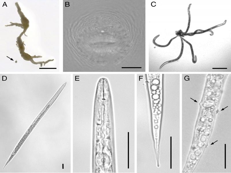
Figure 1:
Root-knot nematodes (Meloidogyne naasi Franklin, 1965) extracted from soil and root samples collected from a football field in Portugal. A: turfgrass root showing galling by root-knot nematodes and excised M. naasi female (arrow); B: perineal pattern; C-G: second-stage juveniles: C: worm-star aggregation; D: whole body; E: pharyngeal region; F: tail; G: Pasteuria penetrans (ex Thorne, 1940) Sayre and Starr, 1985 endospores (arrows) attached to cuticle. Bars = 2000 µm (A); 125 µm (C); 25 µm (B, D-G).
Active soil nematodes were extracted from 100-ml subsamples by means of the tray method (Whitehead and Hemming, 1965). Meloidogyne second-stage juveniles (J2) detected in soil suspensions with an inverted microscope were picked and transferred to distilled water in staining glass blocks. After a few hours, these J2 formed ‘worm-star’ aggregates seemingly caused by attachment of several juveniles by their tails (Fig. 1C). In Caenorhabditis elegans (Maupas, 1900) Dougherty, 1955, this aggregation may be caused by a Leucobacter pathogen, that causes adhesion at the tail spikes within minutes of exposure, followed by bacterial proliferation in the host and death of all trapped nematodes in 24 to 28 hr (Hodgkin et al., 2013). Artificial exposure of C. elegans to other coryneform bacteria of the genus Corynebacterium has resulted in a similar aggregation, leading to nematode lysis and death after 2 to 4 days incubation (Antunes et al., 2016). Although we cannot deny or confirm the cause since it was not an objective of this investigation, we cannot exclude the action of a similar organism. To our knowledge, worm-star formation has not been previously reported on Meloidogyne or on any plant-parasitic nematode, although aggregation in ‘rosettes’ of entomopathogenic nematodes (Pye and Burman, 1981; Stock et al., 2005) and ‘medusa-heads’ or ‘sunflowers’ of human parasites (Yoeli, 1957) have been reported.
Seventeen J2 specimens were heat-killed, mounted in water in temporary slides, and photographed using a Leica DM 5000B + CTR 5000 microscope system (Fig. 1D-G). The J2 were slender and had pointy tails (Fig. 1D). Heads were round and not offset, with a delicate stylet (Fig. 1E). Tails were long, slightly enlarged, and often irregular at the tip (Fig. 1F). Endospores of Pasteuria penetrans (ex Thorne, 1940) Sayre and Starr, 1985, were frequently detected attached to the cuticle of J2 (Fig. 1G). Digital images were measured with ImageJ v. 1.52a (http://imagej.nih.gov/ij) to obtain J2 morphometric characters, presented in Table 1. Second-stage juveniles measured fresh in water were longer than those originally described by Franklin (1965) in lactophenol, but with a similar body length-maximum width proportion (a), and similar stylet size. Tails were longer than originally described, representing a larger part of the body, thus with a smaller c than described.
Table 1.
Morphometric characters of second-stage juveniles in the Portuguese population of Meloidogyne naasi Franklin, 1965.
| M. naasi Portuguese population | M. naasi Original description (Franklin, 1965) | |||||
|---|---|---|---|---|---|---|
| Characters | n | Mean | Range | n | Mean | Range |
| Body length | 17 | 457.3 | 440.6-487.2 | 25 | 435 | 418-465 |
| Maximum body width | 17 | 15.5 | 14.5-16.7 | 25 | 15 | 14-18 |
| Stylet length | 17 | 14.4 | 13.6-15.2 | 25 | 14 | 13-15 |
| Tail length | 13 | 82.8 | 76.4-89.9 | 25 | 70 | 52-78 |
| Hyaline tail terminus | 14 | 26.6 | 23.2-32.0 | 25 | n.d. | n.d. |
| a | 17 | 29.6 | 27.8-32.6 | 25 | 28 | 25-32 |
| c | 13 | 5.5 | 5.2-5.8 | 25 | 6.2 | n.d. |