The genus Deladenus (Thorne, 1941) belongs to the family Neotylenchidae (Thorne, 1941) with D. durus (Cobb, 1922; Thorne, 1941) as type species. It is characterized by the location of the esophageal-intestinal junction immediately behind the nerve ring, a median esophageal chamber sometimes present, and absence of a post-uterine sac. Two biological forms have been observed in this genus: the free-living form (mycetophagous stage) and the insect-parasitic form (infective stage). The mycetophagous stage has been observed in all the members of the genus. Bedding (1968) identified two species (D. wilsoni and D. siricidicola) with dimorphic females and proved a two-part life cycle, the free-living mycetophagous and the insect-parasitic phases. Later he identified other five entomophagous-mycetophagous species (Bedding, 1974). Blinova and Korenchenko (1986) proposed the genus Beddingia and family Phaenopsitylenchidae for the species with a known infective stage. Chitambar (1991) reviewed the mycetophagous stage of this genus, synonymized the genus Beddingia with Deladenus, and provided a diagrammatic classification in mycetophagous females of this genus based on the position of the excretory and hemizonid relative to each other (either posterior or anterior). According to this classification, all species except D. norimbergensis (Rühm, 1956), D. aenea (Rao and Reddy, 1982; Ebsary, 1991) and D. ulani (Sultanalieva, 1983) without information about the position of excretory pore relative to the hemizonid can be grouped into two categories: those with the hemizonid posterior to the excretory pore and species with it in anterior position. Currently, the genus Deladenus has 30 valid species. Three species have been reported from Iran: D. durus (Jahanshahi Afshar et al., 2014), D. persicus (Miraeiz et al., 2017), and D. apopkaetus (Chitambar, 1991) (Miraeiz et al., 2017).
During our surveys on plant-parasitic and free-living nematodes in Northern Iran, a new species of Deladenus was recovered from a wood sample of a dead pine tree (Pinus niger L.) in Siahkal region, Guilan Province, Northern Iran. The observations revealed that this population appeared to be morphologically and morphometrically distinct from any existing Deladenus species and it is herein described as D. gilanica n. sp.
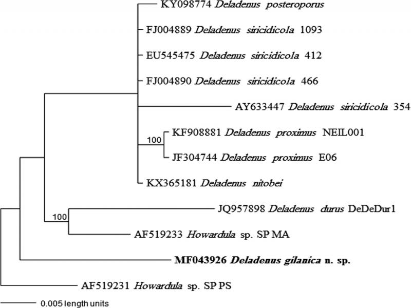
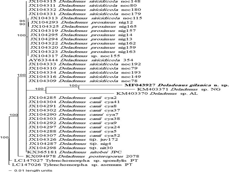
The objectives of the present study were: (i) to provide an accurate description of the new species by an integrative approach to morphological and molecular characterization using the partial 18S and 28S D2 to D3 rRNA gene sequences, and (ii) to investigate the phylogenetic relationships of this neotylenchid nematode within the superfamily Sphaerularioidea.
Materials and methods
Sampling, extraction, mounting, and drawing
Soil, root, and wood samples were randomly collected from different regions of eastern forests in Guilan Province, Northern Iran during 2015. Nematodes were recovered directly from the wood samples by the Whitehead tray method (Whitehead and Hemming, 1965). The extracted nematodes were observed and handpicked under a stereomicroscope. Adult specimens for microscopic observation were killed with gentle heat and fixed in a solution of FGA 4:1:1 (formaldehyde, glycerin, and acetic acid) and then processed to anhydrous glycerin (De Grisse, 1969). Permanent slides were made and examined with a Nikon E200 light microscope. Morphometric data were obtained with the aid of a drawing tube attached to an Olympus BH2 light microscope. Photomicrographs were taken with a digital camera attached to an Olympus BH2 light microscope. Line drawings were made and photographs were taken with a digital camera attached to an Olympus BH2 microscope.
DNA extraction, PCR, and sequencing
Single nematode specimens were handpicked and with light microscopy and then individually transferred to 10 μl distilled water on a glass microscope slide, crushed with a pipette tip and collected in 50 μl AE buffer (10 mM Tris-Cl, 0.5 mM EDTA; pH 9.0; Qiagen, Valencia, CA, USA) by pipette. DNA extracts were stored at −20°C until used as template for PCR amplification. The D2/D3 expansion segment of 28S rRNA gene was amplified using the forward D2A (5´–ACAAGTACCGTGAGGGAAAGTTG–3´) and reverse D3B (5´–TCGGAAGGAACCAGCTACTA–3´) primers (Nunn, 1992) and the partial 18S was amplified using primers 1096F (5´–GGTAATTCTGGAGCTAATAC-3´) and 1912R (5´–TTTACG GTCAGAACTAGGG–3´) (Holterman et al., 2006).
PCR cycle conditions for all rDNA regions were as follows: one cycle of 94°C for 2 min, followed by 35 cycles of 94°C for 30 s, annealing temperature of 55°C for 45 s, 72°C for 3 min, and finally one cycle of 72°C for 10 min. PCR products were purified after amplification using ExoSAP-IT (Affmetrix, USB Products), quantified using a Nanodrop spectrophotometer (Nanodrop Technologies) and used for direct sequencing in both directions using the primers referred to above.
Phylogenetic analyses
The sequences were deposited into the GenBank database. DNA sequences were edited with ChromasPro1.5 2003-2009 (Technelysium Pty Ltd, Helensvale, Australia) and aligned using ClustalW (http://workbench.sdsc.edu; Bioinformatics and Computational Biology Group, Department of Bioengineering, UC San Diego, CA). All available species of Deladenus and some other Hexatylina species from GenBank were also selected for phylogenetic analysis. Outgroup taxa for each data set was chosen according to previous published data (Esmaeili et al., 2016). The model of base substitution in the sequences data were evaluated using MODELTEST version 3.06 (Posada and Criandall, 1998) based on the Akaike-supported model (Arnold, 2010). Bayesian analysis was performed to confirm the tree topology for each gene separately using MrBayes 3.1.0 (Huelsenbeck and Ronquist, 2001) running the chain for 1,000,000 generations and setting the ‘burnin’ at 1,000. Markov Chain Monte Carlo methods were used within a Bayesian framework to estimate the posterior probabilities (pp) of the phylogenetic trees (Larget and Simon, 1999) using the 50% majority rule. The λ 2 test for homogeneity of base frequencies and phylogenetic trees was performed using PAUP* version 4.0 (Sinauer Associates, Inc., Publishers, Sunderland, MA).
Results
Deladenus gilanica n. sp.
Figs. 1 and 2 show line drawings of D. gilanica n. sp.
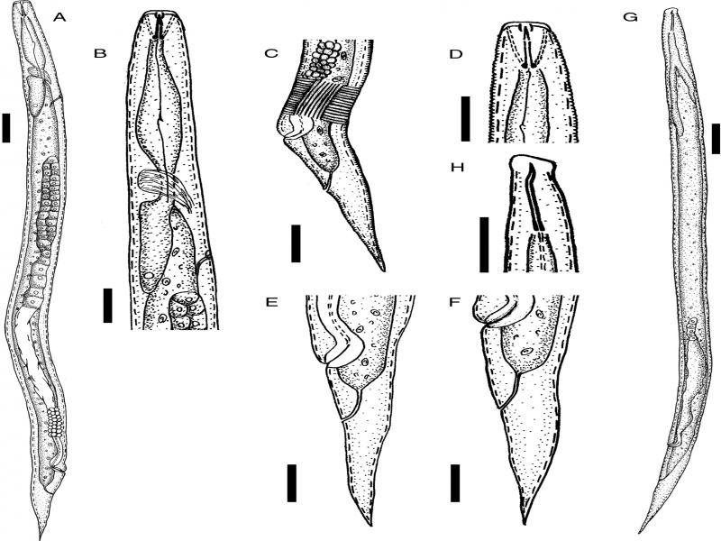
Figure 1:
Line drawings of Deladenus gilanica n. sp. (A-F) Mycophagous female − (A) entire body; (B) pharyngeal region; (C) vulva region; (D) anterior end; (E, F) posterior body (tail). (G and H) Infective female − (G) entire body; (H) anterior end (Scale bars: A, G = 20 µm, B-F, H = 10 µm).
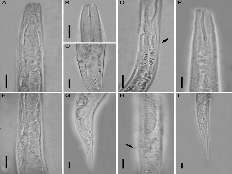
Figure 2:
Photomicrographs of Deladenus gilanica n. sp. (A-D, F and G) Mycophagous female − (A) pharyngeal region; (B) anterior end; (C) vulva region; (D) pharyngeal region showing excretory pore and hemizonid; (F) end of female pharyngeal region; (G) female posterior body (tail). (E, H, and I) Infective female − (E) anterior end; (H) vulva region; (I) posterior body (tail) (All scale bars = 10 µm).
Measurements
Specific measurements are provided in Table 1.
Table 1.
Morphometrics of female of Deladenus gilanica n. sp. from Iran.
| Mycetophagous stage | Infective stage | |||||||||||
|---|---|---|---|---|---|---|---|---|---|---|---|---|
| Characters | Holotype female | Paratype females | Paratype females | |||||||||
| n | − | 10 | 4 | |||||||||
| L | 367 | 364.8 ± 33.7 (314-422) | 330.3 ± 17.6 (310-341) | |||||||||
| L' | 361 | 335.4 ± 33.3 (285-393) | 294.7 ± 15.3 (277-304) | |||||||||
| a | 18.6 | 21.1 ± 2.6 (18.1-26.5) | 28.3 ± 0.1 (28.2-28.4) | |||||||||
| b | 8.5 | 7.1 ± 0.9 (5.9-8.5) | 6.2 ± 1.8 (5.1-8.3) | |||||||||
| b' | 5.7 | 5.0 ± 0.9 (4.1-6.9) | 3.7 ± 0.4 (3.4-4.1) | |||||||||
| c | 13.4 | 12.4 ± 1.1 (10.8-14.6) | 9.3 ± 0.3 (9.0-9.4) | |||||||||
| c' | 2.4 | 3.3 ± 0.4 (2.4-3.8) | 5.1 ± 0.4 (4.7-5.4) | |||||||||
| V | 87.4 | 86.6 ± 0.7 (85.4-87.4) | 82.6 ± 0.6 (82.1-83.2) | |||||||||
| Head height | 1 | 1.4 ± 0.2 (1-1.5) | 2.1 ± 0.3 (2-2.5) | |||||||||
| Head width | 6 | 6.5 ± 0.5 (6-7) | 4.8 ± 0.3 (4.5-5) | |||||||||
| Stylet | 8 | 8.0 ± 0.2 (7.5-8.0) | 13.9 ± 0.9 (13-15) | |||||||||
| Excretory pore | 71 | 68.4 ± 5.2 (61-76) | 69 ± 6.5 (63-76) | |||||||||
| Hemizonid | 71 | 68.4 ± 5.2 (61-76) | 68.5 ± 6.6 (63-76) | |||||||||
| Pharynx | 46 | 51.4 ± 4.9 (46-63) | 54 ± 10.6 (41-64) | |||||||||
| Overlapping | 22 | 23.1 ± 7.5 (11-33) | 32.3 ± 5.9 (29-41) | |||||||||
| Body width | 21 | 17.5 ± 2.8 (14-22) | 11.8 ± 0.5 (11-12) | |||||||||
| Head-vulva | 341 | 316.0 ± 30.8 (268-367) | 272.7 ± 12.7 (258-281) | |||||||||
| Vulva-anus | 20 | 19.4 ± 3.4 (15-26) | 22 ± 3.0 (19-25) | |||||||||
| Tail | 29 | 29.4 ± 1.3 (27-32) | 35.7 ± 2.5 (33-38) | |||||||||
| Species | L | a | b | c | V | Stylet | Median corporeal chamber | Excretory pore relative to hemizonida | Tail | Lib | Male | Infective stage |
|---|---|---|---|---|---|---|---|---|---|---|---|---|
| D. gilanica n. sp. | 314-422 | 18.1-26.5 | 5.9-8.5 | 10.8-14.6 | 85.4-87.4 | 7.5-8 | – | SL | 27-32 | 8 at level of vulva | – | + |
| D. durus | 620-13,60 | 21-50 | 7.7-16 | 19.5-39.3 | 90-95 | 6-11 | + | P | 21-43 | 6-7 | + | – |
| D. aridus | 693 | 31.2 | 19.8 | 91.9 | 9.4 | – | P | 5 | – | – | ||
| D. obesus | 970-1,400 | 18-26 | 8-16.9 | 32-33.6 | 92-93 | 7.3-9 | – | P | 30-47 | 8-10 | – | – |
| D. processus | 760-990 | 34-49 | 13.3-17.7 | 19.6-22.8 | 92.2-93.5 | 6-7 | – | P | 35-44 | 6 | + | – |
| D. oryzae | 660-960 | 19-26 | 7.9 | 29 | 92-93.5 | 7-9 | + | A | 29-34 | Indistinct | + | + |
| D. wilsoni | 1,490-2,700 | 34.6-62.6 | 15.5-26.5 | 46.6-66.9 | 94.6-96.2 | 10-11 | – | A or SL | 7-9 at level of vulva | + | + | |
| D. proximus | 1,760-2,200 | 40-53.7 | 16.3-21.3 | 44-60 | 95.1-95.9 | 11-12 | – | A | 31-47 | 7-8 at level of vulva | + | + |
| D. posteroporus | 889-1,026 | 38-43 | 10.1-12.8 | 31.3-37.1 | 93.5-95.9 | 8-11 | – | A | 27-29 | 11-12 at midbody | + | + |