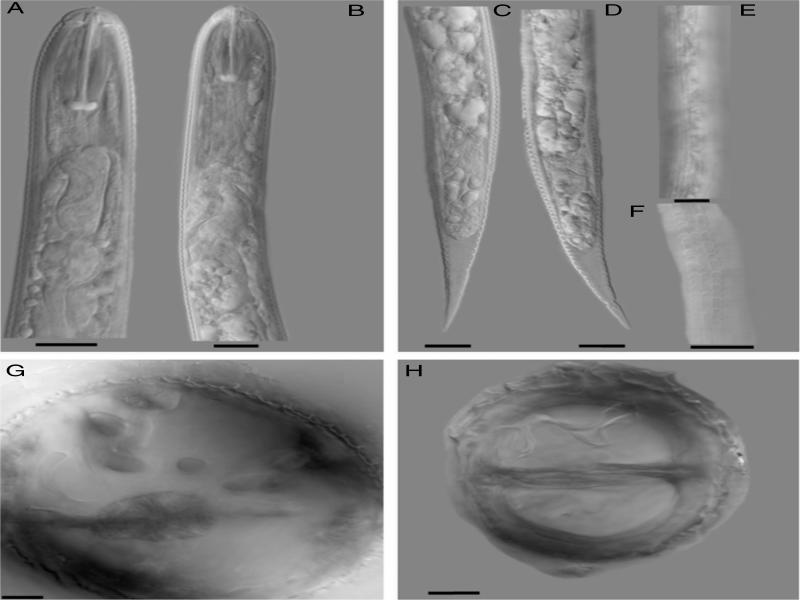
Figure 1:
Photomicrographs of second-stage juveniles (A-F) and vulva cones (G and H) of Heterodera medicaginis. A-B heads; C-D tails; E-F lateral field; G-H cone mounts, G showing the bullae and H showing the underbridge. The scale bar=10 µm.

© 2020 Zafar A. Handoo, Andrea M. Skantar, Saad L. Hafez, Mihail R. Kantor, Maria N. Hult, Stephen A. Rogers, published by Society of Nematologists, Inc.
This work is licensed under the Creative Commons Attribution 4.0 License.