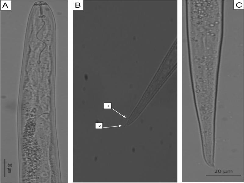
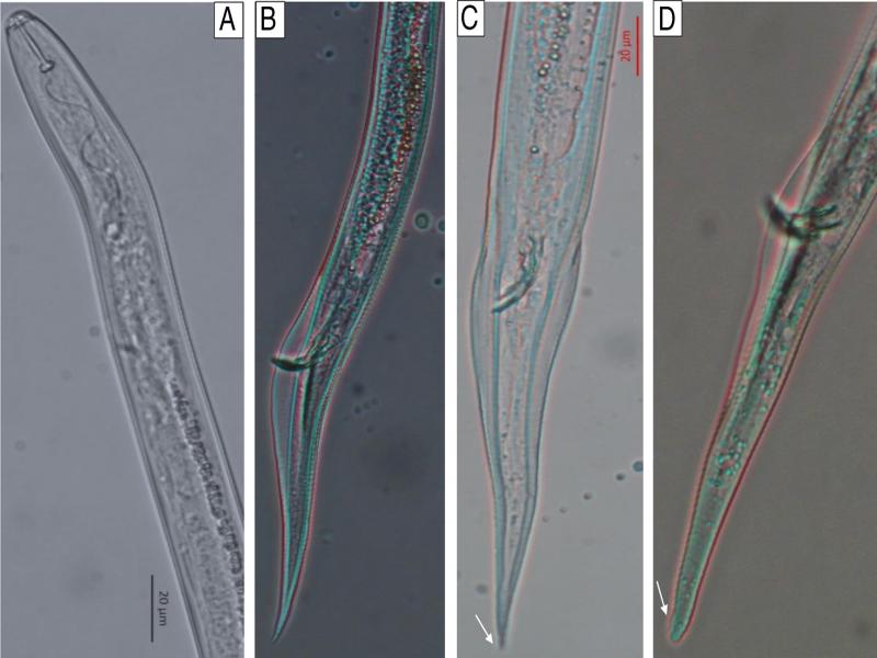
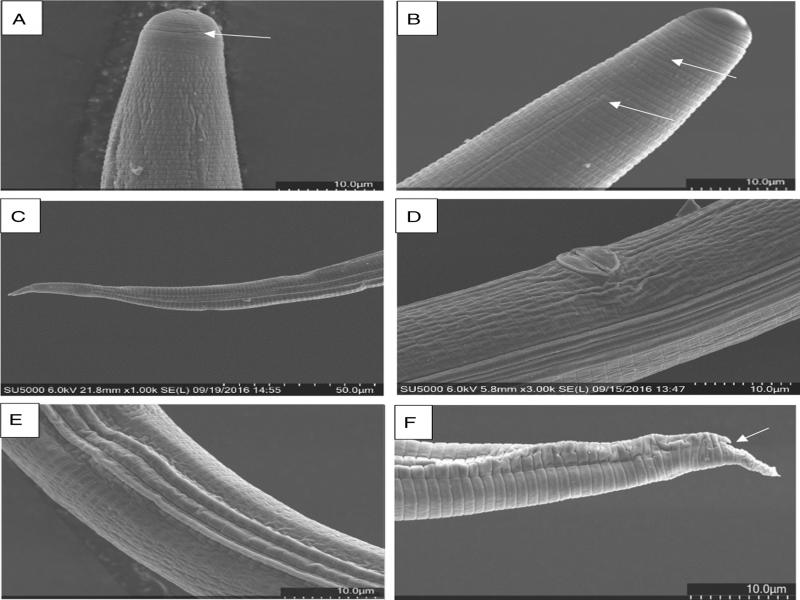
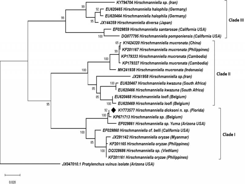
The University of Florida Nematode Assay Laboratory (NAL) received a rhizosphere soil sample for a routine diagnosis purpose from Beef Farm Prairie, Osceola County in Florida. The sample was taken from a site dedicated to limpograss (Hemarthria altissima). Limpograss is a warm-season perennial grass used by Florida beef cattle producers because of its high digestibility, adaptation to seasonally flooded soils, cool-season growth, and tolerance of light frost (Newman, 2014). This grass is well adapted to flatwoods sites and to similar areas characterized by a flat to gently sloping topography and poorly drained, sandy soils, where standing water is common during wet weather (Newman et al., 2014).
The owner of this property had recognized a patchy appearance of the field and yield reduction on the limpograss, and sent soil samples for plant-parasitic nematode diagnosis to the NAL in November 2015. We found several individuals belonging to the genus Hirschmanniella Luc and Goodey (1964). The genus Hirschmanniella contains 29 valid species as revised by Khun et al. (2015). With the exception of H. gracilis (Luc and Goodey, 1964; de Man, 1880), the genus Hirschmanniella is considered a harmful pest and is a quarantine taxon for the European Union (Karssen, 2009). These species occur in diverse habitats and hosts in all continents except for Antarctica, and they appear to be well adapted to a broad range of moisture conditions, with a preference for periodically or permanently saturated soils and for graminaceous root systems (Tandingan De Ley et al., 2007).
Hirschmanniella parasitizes diverse plant groups ranging from paddy rice (Maung et al., 2010; Pascual et al., 2014; Khun et al., 2015), crops such as taro (Bridge et al., 1983), cabbage (Duan et al., 1996), maize, tomato, and sugarcane (Bridge et al., 1990) to a number of aquatic plants (Sher, 1968; Gerber and Smart, 1987; Tandingan De Ley et al., 2007). Several species of Hirschmanniella are a serious threat to lowland rice fields in Asia (Bridge et al., 1990; Prot et al., 1994; Maung et al., 2010; Win et al., 2013).
Only a handful of species in this genus have been reported in the USA, mainly from Florida and California. In Florida, Sher (1968) reported H. marina from a marine plant (Diplanthera wrightii), H. microtyla from eelgrass (Vallisneria americana), and H. mexicana (Chitwood, 1951, Sher, 1968) from aquatic rootless plants (Ceratophyllum demersum). Hirschmanniella caudacrena (Sher, 1968) was also recovered from a tissue of a submerged plant Ceratophyllum demersum by Gerber and Smart (1987) and the rice root nematode, H. oryzae (Van Breda de Haan, 1902; Luc and Goodey, 1964), was also reported in Florida (www.cabi.org/isc/datasheet/27867#20127201272).
In California, Golden (1957) reported H. gracilis from Greene’s bur-reed (Sparganium greenei), prairie bulrush (Scirpus paludosus), and three-square bulrush (S. americanus). Hirschmanniella spinicaudata (Schuurmans Stekhoven, 1944; Luc and Goodey, 1964) from cattail (Typha sp.) and H. belli (Sher, 1968) from both cattail and spike rush (Eleiocharis spp.) were reported by Sher (1968). Hirschmanniella pomponiensis (Abdel-Rahman and Maggenti, 1987) was recorded from saltmarsh bulrush (Scirpus robustus) (Abdel-Rahman and Maggenti, 1987). Recently, Tandingan De Ley et al. (2007) reported H. santarosae (Tandingan De Ley et al., 2007) from cattail in vernal pools in the Santa Rosa Plateau Ecological Reserve, California.
There were very limited reports of Hirschmanniella species from other states in the USA. H. caudacrena and H. oryzae were isolated from rice fields in Louisiana (Atkins et al., 1955; Sher, 1968). In Hawaii, H. diversa (Sher, 1968) was reported from lotus (Nelumbium nelumbo). In Texas, H. mexicana and H. oryzae were recovered from an aquatic rootless plant and a rice field, respectively (Chitwood, 1951; Atkins et al., 1955). In Arkansas, H. oryzae and H. spinicaudata were isolated from pasture and cattails, respectively (Wehunt et al., 1989). Although it was not identified at the species level, one Hirschmanniella sp. was isolated from cattail (Typha spp.) along the Colorado River in Yuma, Arizona (Tandingan De Ley et al., 2007).
In this study, we report the results of our morphological, morphometric, and molecular characterization of the new species Hirchmanniella dicksoni n. sp. from the rhizosphere soil of limpograss (Hemarthria altissima) from Florida.
Materials and methods
Nematode extraction
Soil samples were collected from Beef Farm Prairie, Osceola County in Florida, with latitude 28.087400 and longitude 80.897847, and submitted to the NAL for nematode diagnosis in December 2015. Nematodes were extracted from 100 cm3 of soil using centrifugation and sugar floatation techniques (Harrison and Green, 1976).
Morphological and morphometric characterization
In total, 20 females and 20 males were used for morphological and morphometric characterization. Nematodes were processed for permanent mounting according to Ryss (2003). Nematodes were picked and transferred to a PCR tube and killed with hot (95°C) 4% formalin. The PCR tube with heat-killed nematodes was immediately placed in a thermo cycler: 95°C for 2 min, 65°C for 10 min, 75°C for 10 min, 85°C for 10 min, and 95°C for 10 min. After the tube reached room temperature, the tube was rinsed with distilled water and the content was transferred to a staining dish. Nematodes were picked out and transferred to a glass slide with cavity filled with a mixture of glycerol and distilled water in proportion of 1:20. Nematodes were infiltrated in glycerin for permanent slide preparation by placing the cavity slide with the nematodes on a hot plate at 70°C for 15 to 20 min and mounted on glass slides following the method described by De Grisse (1969). The specimens were examined for morphological features and morphometrics using a camera installed on ZEISS AXIO microscope (Carl Zeiss MicroImaging GmbH, Gottingen, Germany). Measurements and pictures were taken using the ZEN lite software on ZEISS Axiocam ERc5s digital camera. Morphological and morphometric characteristics were compared with identification keys for genus Hirschmanniella revised by Khun et al. (2015).
Scanning electron microscopy
Adults and juveniles of the new species were collected individually by hand and washed with sodium phosphate buffered in 3% glutaraldehyde solution for 24 hr at 4°C. The specimens were then post fixed overnight in 3% Osmium tetraoxide solution and dehydrated in a concentration gradient of ethanol. Dehydrated nematodes were dried with a critical point dryer, attached to a self-adhesive stub, and coated with gold/palladium auto sputter coater. Specimens were studied with a Hitachi SU5000 electron microscope at the Interdisciplinary Center for Biotechnology Research (ICBR), University of Florida, Gainesville, FL.
Molecular characterization
Total genomic DNA was extracted following the sodium hydroxide method (Wang et al., 1993). A single female was transferred to 20 µl of 0.25 NaOH in 1.5 ml eppendorf tubes, crushed in a sterile glass rod, and incubated overnight at 25°C. The tubes were then heated at 99°C for 3 min and cooled down to room temperature. In total, 5 µl of 1 M HCl, 10 µl of 0.5 M Tris-HCl (pH 8.0), and 5 µl of 2% Triton X-100 were added to the lysate and mixed briefly, and heated at 99°C for 3 min. The digest was cooled to room temperature and 1.5 µl of the digest was immediately used for polymerease chain reaction (PCR). DNA amplification was performed in 25 µl of reaction mix containing 12.5 µl 2× Hot Start Master mix (New England BioLabs, Ipswich, MA), 1.25 µl forward and reverse primers, 8.5 µl sterile nuclease free water (Thermo Fisher Scientific, Gainesville, FL), and 1.5 µl template DNA. For amplification of the ribosomal region spanning the internal transcribed spacer (ITS)1, 5.8 S gene, and ITS2, the primers TW81R (5′-GTTTCCGTAGGTGAACCTGC-3′) and AB28F (5′-ATATGCTTAAGTTCAGCGGGT-3′) were used. D2–D3 expansion segment of the large subunit (LSU) rDNA was amplified using the primers sets D2A (5′-CAAGTACCGTGAGGGAAAGTTG-3′) and D3B (5′-TCGGAAGGAACCAGCTACTA-3′). The thermocycling was carried out using a Master cycler nexus (Eppendorf AG, Hamburg, Germany). The thermocycling reactions for both DNA regions were as follows: 95°C for 15 min, followed by 40 cycles of 94°C for 30 sec, 50°C for 45 sec, and 72°C for 2 min, and a final extension step of 72°C for 7 min.
DNA fragments were separated by electrophoresis in 1XTAE buffer and 1.2% agarose gel, stained with ethidium bromide, and visualized under UV light using the ChemiDOC XRS One 4.5.2 program (Bio-Rad Laboratories, Life Science Group, Hercules, CA). The DNA fragments were purified using QIAquick PCR Purification kit (Qiagen, Valencia, CA), according to the manufacturer’s instructions. The purified DNA was quantified using a NanoDrop 1000 (Thermo Science, Grand Island, NY) spectrophotometer and sequenced at the ICBR, University of Florida, Gainesville, FL. To identify the species, raw sequences were checked and edited manually using BioEDIT, v. 7.2.5 (Hall, 1999). Consensus sequences obtained were compared with those deposited in the GenBank database using BLAST engine search for sequence homology. The newly obtained consensus sequences were submitted to the GenBank database under accession numbers KY773577 for D2–D3 of 28S gene and KY773578 for ITS rRNA.
Phylogenetic analysis
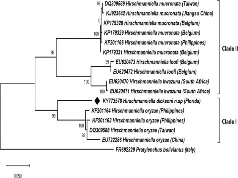
To perform the phylogenetic analysis, D2–D3 LSU, ITS1-5.8S-ITS2 rDNA sequences obtained from H. dicksoni n. sp. identified in this study and those retrieved from the GenBank and NCBI databases (Tables 1, 2) were aligned over the same length in MUSCLE using MEGA v. X (Kumar et al., 2018). The alignment was analyzed to get the base substitution model for these sequences using MEGA X (Kumar et al., 2018). For D2–D3 LSU sequences, the evolutionary history was inferred by using the Kimura 2-parameter model (Kimura, 1980). For ITS1-5.8S-ITS2 rDNA sequences, the evolutionary history was inferred by using the Tamura 3-parameter model (Tamura, 1992). Initial tree(s) for the heuristic search were obtained automatically by applying neighbor-joining and BioNJ algorithms to a matrix of pairwise distances estimated using the maximum composite likelihood (MCL) approach, and then selecting the topology with superior log-likelihood value. A discrete Gamma distribution was used to model evolutionary rate differences among sites. The phylogram was generated using base substitution model selected for each alignment and running maximum likelihood model with 1,000 bootstrap replicates using MEGA X (Kumar et al., 2018) to assess the degree of support for each branch on the tree (Landa et al., 2008). Unique data sequences obtained from this study were submitted to GenBank under accession numbers KY773577 and KY773578 for D2–D3 LSU and ITS1-5.8S-ITS2 rDNA sequences, respectively. The D2–D3 sequence from Pratylenchus vulnus was used as outgroup for constructing the phylogenetic tree based on the sequences form D2–D3 expansion segment of 28S rRNA, whereas P. bolivianus was used as outgroup in the case of phylogenetic tree based on ITS1-5.8S-ITS r DNA sequence.
Table 1.
List of 28S ribosomal RNA gene partial sequences (D2–D3 segment) of Hirschmaniella and outgroup species used for phylogenetic analysis.
| Species | Accession No. | Location | |
|---|---|---|---|
| H. dicksoni n. sp. | KY773577 | Florida | |
| Hirschmanniella sp. | EU620464 | South Africa | |
| Hirschmanniella sp. | KY794704 | Iran | |
| Hirschmanniella sp. | JX261958 | Iran | |
| Hirschmanniella sp. | EU620465 | South Africa | |
| H. diversa | JX144359 | Japan | |
| H. santarosae | EF029859 | California | |
| H. pomponiensis | DQ077795 | California | |
| H. kuazuna | EU620466 | South Africa | |
| H. kuazuna | EU620467 | South Africa | |
| H. loofi | EU620468 | Belgium | |
| H. loofi | EU620469 | Belgium | |
| H. mucronata | KF201167 | The Philippines | |
| H. mucronata | KP179327 | Cambodia | |
| H. mucronata | KP179333 | Cambodia | |
| H. mucronata | KY424320 | China | |
| H. mucronata | MK241938 | Indonesia | |
| Hirschmanniella sp. Yuma | EF029861 | Arizona | |
| H. belli | EF029860 | California | |
| H. oryzae | JX291142 | Myanmar | |
| H. oryzae | KF201165 | The Philippines | |
| Hirschmanniella sp. | DQ328686 | Vietnam | |
| Hirschmanniella sp | KP671713 | Belgium | |
| Pratylenchus vulnus | JX047010 | USA | |
| Species | Accession No. | Location | |
|---|---|---|---|
| H. dicksoni n. sp. | KY773578 | Florida | |
| H. mucronata | KP179328 | Belgium | |
| H. mucronata | KF201166 | The Philippines | |
| H. mucronata | DQ309589 | Taiwan | |
| H. mucronata | KP179329 | Belgium | |
| H. mucronata | KJ923642 | China | |
| H. mucronata | KP179331 | Belgium | |
| H. loofi | EU620472 | South Africa | |
| H. loofi | EU620473 | South Africa | |
| H. kuazuna | EU620471 | South Africa | |
| H. kuazuna | EU620470 | South Africa | |
| H. oryzae | KF201164 | The Philippines | |
| H. oryzae | KF201163 | The Philippines | |
| H. oryzae | DQ309588 | Taiwan | |
| Pratylenchus bolivianus | FR692329 | Italy | |
| Female | Male | ||
|---|---|---|---|
| Character | Holotype | Paratypes | Paratype |
| n | – | 19 | 20 |
| L | 1,906 | 1714 ± 152 | 1506 ± 143 |
| (1,582–1,929) | (1,235–1,772) | ||
| a | 61 | 59 ± 4.9 | 60 ± 6.3 |
| (50–69) | (47–72) | ||
| b | 11 | 11 ± 1.1 | 9.6 ± 0.9 |
| (10–14) | (8.6–11.3) | ||
| b′ | 4.1 | 4.2 ± 0.4 | 4.3 ± 0.4 |
| (3.8–4.8) | (3.8–5.1) | ||
| c | 13 | 14 ± 0.9 | 15 ± 1.5 |
| (13–15) | (13–18) | ||
| c′ | 6.7 | 6.2 ± 0.5 | 6.2 ± 0.8 |
| (5.1–6.7) | (4.8–7.2) | ||
| G1 | 22 | 23 ± 2.3 | – |
| (20–26) | |||
| G2 | 20 | 20 ± 1.8 | – |
| (17–24) | |||
| T | – | – | 37 ± 5 |
| (29–45) | |||
| V | 55 | 53 ± 1.7 | – |
| (50–56) | – | ||
| M | 51 | 52 ± 1.7 | 54 ± 1.9 |
| (51–55) | (51–57) | ||
| o | 23 | 22 ± 2.2 | 21 ± 2.9 |
| (19–27) | (17–27) | ||
| DGO | 4.5 | 4.3 ± 0.4 | 4 ± 0.6 |
| (3.8–5.1) | (3.3–5.1) | ||
| Lip region diameter | 10 | 10 ± 0.7 | 9.9 ± 0.4 |
| (9.1–11.2) | (9.2–11) | ||
| Lip region height | 3.7 | 4.1 ± 0.3 | 4.3 ± 0.3 |
| (3.7–4.8) | (4.0–5.1) | ||
| Stylet length | 19 | 20 ± 0.6 | 19 ± 0.5 |
| (19–21) | (18–20) | ||
| Conus length | 9.8 | 10 ± 0.5 | 10 ± 0.4 |
| (9.7–11) | (9.6–11) | ||
| Shaft length | 9.5 | 9.3 ± 0.4 | 8.7 ± 0.4 |
| (8.6–9.9) | (8.2–9.6) | ||
| Stylet knob height | 3 | 2.7 ± 0.4 | 2.7 ± 0.2 |
| (2–3.4) | (2.4–3.2) | ||
| Stylet knob width | 4.6 | 4.5 ± 0.4 | 4.3 ± 0.5 |
| (3.8–5.3) | (3.4–5.0) | ||
| Procorpus length | 80 | 63 ± 13.8 | 70 ± 7.2 |
| (50–87) | (54–80) | ||
| Median bulb length | 17 | 17 ± 1.4 | 16 ± 1.3 |
| (15–20) | (14–18) | ||
| Median bulb diam. | 13 | 12 ± 0.8 | 11 ± 1.4 |
| (11–13) | (7.5–13) | ||
| Median bulb valve length | 5.8 | 5 ± 0.7 | 4.7 ± 0.7 |
| (3.5–6.3) | (3.8–6) | ||
| Median bulb valve width | 3.6 | 2.9 ± 0.4 | 2.9 ± 0.5 |
| (2.3–3.6) | (2.1–3.6) | ||
| Anterior end to oesophageal-intestinal junction | 177 | 154 ± 12 | 158 ± 13 |
| (142–177) | (129–179) | ||
| Anterior end to excretory (EP) pore | 146 | 133 ± 7 | 119 ± 11 |
| (124–146) | (87–134) | ||
| EP pore to oesophageal-intestinal junction | 31 | 25 ± 4.3 | 39 ± 8.9 |
| (20–32) | (24–50) | ||
| Metacorpus valve from anterior end | 89 | 85 ± 4.1 | 80 ± 4.7 |
| (80–94) | (72–87) | ||
| Nerve ring from anterior end | 116 | 113 ± 5 | 102 ± 10 |
| (105–122) | (73–114) | ||
| Oesophageal gland overlap | 294 | 257 ± 34 | 193 ± 23 |
| (204–311) | (150–223) | ||
| Oesophagus length | 470 | 411 ± 38 | 351 ± 30 |
| (330–470) | (303–392) | ||
| Max body length | 31 | 29 ± 1.6 | 26 ± 3.1 |
| (26–31) | (21–30) | ||
| Length from phasmids to terminus | 32 | 29 ± 2 | 32 ± 2.9 |
| (25–32) | (27–35) | ||
| Tail length | 149 | 125 ± 11 | 102 ± 11 |
| (103–149) | (84–119) | ||
| Anal body width | 22 | 20 ± 1 | 17 ± 1.8 |
| (18–22) | (15–20) | ||
| From cloaca to anterior most part of testis | – | 559 ± 92 | |
| (420–708) | |||
| Vulva to anterior end | 1045 | 912 ± 90 | – |
| (803–1056) | |||
| Anterior genital branch | 420 | 391 ± 57 | – |
| (305–472) | |||
| Posterior genital branch | 385 | 333 ± 39 | – |
| (289–433) | |||
| Ant. Spermatheca length | 26 | 28 ± 4.2 | – |
| (20–35) | |||
| Ant. Spermatheca diam | 15 | 16 ± 2.6 | – |
| (9.3–19) | |||
| Post. Spermatheca length | 24 | 24 ± 3.9 | – |
| (17–30) | |||
| Post. Spermatheca diam | 14 | 14 ± 2 | – |
| (8.9–17) | |||
| Bursa | – | – | 70 ± 15 |
| (45–94) | |||
| Cloaca to anterior tip of bursa | – | – | 25 ± 5.8 |
| (15–33) | |||
| Cloaca to posterior tip of bursa | – | – | 45 ± 9.5 |
| (27–61) | |||
| Gubernaculum | – | – | 8.9 ± 1.1 |
| (7.4–11) | |||
| Spicules | – | – | 25 ± 2.9 |
| (19–30) | |||