Yellow nutsedge (Cyperus esculentus L.) and purple nutsedge (C. rotundus L.) are perennial weeds of global importance that can enhance survival and population densities of Meloidogyne incognita (Kofoid and White, 1919) Chitwood, 1949 and result in injury to crops (Schroeder et al., 1993, 1994, 2004; Thomas et al., 2004). Shoot growth of both nutsedges is not affected by M. incognita parasitism; numbers and size of reproductive tubers increase as nutrient resources are reallocated to roots, and galls do not develop on roots (Mauk et al., 1999; Schroeder et al., 1999). During routine bioassays of nutsedge cultures maintained in the greenhouse, galling was observed on roots of purple nutsedge, but was not present on any comingled roots of M. incognita-susceptible ‘Rutgers’ tomato (Solanum lycopersicum L.). Subsequent dissection of the galls revealed small, mature Meloidogyne females and egg masses that were primarily contained inside the root tissue. Eggs were recovered from roots of purple nutsedge, but not that of tomato following extraction with sodium hypochlorite. Additional pots of purple nutsedge and Rutgers tomato that were established in pasteurized soil using surface-sterilized nutsedge tubers and inoculated with eggs recovered from galled purple nutsedge roots showed similar results: root galling and egg recovery from nutsedge, but no galling or egg recovery from tomato.
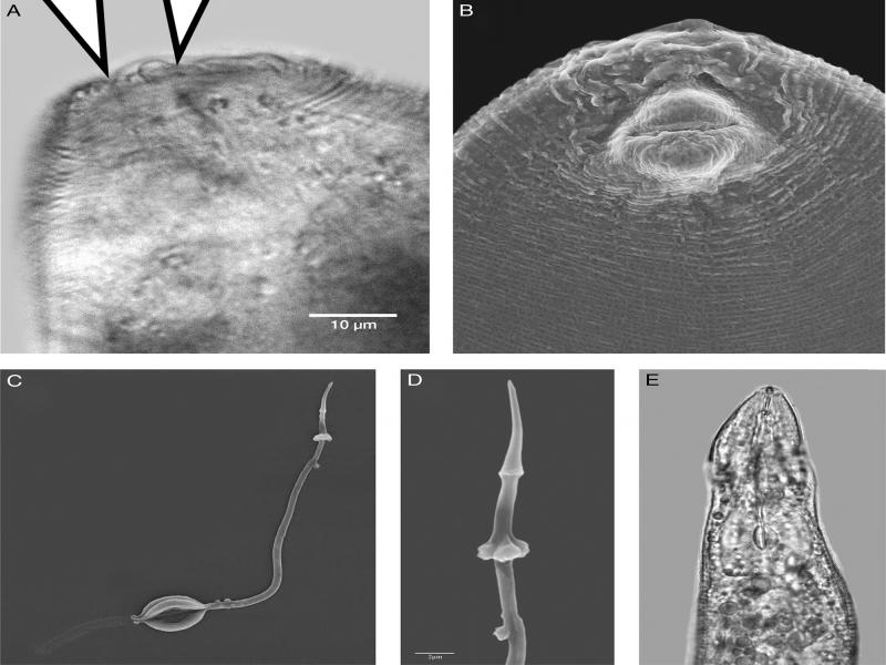
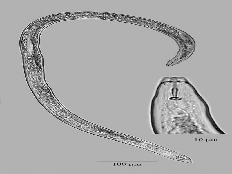
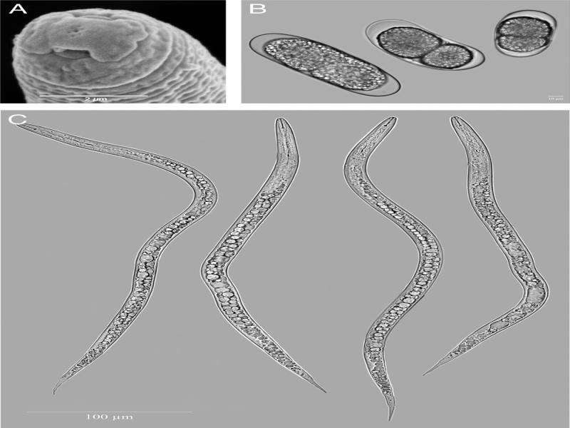
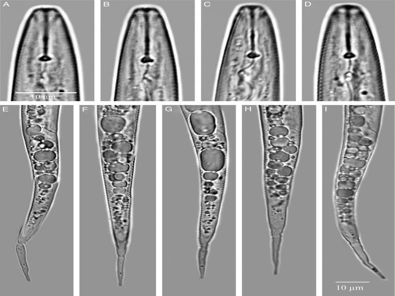
Additional research on the morphology and host range conducted at Virginia Tech and New Mexico State University revealed several unusual morphological characters and a unique host range that indicated it was a new species. The perineal pattern, shape of the female stylet, and shape of the male head and stylet were unique and different from those of any other described species. Meloidogyne aegracyperi n. sp. is described herein, and the common name ‘nutsedge root-knot nematode’ is proposed. The specific epithet was derived from the Latin word ‘aegra’ and the host plant name, meaning ‘diseased Cyperus.’
Materials and methods
Meloidogyne aegracyperi n. sp. was established from field collected soil and plants from the type locality and propagated on purple nutsedge to maintain stock cultures that were kept in a greenhouse at 22 to 28°C. All nematodes used in morphologic, morphometric, and host range studies were from these cultures.
Morphological studies
Males and second-stage juveniles (J2) were extracted from washed galled roots incubated in a moist chamber. Light microscopy (LM) observations were made from specimens mounted on 5% water agar pads and paralyzed with 0.1 m sodium azide (Eisenback, 2012). Females and J2 were prepared for scanning electron microscopy (SEM) according to Eisenback (1985). Perineal patterns were prepared for SEM according to Charchar and Eisenback (2001). They were observed and photographed by LM with negative contrast as reported by Eisenback (2012). Eggs were measured in fresh tap water mounted on agar pads. All LM observations except for perineal patterns were made with a bright field microscope, and at least 100 specimens were observed. However, only two males were recovered from hundreds of infected nutsedge plants. Measurements were made of females in 2% glutaraldehyde in 0.1 M cacodylic acid buffer, pH 7.2; perineal patterns were mounted in glycerin, and extracted stylets (Eisenback et al., 1980) were measured with the SEM. In total, 30 specimens of J2 and females were randomly selected for measurements along with the two males.
Host range test
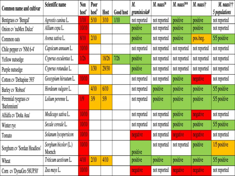
Seedlings of alfalfa (Medicago sativa L.) cv. Doña Ana, chile pepper (Capsicum annuum L.) cv. NM 6-4, corn (Zea mays L.) DynaGro 58UP30, cotton (Gossypium hirsutum L.) cv. Deltapine 393, onion (Allium cepa L.) cv. nuMex Dulce, sorghum (Sorghum bicolor (L.) Moench.) cv. Sordan Headless, common oats (Avena sativa L.), winter rye (Secale cereale L.), perennial ryegrass (Lolium perenne L.) cv. Barlennium, wheat (Triticum aestivum L.), barley (Hordeum vulgare L.) cv. Robust, tomato (Solanum lycopersicum L.) cv. Rutgers, purple nutsedge (Cyperus rotundus L.), yellow nutsedge (Cyperus esculentus L.) were transplanted as single plants into 11 cm diameter clay pots containing 500 cm3 of sterilized sandy loam soil. They were inoculated with a suspension of 3,000 freshly hatched juveniles in 50 ml of water in holes around the root system. Each treatment was replicated 10 times and the plants were kept in a greenhouse at 22 to 28°C. Roots were washed with tap water and stained with phloxine B (Dickson and Struble, 1965) to aid in the counting of egg masses.
Molecular studies
Sample isolation
In total, 24 individual females were isolated from nutsedge roots and transferred into separate polymerase chain reaction (PCR) tubes containing 20 μl of lysis buffer (0.2 M Tris-Cl, pH 7.8) and stored at −20°C.
Lysis, PCR, and sequencing
Lysis was performed using a rapid single tube lysis procedure (Solano and Hanson, unpubl. data). Briefly, samples were removed from −20°C and incubated at 90°C for 10 min. After heating, 30 μl of Proteinase K digestion mixture was added to each sample (5 μl Proteinase K (Qiagen Inc., Valencia, CA), 3 μl of 10× Platinum Taq DNA Polymerase PCR Buffer (Invitrogen, Carlsbad CA), and 22 μl water per sample). Samples were then sonicated for 8 min in a Branson 2510 ultrasonic cleaner then incubated for 30 min at 60°C. After Proteinase K digestion samples were frozen at −80°C for 10 min then incubated at 90°C for 10 min. After heating, 50 μl of water was added to each tube and samples were well mixed then stored at −20°C prior to PCR.
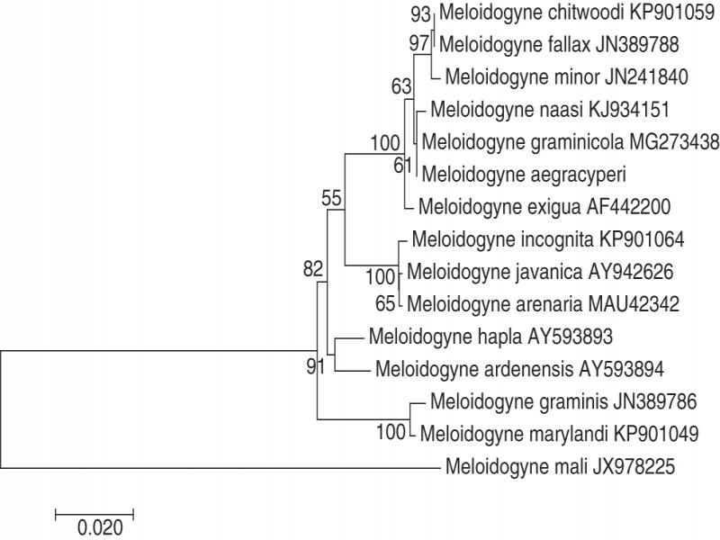
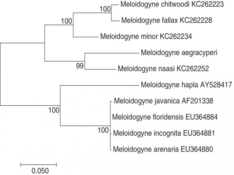
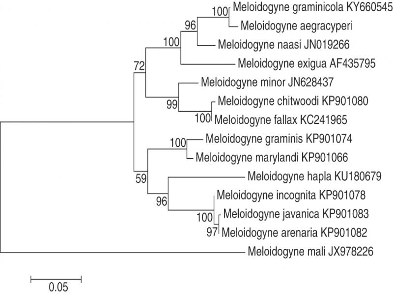
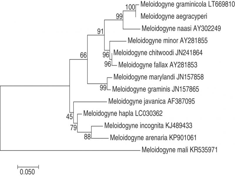
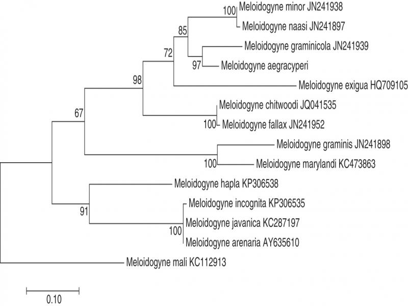
Sequence-based identification of individual nematodes was performed using 18S rRNA and heat shock protein 90 (Hsp90) markers with primers used for amplification and sequencing listed in Table 1. Amplification of the 18S rRNA was performed using two primer sets; 79 F deg plus 1629 R deg which amplifies the majority of the 18S rRNA gene and 983 F deg plus Nema 28S R AG which amplifies the 3′ ~1/2 of the 18S rRNA gene and the ITS region. The previously described primer set RKN-d1F plus RKN-5R was used for amplification and sequencing of the Hsp90 gene (Skantar and Carta, 2004). All amplification reactions were performed in 40 μl PCR reactions using NEBNext Q5 Hot Start HiFi PCR Master Mix (New England BioLabs, Ipswich, MA). PCR reactions contained: 2 μl of nematode lysate, 4 μl of each primer, 17.2 μl of water, and 20 μl of the 2× PCR master mix. The cycling conditions for the 18S rRNA reactions were 94°C for 2 min, followed by 34 cycles of 94°C for 20 sec, 58°C for 30 sec, and 65°C for 80 sec, with a final extension at 68°C for 10 min. Cycling conditions used for the Hsp90 gene were 94°C for 2 min, followed by 40 cycles of 94°C for 30 sec, 55°C for 20 sec, and 68°C for 90 sec, with a final extension at 68°C for 5 min. PCR products were run on a 1% agarose gel in SB buffer (Brody and Kern, 2004) and stained with SYBR Gold according to manufacturer’s instructions (Invitrogen Inc., Carlsbad, CA). Gels were visualized on a digital gel imager (UVP EC3 Imaging System, UVP Inc). PCR reactions that contained the expected size amplicons were treated with ExoSAP-IT (Affymatrix Inc., Santa Clara, CA) according to manufacturer’s instructions. Amplicon concentrations in ExoSAP-IT-treated reactions were determined using a commercial SYBR green-I based DNA quantification kit (Invitrogen Inc., Carlsbad, CA) and read on a fluorescent plate reader (Synergy HTX Multi-Mode Microplate Reader). Automated dideoxy sequencing was performed by Genewiz Inc. (South Plainfield, NJ). Sequence editing, assembly, and analysis were performed using the integrated sequence analysis package, Genious 9.0.2 (Kearse et al., 2012) with the MAFFT aligner being used to generate multiple sequence alignments. Maximum likelihood phylogenetic trees were generated using Mega 6.06 with default parameters and 500× bootstrapping (Tamura et al., 2013).
Table 1.
Primers used to compare M. aegracyperi n. sp. with its closest relatives.
| Primer | Sequence (5′-3′) | Use | Marker | ||
|---|---|---|---|---|---|
| D2A | ACAAGTACCGTGAGGGAAAGTTG | PCR and sequencing | 18S rRNA | ||
| D2B | TCGGAAGGAACCAGCTACTA | PCR and sequencing | 18S rRNA | ||
| ITS1 | CGTAACAAGGTAGCTGTAG | PCR and sequencing | ITS rRNA | ||
| ITS2 | TTTCACTCGCCGTTACTAAGG | PCR and sequencing | ITS rRNA | ||
| 1618 F | TTT GTA CAC AC GCC CGT CG | Sequencing | 18S rRNA | ||
| 1421 F deg | GGT CTG TGA TGC CCT WRG ATG T | Sequencing | 18S rRNA | ||
| 546 F | GGG CAA GTC TGG TGC CAG CAG | Sequencing | 18S rRNA | ||
| Nema 28 S R AG | ACT CCT TGG TCC GTG TTT CAA GA | PCR | 18S rRNA | ||
| 983 F deg | CGA MRG YGA TYA GAT ACC GCY | PCR | 18S rRNA | ||
| 1629 R deg | GGT GTG TAC AAA KSR CAG GGA | PCR | 18S rRNA | ||
| 79 F deg | GDG AAACYG CGWACG GCT | PCR | 18S rRNA | ||
| RKN-5R | TCG AAC ATG TCA AAA GGA GC | PCR and sequencing | HSP90 | ||
| RKN-d1F | GCY GAT CTT GTY AAC AAC CYT GGA AC | PCR and sequencing | HSP90 | ||
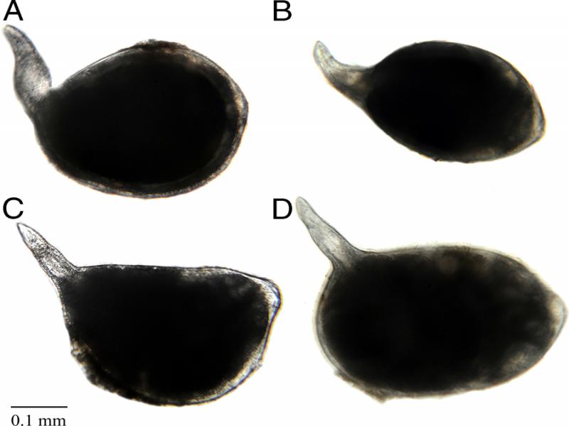
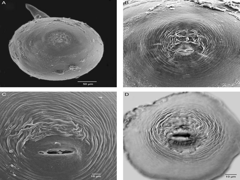
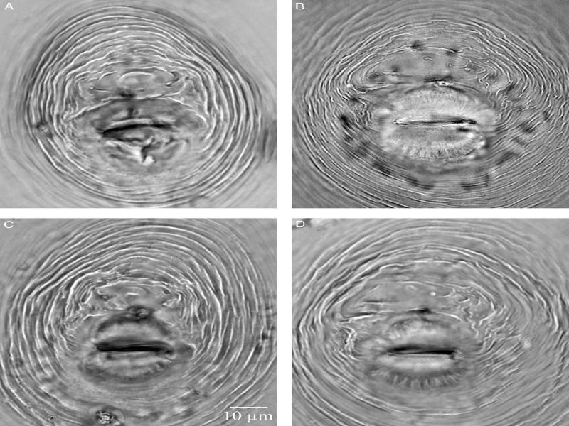
| Female | Male | Second-stage Juv. | |||
|---|---|---|---|---|---|
| Character | Holotype | Paratypes | Paratypes | Paratypes | |
| N | – | 30 | 2 | 30 | |
| L | 360 | 373 ± 44 | 1124 ± 10.5 | 426 ± 24.7 | |
| – | (310–460) | (1113–1134) | (388–484) | ||
| Body diam. | 292 | 306 ± 46 | 30.9 ± 1.4 | 15 ± 0.9 | |
| – | (210–420) | (29.5–32.3) | (13.6–17.4) | ||
| Neck length | 148 | 153 ± 27 | – | – | |
| – | (100–210) | – | – | ||
| Stylet length | 12 | 12 ± 0 | 15.6 ± 1.0 | 10.9 ± 0.4 | |
| – | (12–12) | (14.6–16.5) | (10.1–11.8) | ||
| Stylet knob height | 1.5 | 1.5 ± 0.2 | 2.3 ± 0.2 | 1.4 ± 0.2 | |
| – | (1.2–1.9) | (2–2.5) | (1.1–1.9) | ||
| Stylet knob width | 2.4 | 2.6 ± 0.3 | 4.1 ± 0.1 | 2.1 ± 0.2 | |
| – | (2–3) | (4–4.2) | (1.7–2.5) | ||
| DGO | 4.8 | 4.8 ± 0.6 | 3.1 ± 0.1 | 3.7 ± 0.5 | |
| – | (4–6.1) | (3.0–3.3) | (2.7–4.8) | ||
| Stylet tip to metacorpus center | 60.1 | 63.8 ± 2.2 | – | – | |
| – | (59–67) | – | – | ||
| Interphasmidial distance | – | 18 ± 2.7 | – | – | |
| – | (14.2–24.4) | – | – | ||
| Vulva length | – | 21 ± 2.9 | – | – | |
| – | (13.5–26) | – | – | ||
| Vulva-anus distance | – | 15.7 ± 2.3 | – | – | |
| – | (10.9–21) | – | – | ||
| Ant. end to excretory pore | – | – | 94.7 ± 1.6 | 68.5 ± 7.9 | |
| – | – | (93.1–96.3) | (52–80.4) | ||
| Tail length | – | – | 12.7 ± 1.9 | 73.1 ± 4.9 | |
| – | – | (10.8–14.5) | (63.6–88.7) | ||
| Body width at anus | – | – | – | 11 ± 0.6 | |
| – | – | – | (10.2–11.9) | ||
| a | 1.3 | 1.2 ± 0.2 | – | 28.4 ± 2.8 | |
| – | (0.8–1.7) | – | (23.6–34.6) | ||
| c | – | – | – | 5.8 ± 0.4 | |
| – | – | – | (5.1–6.5) | ||
| Spicule length | – | – | 23.6 ± 1.8 | – | |
| – | – | (21.8–25.4) | – | ||
| Hyaline tail terminus | – | – | – | 22 ± 2.0 | |
| – | – | – | (18.5–26.6) | ||
| Female | |||||
| Species | Length | Width | Stylet | DGO | Vulva |
| M. chitwoodi (n=60)a | (430–750) 591 | (344–518) 422 | (11.2–12.5) 11.9 | (3.4–5.5) 4.2 | |
| M. fallax (n=30)b | (404–720) 491 | (256–464) 362 | (13.9–15.2) 14.5 | (3.8–6.3) 4.3 | (20.2–28.4) 24.7 |
| M. minor (n=25)c | (416–608) 526 | (240–464) 339 | (12.6–15.2) 14.2 | (3.2–6.3) 4.1 | (22.8–29.1) 25.8 |
| M. naasi (n=25)d | (455–705) 557 | (227–398) 330 | (11–15) 13 | (2–4) 3 | (17–25) 22 |
| M. graminicola (n=20)e | (455–765) 573 | (275–520) 419 | (10.6–11.2) 11.1 | (2.8–3.9) 3.2 | |
| M. aegracyperi n. sp. (n=30) | (310–460) 373 | (210–420) 306 | (12–12) 12 | (4–6.1) 4.8 | (13.5–26) 21 |
| Juvenile | |||||
| Species | Length | Stylet | DGO | Tail | Terminus |
| M. chitwoodi (n=60)a | (336–417) 390 | (9.0–10.3) 9.9 | (2.6–3.9) 3.2 | (39–47) 43 | (8.6–13.8) 11 |
| M. fallax (n=30)b | (381–435) 403 | (10.1–11.4) 10.8 | (3.2–3.8) 3.5 | (46.1–55.6) 49.3 | (12.2–15.8) 13.5 |
| M. minor (n=25)c | (310–416) 377 | (7.6–10.1) 9.2 | (3.2–4.4) 3.8 | (58.1–77.1) 54 | (12.0–22.1) 16.1 |
| M. naasi (n=25)d | (418–465) 435 | (13–15) 14 | (2–3) 2.4 | (52–78) 70 | 17.5 |
| M. graminicola (n=20)e | (415–484) 441 | (11.2–12.3) 11.4 | (2.8–3.4) 2.8 | (67.0–76.0) 70.9 | (14.0–21.2) 17.9 |
| M. aegracyperi n. sp. (n=30) | (388–484) 426 | (10.1–11.8) 10.8 | (2.7–4.8) 3.7 | (63.6–88.7) 73.1 | (18.5–26.6) 22 |
| Male | |||||
| Species | Stylet | DGO | |||
| M. chitwoodi (n–30)a | (18.1–18.5)18.3 | (2.2–3.4) 3 | |||
| M. fallax (n=30)b | (18.9–20.9) 19.6 | (3.2–5.7) 4.4 | |||
| M. minor (n=25)c | (17.1–19.0) 17.8 | (3.2–4.4) 3.8 | |||
| M. naasi (n=25)d | (16–19) 18 | (2–4) 3 | |||
| M. graminicola (n=20)e | (16.2–17.4) 16.8 | (2.8–3.9) 3.3 | |||
| M. aegracyperi n. sp. (n= 2) | (14.6–16.5) 15.6 | (3.0–3.3) 3.1 | |||