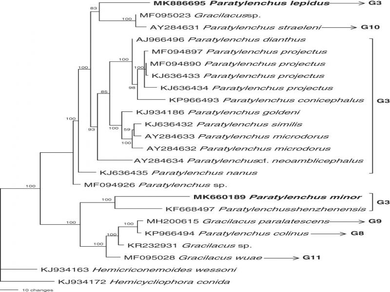
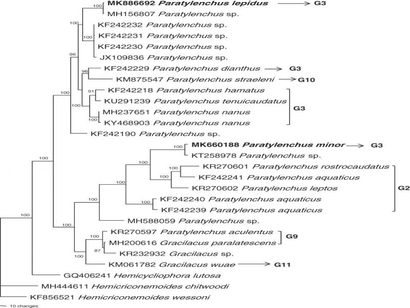
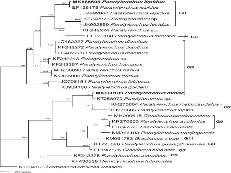
The species of pin nematodes are commonly distributed in China, more than 30 species have been reported from 21 provinces (Maria et al., 2018a). The host range varies from horticultural to agronomic crops as well as ornamental and forestry plants (Raski, 1991). The species of pin nematodes were divided into two genera, i.e. Gracilacus and Paratylenchus based on stylet length (Raski, 1962). The species having longer stylet are cortical feeders while the short stylet species are epidermal feeder (Siddiqi, 2000). The morphological identification of pin nematodes is complicated due to their smaller size and variable characters (Raski, 1962; Brzeski and Hanel, 2000). However several robust characters such as number of lateral lines, stylet length and the presence/absence of vulval flap can sufficiently be used to differentiate pin nematode species (Ghaderi et al., 2014). For precise species identification, rRNA sequences of 28S (Subbotin et al., 2005), 18S (Van Megen et al., 2009), and ITS (Chen et al., 2008, 2009; Van den Berg et al., 2014) have been successfully utilized in pin nematode taxonomy. Recently, several new and known species of nematodes were described based on combined morphological, molecular, and SEM observation (Maria et al., 2018b; Zhuo et al., 2018). Moreover, DNA studies have significant advantages to precisely identify morphologically similar species, such as cryptic species. In an attempt to document the ectoparasitic nematodes from Zhejiang Province, China, two population of Paratylenchus species were detected in a soil sample of Elaeocarpus sp. and Chinese red pine (Pinus tabuliformis Carr). The preliminary studies revealed the status of these species as P. lepidus Raski, 1975a, 1975b and P. minor (Sharma et al., 1986). P. lepidus was originally described from Srilanka and has been reported from China (Chen et al., 2007) whereas P. minor was described from India in the rhizosphere of peach plants and has not been reported thereafter. As these species were reported decades ago, there was no molecular sequencing data and SEM observations. In order to fill these gaps, the objectives of this study were (i) to provide the updated description with morphological and molecular characterization, (ii) to elucidate important morphological details through SEM observations, (iii) to investigate the phylogenetic position and to locate the closely related Paratylenchus species.
Materials and methods
Nematode samplings, extraction, and morphological study
Nematodes were extracted from soil and root samples using the modified Cobb sieving and flotation-centrifugation method (Jenkins, 1964). For morphometric studies, nematodes were killed and fixed in hot Formalin (4% with 1% glycerol) and processed to glycerin (Seinhorst, 1959). The measurements and light micrographs of nematodes were made with a Nikon Eclipse Ni-U 931845 compound microscope. For the SEM examination, the nematodes were fixed in a mixture of 2.5% paraformaldehyde and 2.5% glutaraldehyde, washed three times in 0.1 M cacodylate buffer, post-fixed in 1% osmium tetroxide, dehydrated in a series of ethanol solutions and critical point-dried with CO2. After mounting on stubs, the samples were coated with gold at 6 to 10-nm thickness and the micrographs were made at 3 to 5 kV operating system (Maria et al., 2018c).
Molecular analyses
DNA was extracted by transferring individual nematodes into an Eppendorf tube containing 16 μ L ddH2O. Nematodes were crushed using a sterilized pipette tip, the tubes were centrifuged at 12,000 rpm for 1 min and frozen at -68°C for at least 30 min. Tubes were heated to 85°C for 2 min, then 2 μ L proteinase K was added and PCR buffer solution. The tubes were incubated at 56°C for 1 to 2 hr, followed by 10 min at 95°C. After incubation, these tubes were cooled to 4°C and used for conducting PCR (Zheng et al., 2003). Several sets of primers (synthesized by Invitrogen, Shanghai, China) were used in the PCR analyses to amplify the partial 18S, ITS region and D2-D3 of 28S of rDNA. Primers for amplification of partial 18S were 18s39F-18s977R and 18s900-18s1713 (Olson et al., 2017). Primers for amplification of ITS were TW81-AB28 (Joyce et al., 1994). The primers for amplification of D2-D3 of 28S were D2A and D3B (De Ley et al., 1999). PCR conditions were as described by Ye et al. (2007) and Powers et al. (2010). PCR products were evaluated on 1% agarose gels stained with ethidium bromide. PCR products of sufficiently high quality were sent for sequencing by Invitrogen (Shanghai, China).
Phylogenetic analysis
The sequences were deposited into the GenBank database. DNA sequences were aligned by MEGA7 (Kumar et al., 2016) using default settings. The DNA sequences were compared with those of the other pin nematode species available at the GenBank sequence database using the BLAST homology search program. The model of base substitution was evaluated using MODELTEST (Posada and Crandall, 1998; Huelsenbeck and Ronquist, 2001). The Akaike-supported model, the base frequencies, the proportion of invariable sites, and the gamma distribution shape parameters and substitution rates were used in phylogenetic analyses. Bayesian analysis was performed to confirm the tree topology for each gene separately using MrBayes 3.1.0 (Huelsenbeck and Ronquist, 2001) running the chain for 1 × 10⁶ generations and setting the “burnin” at 2,500. The Markov Chain Monte Carlo (MCMC) method was used within a Bayesian framework to estimate the posterior probabilities of the phylogenetic trees (Larget and Simon, 1999) using 50% majority rule.
Results and description
Systematics
Paratylenchus lepidus Raski, 1975a, b
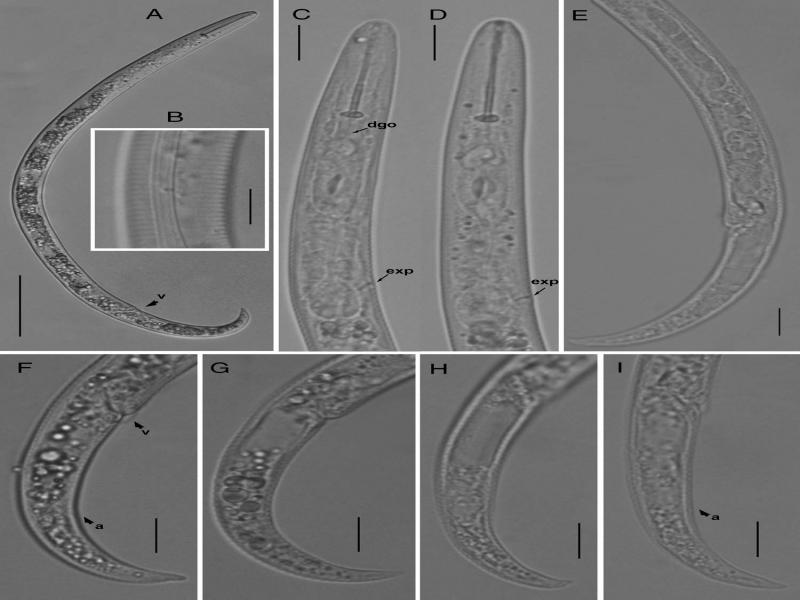
Figure 1:
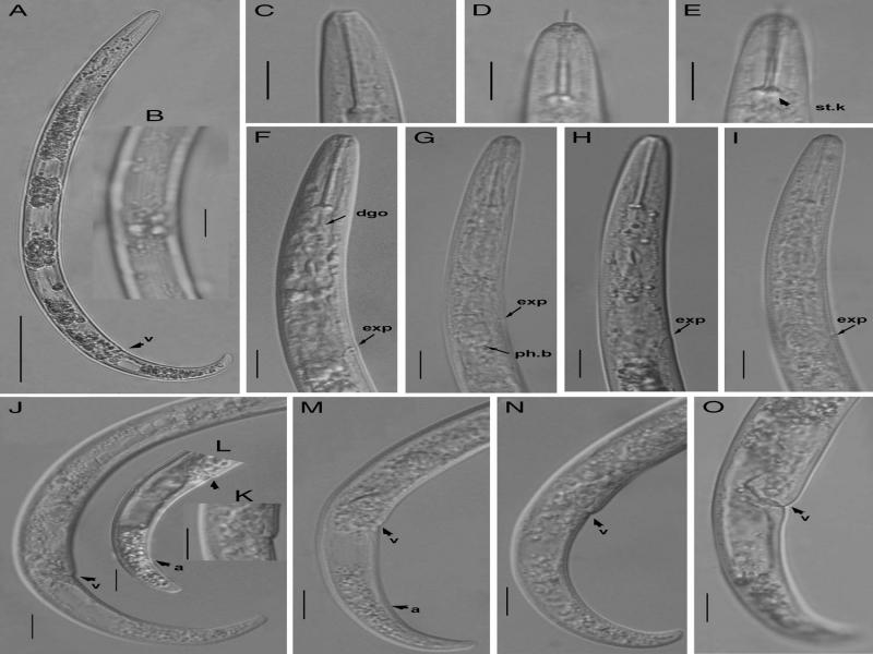
Light photomicrographs of Paratylenchus lepidus (Raski, 1975a, b). (A) Entire female; (B) lateral lines; (C-D) pharyngeal regions, arrow pointing on the excretory pore (exp); (E) posterior region showing gonad; (F-I) tail regions, arrows pointing on vulva and anus (a) (Scale bars = A = 50 μm, B-I = 10 μm).
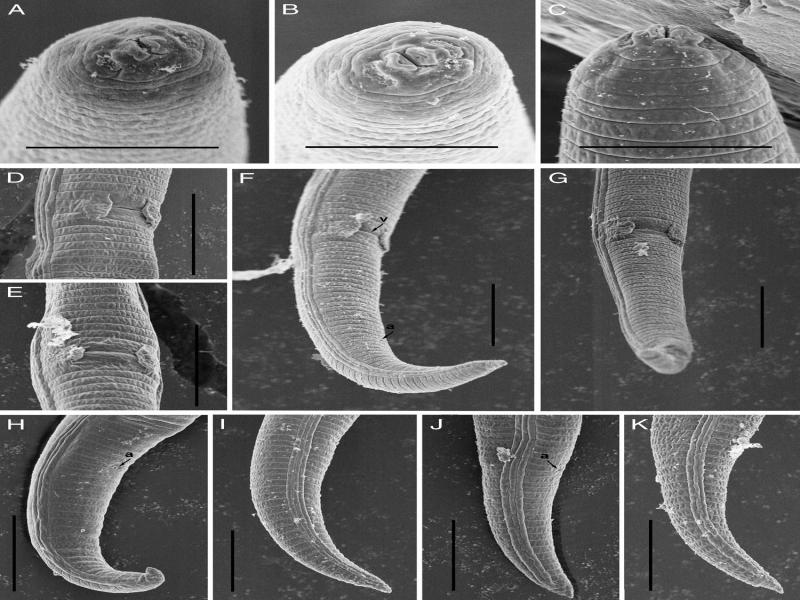
Figure 2:
Scanning electron microscopy of Paratylenchus lepidus (Raski, 1975a, 1975b). (A-C) Lip regions; (D, E) vulval regions; (F, G) posterior region arrows showing position of vulva; (H-K) female tails (Scale bars, A-C = 5 μm; D-K = 10 μm).
Measurements
Measurements of the females of P. lepidus Taizhou population are given in Table 1.
Table 1.
Morphometric data for Paratylenchus lepidus (Raski, 1975a, 1975b).
| Type | Additional type pop. | ||||
|---|---|---|---|---|---|
| This study | Raski (1975b) | Raski (1975b) | Chen et al. (2007) | Yu et al. (2014) | |
| Elaeocarpus sp. | Thea sinensis L. | Thea sinensis L. | a | Boehmeria nivea L. | |
| Characters/ratios | Taizhou | Sri Lanka | Sri Lanka | Taiwan | Hunan |
| n | 23 | 21 | 13 | 26 | |
| Body length | 324.2 ± 21.0 (288.4–359) | 330 (280–400) | 320 (290–370) | 270–400 | 260–420 |
| a | 27.4 ± 2.5 (22.2–31.6) | 25 (23–31) | 27 (23–31) | 19.4–31 | 13.1–23.1 |
| b | 4.0 ± 0.3 (3.5–4.4) | 4.0 (3.4–4.0) | 3.8 (3.5–4.6) | 3.2–4.6 | 3.3–5.1 |
| c | 10.4 ± 0.6 (9.2–11.6) | 14 (11–16) | – | 11–16 | 5.3–10.3 |
| c' | 3.8 ± 0.2 (3.3–4.3) | – | – | – | |
| V | 81.4 ± 0.9 (80.0–83.3) | 82 (80–84) | 82 (80–83) | 78.5–84 | 78–84 |
| Lip height | 3.1 ± 0.2 (2.6–3.4) | – | – | – | – |
| Lip width | 5.5 ± 0.3 (5.1–6.2) | – | – | – | – |
| Stylet | 27 ± 1.0 (24.8–28.7) | 25 (22–27) | 25 (23–28) | 21.3–27 | 13.7–27.9 |
| Stylet percentage | 8.3 ± 0.4 (7.6–9.2) | – | – | – | – |
| Median bulb Length | 18.0 ± 1.4 (15.2–19.9) | – | – | – | – |
| Median bulb width | 6.4 ± 0.5 (5.3–7.2) | – | – | – | – |
| SE pore | 71.4 ± 2.9 (65.2–76.4) | – | – | – | – |
| Pharynx | 80.2 ± 2.9 (73.7–85.4) | 75 (69–82) | 68 (66–75) | 58–85 | |
| Body width | 12.0 ± 1.0 (10.2–13.5) | – | – | – | – |
| Vulval body diam | 10.6 ± 0.8 (8.9–12.1) | – | – | – | – |
| Anal body diam | 8.3 ± 0.6 (7.3–9.4) | – | – | 8–15 | – |
| Distance from vulva to tail terminus | 60.3 ± 4.6 (51.7–69) | – | – | 79–84 | – |
| Tail length | 31.2 ± 2.3 (26.5–34.8) | – | – | – | – |
| Characters/ratios | Zhejiang population | Type species |
|---|---|---|
| n | 23 | 10 |
| L | 314.0 ± 17.0 (284.4–352.0) | 260 (230–280) |
| a | 25.0 ± 1.8 (21.6–28.0) | 21 (18–24) |
| b | 4.3 ± 0.2 (3.8–4.7) | 3.8 (3.4–4.3) |
| c | 13.0 ± 2.2 (9.6–17.3) | 12.0 (10–16) |
| c' | 2.9 ± 0.3 (2.3–3.4) | – |
| V | 81.7 ± 0.9 (80.0–83.6) | 81 (73–84) |
| Lip height | 2.8 ± 0.3 (2.2–3.3) | – |
| Lip width | 5.6 ± 0.3 (5.1–6.3) | – |
| Stylet | 23.2 ± 0.9 (21.8–24.8) | 22 (20–24) |
| Stylet percentage | 7.4 ± 0.4 (6.6–7.9) | – |
| Median bulb length | 15.0 ± 1.1 (12.4–16.9) | – |
| Median bulb diam. | 6.6 ± 0.6 (5.7–8.0) | – |
| Ant. end to excretory pore | 66.7 ± 3.1 (61.7–72.5) | – |
| Pharynx | 73.0 ± 2.4 (68.6–77.3) | – |
| Max. body diam. | 12.6 ± 0.9 (10.6–14.3) | – |
| Vulval body diam | 11.0 ± 0.8 (8.4–12.1) | – |
| Anal body diam. | 8.7 ± 1.1 (7.0–11.3) | – |
| Vulva to tail term. | 57.5 ± 4.2 (51.0–65.3) | – |
| Tail length | 25.0 ± 3.9 (18.4–32.5) | – |