Andrássy (1976) proposed the new genus Oscheius under Rhabditidae (Örley, 1880), Rhabditinae (Örley, 1880), with O. insectivorus (=Rhabditis insectivora Körner in Osche, 1952) as its type and only species. Oscheius was distinguished from other Rhabtidinae by its unusually short buccal tube, about as long as wide, and the absence of median pharyngeal swelling. Later, the same author (1983, 1984) transferred a second species, O. koerneri (=Rhabditis koerneri Osche, 1952), to the genus and provided its diagnosis.
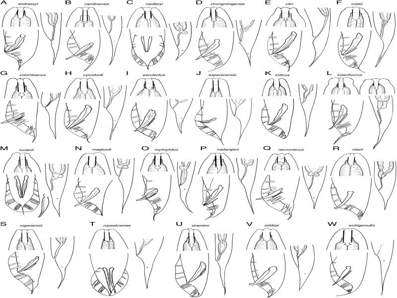
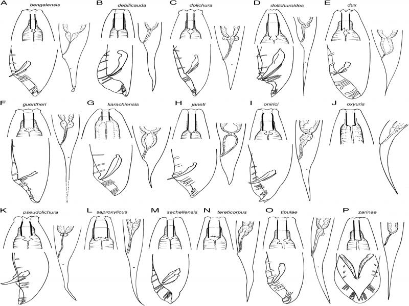
Sudhaus and Hooper (1994) provided new ideas about the taxonomy and the phylogeny of several rhabditid species: (i) accepted Oscheius as a subgenus of Rhabditis, (ii) regarded it as a monophyletic taxon based on three synapomorphies (long female rectum, terminal duct of the excretory system forwards coiled and with heavily sclerotized wall, and several features of spicule shape), (iii) considered Dolichorhabditis (Andrássy, 1983) as a junior synonym of Oscheius, and (iv) distinguished two species groups within the subgenus. One of these groups, the Insectivorus-group, included seven species with leptoderan or pseudopeloderan bursa (male tail with a filiform part standing out behind the bursa, a plesiomorphic state), and spicules with crochet needle shaped tip (apomorphic state). The second group, the Dolichura-group, with five species previously classified under Dolichorhabditis and having peloderan bursa (lacking the filiform part, an apomorphic condition) and spicules with thin tubular tip (plesiomorphic condition). Andrássy (2005) reinstated the generic range for Oscheius, listed a total of eight species under it, and distinguished it from Dolichorhabditis, with ten valid species, by several differences in stomatal teeth, bursa and spicules. The separation of both genera has been accepted in several contributions (Abolafia and Peña-Santiago, 2010; Gorgadze, 2010), but Sudhaus (2011) and Tabassum et al. (2016) maintained Dolichorhabditis as junior synonym of Oscheius as well the two monophyletic species groups within the latter.
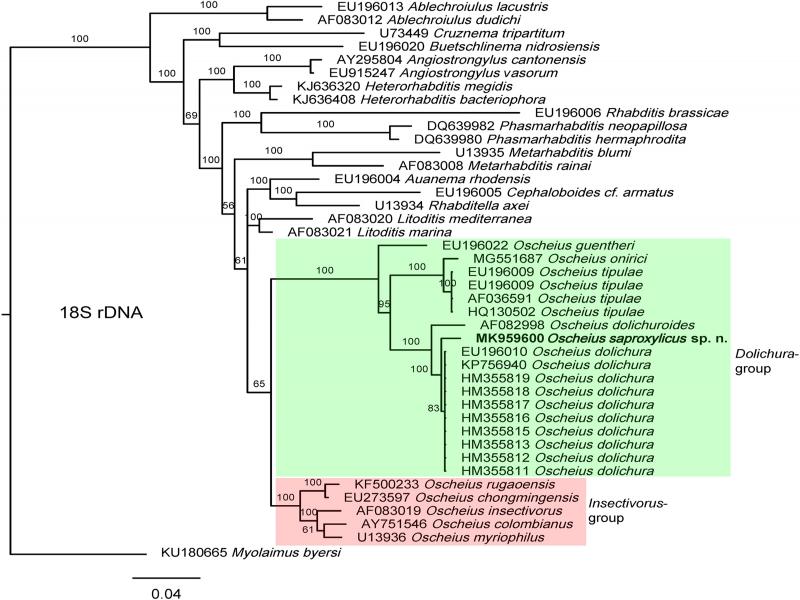
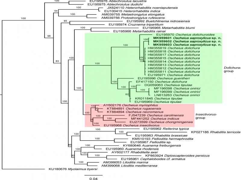
Molecular data of Oscheius sensu lato species, many of them described during the last years, have been matter of analyses by several authors (Félix et al., 2001; Darby et al., 2011; Darsouei et al., 2014; Campos-Herrera et al., 2015; Torrini et al., 2015; Tabassum et al., 2016; Lima de Brida et al., 2017; Valizadeh et al., 2017; Zhou et al., 2017), resulting in the confirmation of the monophyly of the Insectivorus- and the Dolichura-group. Nonetheless, their nature as sister groups was not always corroborated (van Megen et al., 2009; Darsouei et al., 2014; Zhou et al., 2017).
An Oscheius population was collected in the course of a nematological survey conducted in southern Iberian soils. Its study revealed it belonged to a non-described form. The aims of this contribution are to characterize this material, to provide new insights on the phylogeny of the group, and to update its taxonomy.
Materials and methods
Nematode extraction and processing
Nematodes were collected from dead wood using a modified trays technique (Whitehead and Hemming, 1965), killed by heat, fixed in 4% formalin, transferred to pure glycerine following the Siddiqi’s (1964) method, and mounted on permanent glass slides. Moist, dead wood was maintained as a culture to extract specimens every several months.
Light microscopy (LM)
Observations were made using a Leitz Laborlux S (Leitz, Wetzlar, Germany) and Nikon Eclipse 80i (Nikon, Tokio, Japan) microscopes. Measurements were taken with the Leitz microscope, which has a drawing tube (camera lucida) attached to it, and Demanian indices and other ratios calculated. Drawings were made using the Leitz microscope. Images were taken with the Nikon microscope that was provided with differential interference contrast (DIC) optics and Nikon Digital Sight DS-U1 camera. Micrographs were edited using Adobe® Photoshop® CS. The terminology used for the morphology of stoma and spicules follows the proposals by De Ley et al. (1995) and Abolafia and Peña-Santiago (2017), respectively.
Scanning Electron Microscopy (SEM)
Specimens preserved in glycerine were selected for observation under SEM according to Abolafia (2015). They were hydrated in distilled water, dehydrated in a graded ethanol-acetone series, critical point dried, coated with gold, and observed with a Zeiss Merlin microscope (5 kV) (Zeiss, Oberkochen, Germany).
DNA Extraction, PCR and Sequencing
Nematode DNA was extracted from single fresh individuals using the proteinase K protocol and PCR assays as described Castillo et al. (2003) somewhat modified. Specimen was cut in small pieces using a sterilized dental needle on a clean slide with 18 ml of AE buffer (10 mM Tris-Cl + 0.5 mM EDTA; pH 9.0), transferred to a microtube and adding 2 μl proteinase K (700 μg/ml) (Roche, Basel, Switzerland), and stored to –80°C within 15 min (for several days). The microtubes were incubated at 65°C (1 hr), then at 95°C (15 min). The microtube was centrifuged to 13,000 r.p.m. (or 15,900 × g) for 3 min. and 2 μl of the supernatant extracted DNA was transferred to a microtube containing: 2.5 μl ×10 PCR reaction buffer, 5 μl Q-solution ×5, 0.5 μl dNTPs mixture (10 mM each), 1 μl of each primer (10 mM), 0.2 μl Taq DNA Polymerase (Qiagen, Venlo, The Netherlands) and ddH2O to a final volume of 25 μl. The primers used for amplification of the D2-D3 region of 28S rRNA gene were the D2A (5′-ACAAGTACCGTGAGGGAAAGTTG-3′) and the D3B (5′-TCGGAAGGAACCAGCTACTA-3′) primers (De Ley et al., 1999). PCR cycle conditions were as follows: one cycle of 94°C for 3 min., followed by 35 cycles of 94°C for 1 min. + annealing temperature of 55°C for 45 s + 72°C for 2 min., and finally one cycle of 72°C for 10 min. After DNA amplification, 5 μl of product was loaded on a 1% agarose gel in 0.5% Tris-acetate-EDTA (40 mM Tris, 20 mM glacial acetic acid and 2 mM EDTA; pH = 8) to verify the amplification using a electrophoresis system (Labnet Gel XL Ultra V–2, Progen Scientific, London, UK). The bands were stained with RedSafe (×20,000) previously added to the agarose gel solution. PCR products were purified using the QIAquick PCR purification kit (Qiagen, Venlo, The Netherlands), quantified using a spectrophotometer (Synergy HT, BioTek, Winooski, USA) and used for direct sequencing in both directions using the primers referred to above. The sequencing reactions were performed at “Centro de Instrumentación Científico-Técnica (CICT)” of the University of Jaén (Spain) using an Applied Biosystems Hitachi 3500 Genetic Analyzer. The sequences obtained were submitted to the GenBank database.
Phylogenetic analyses
For phylogenetic relationships, analyses were based on 18S and 28S rDNA. The newly obtained sequences were manually edited using BioEdit 7.2.6 (Hall, 1999) and aligned with another 18S or 28S rRNA gene sequences available in GenBank using Muscle alignment tool implemented in the MEGA7 (Kumar et al., 2016). The ambiguously aligned parts and divergent regions were known using the online version of Gblocks 0.91b (Castresana, 2000) (http://molevol.cmima.csic.es/castresana/Gblocks_server.html) and were removed from the alignments using MEGA7. The best-fit model of nucleotide substitution used for the phylogenetic analysis was statistically selected using jModelTest 2.1.10 (Darriba et al., 2012). Phylogenetic tree was generated with Bayesian inference method using MrBayes 3.2.6 (Huelsenbeck and Ronquist, 2001; Ronquist and Huelsenbeck, 2003). Myolaimus byersi (KU180665 for 18S and KU180676 for 28S) was chosen as outgroup according to previous results by Kanzaki et al. (2009). The analysis under GTR+I+G model was initiated with a random starting tree and run with the Markov Chain Monte Carlo (MCMC) for 1 × 10⁶ generations. The tree was visualized and saved with FigTree 1.4.3 (Rambaut, 2014).
Descriptions
Oscheius saproxylicus sp. n.1
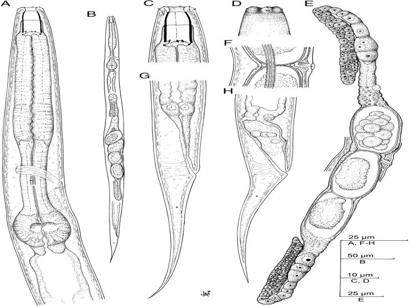
Figure 1:
Oscheius saproxylicus sp. n. (line drawing). (A) Neck; (B): Entire female; (C) Stoma; (D) Lip region; (E) Genital system; (F) Vagina; (G, H) Female tail.
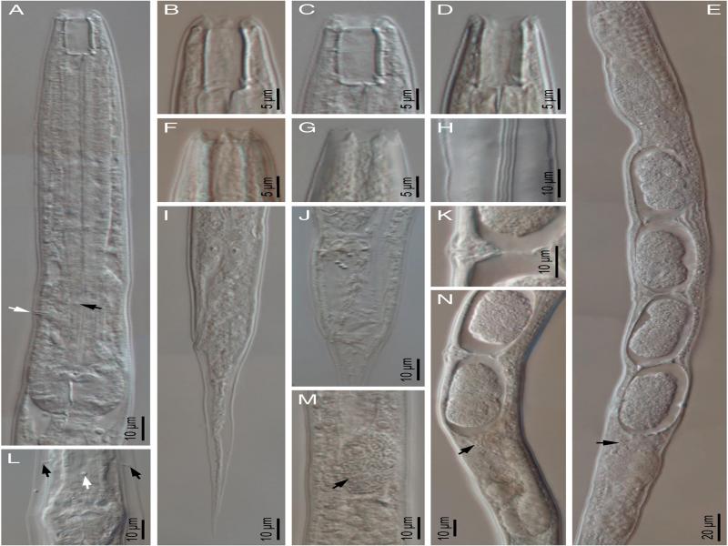
Figure 2:
Oscheius saproxylicus sp. n. (light microscopy, female). (A) Neck; (B-D) Stoma in lateral (B, C) and dorso-ventral (D) views; (E, N) Reproductive system (arrow at spermatozoa); (F, G) Lip region in lateral and ventral views, respectively; (H) Lateral field; (I, J) Posterior end with rectum empty and swollen, respectively; (K) Vagina; (L) Excretory pore in ventral view (white arrow) and deirids (black arrow); (M) Intestine cell with microsporidia (arrow).
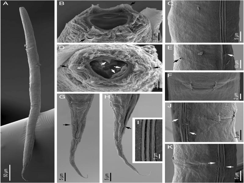
Figure 3:
Oscheius saproxylicus sp. n. (scanning electron microscopy, female). (A) Entire body; (B, D) Lip region in subfrontal and frontal views, respectively (black arrows at amphid and white arrows pointing the metastegostomatal teeth, up: dorsal tooth, down: sublateral teeth); (C) Cuticle at midbody; E: Excretory pore (black arrow) and deirid (white arrow); (F, J, K) Vulval region at ventral, sublateral right and left views, respectively (arrows at lineal warts of the lateral fields); (G, H) Tail in ventral and lateral views, respectively (arrow at phasmid); (I) Lateral field.
Material examined
Fifty one females in generally acceptable state of preservation.
Measurements
See Table 1.
Table 1.
Morphometrics of Oscheius saproxylicus sp. n. Measurements in μm and in the form: mean ± standard deviation (range) where appropriate.
| Locality | Puente de la Sierra | |
|---|---|---|
| Province | Jaén | |
| Habitat | Dead wood | |
| n | Holotype female | Paratypes 50 females |
| Body length | 829 | 837 ± 78.4 (669–994) |
| a | 37.7 | 29.6 ± 3.2 (23.9–37.1) |
| b | 5.1 | 5.0 ± 0.5 (3.9–6.5) |
| c | 11.5 | 11.0 ± 1.2 (8.7–13.8) |
| c' | 5.5 | 5.5 ± 0.5 (5.0–7.0) |
| V | 57 | 54.8 ± 2.4 (48–59) |
| Lip region width | 9 | 9.6 ± 0.5 (9–10) |
| Stoma length | 18 | 18.7 ± 1.0 (17–21) |
| Stoma width | 5 | 5.8 ± 0.6 (5–7) |
| Pharyngeal corpus length | 72 | 75.9 ± 6.5 (68–96) |
| Isthmus length | 48 | 47.7 ± 3.9 (39–54) |
| Bulbus length | 26 | 26.6 ± 1.4 (24–29) |
| Pharynx length | 146 | 149 ± 9.4 (113–165) |
| Neck length | 164 | 168 ± 9.6 (131–182) |
| Body diameter at neck base | 22 | 23.9 ± 1.7 (20–28) |
| Body diameter at midbody | 22 | 28.5 ± 3.4 (21–37) |
| Vulva - anterior end | 469 | 459 ± 49.0 (366–549) |
| Rectum length | 48 | 44.8 ± 3.8 (40–54) |
| Anal body diameter | 13 | 13.9 ± 1.1 (11–16) |
| Tail length | 72 | 76.3 ± 5.0 (67–88) |
| Phasmid - anus distance | 19 | 23.9 ± 3.4 (20–30) |