
The genus Labronema Thorne, 1939 is a worldwide dorylaimid (order Dorylaimida) group with about 41 valid species of intricate taxonomy (cf. Andrássy, 2009) and predator feeding habits (Thorne, 1939, 1974). The study of its representatives in the Iberian fauna has not received too much attention, except for the detailed description of L. vulvapapillatum (Meyl, 1954) Loof & Grootaert, 1981 by Murillo-Navarro and Jiménez-Guirado (2006; see also Jiménez-Guirado, 1989); the available information is nearly restricted to reporting the presence of other seven species (Peña-Santiago et al., 2003).
A nematological survey conducted in November 2017 to explore the dorylaimid diversity in a mountain area of the southern Iberian Peninsula yielded a Labronema population consisting of numerous females and males. An additional study revealed that it was an unknown species of the genus that is described herein.
Material and methods
Nematode extraction and processing
Nematodes were collected from a grassy area with stony soil at 1,800 elevation in Pandera mountain, Jaén province, southern Iberian Peninsula, Spain. They were extracted using Flegg’s (1967) sieving method and a modified Baermann’s (1917) funnel technique; they were killed with heat, fixed into 4% formalin, transferred to pure glycerine following Siddiqi’s (1964) method, and mounted on permanent glass slides.
Light microscopy (LM)
Observations, measurements, line illustrations, and LM photomicrographs were made using a Nikon Eclipse 80i (Nikon, Tokio, Japan) microscope with differential interference contrast (DIC) optics, a drawing tube (camera lucida) and a Nikon Digital Sight DS-U1 camera. Photographs were edited using Adobe® Photoshop® CS software.
Scanning electron microscopy (SEM)
Specimens preserved in glycerine were selected for observation under SEM according to Abolafia (2015). They were hydrated in distilled water, dehydrated in a graded ethanol–acetone series, critical point dried, coated with gold, and observed with a Zeiss Merlin microscope (5 kV) (Zeiss®, Oberkochen, Germany).
DNA extraction, PCR, and sequencing
Nematode DNA was extracted from single fresh individuals using the proteinase K protocol and PCR assays, as described in the study of Castillo et al. (2003). The specimen was cut in small pieces using a sterilized needle on a clean slide, with 18 ml of AE buffer (10 mM Tris-Cl + 0.5 mM EDTA; pH 9.0), it was transferred to a microtube and 2 μl proteinase K (700 μg/ml−1) (Roche®, Basel, Switzerland) was added, and it was stored at −80°C for 15 min (for several days). The microtubes were incubated at 65°C for 1 hr, followed by 95°C for 15 min. The microtube was centrifuged at 13,000 r.p.m. or 15,900× g for 3 min, and 2 μl of the supernatant extracted DNA was transferred to a microtube containing 2.5 μl 10× PCR reaction buffer 5 μl Q-solution 5×, 0.5 μl dNTPs mixture (10 mM each), 1 μl of each primer (10 mM), 0.2 μl Taq DNA Polymerase (Qiagen®, Venlo, the Netherlands), and ddH2O with a final volume of 25 μl. The primers used for amplification of the D2-D3 region of 28S rRNA gene were the D2A (5′-ACAAGTACCGTGAGGGAAAGTTG-3′) and the D3B (5′-TCGGAAGGAACCAGCTACTA-3′) primers (De Ley et al., 1999). PCR cycle conditions were as follows: one cycle of 94°C for 3 min, followed by 35 cycles of 94°C for 1 min + annealing temperature of 55°C for 45 s + 72°C for 2 min, and finally one cycle of 72°C for 10 min. After DNA amplification, 5 μl of product was loaded on a 1% agarose gel in 0.5% Tris-acetate-EDTA (40 mM Tris, 20 mM glacial acetic acid and 2 mM EDTA; pH = 8) to verify the amplification using an electrophoresis system (Labnet Gel XL Ultra V-2, Progen Scientific®, London, UK). The bands were stained with RedSafe (20,000×), which was previously added to the agarose gel solution. The sequencing reactions were performed at Sistemas Genómicos (Valencia, Spain). The sequences obtained (with 769, 772 and 783 pb) were submitted to the GenBank database under accession numbers MK894244, MK894245 and MK894246.
Phylogenetic analyses
For phylogenetic relationships, analyses were based on 28S rDNA. The newly obtained sequences were manually edited using BioEdit 7.2.6 (Hall, 1999) and aligned with another D2–D3 expansion segments of 28S rRNA gene sequences available in GenBank, using MUSCLE alignment tool implemented in the MEGA7 (Kumar et al., 2016). The ambiguously aligned parts and divergent regions were known using the online version of Gblocks 0.91b (Castresana, 2000) (http://molevol.cmima.csic.es/castresana/Gblocks_server.html) and were removed from the alignments using MEGA7. The best-fit model of nucleotide substitution used for the phylogenetic analysis was statistically selected using jModelTest 2.1.10 (Darriba et al., 2012). Phylogenetic tree was generated with Bayesian inference method using MrBayes 3.2.6 (Huelsenbeck and Ronquist, 2001; Ronquist and Huelsenbeck, 2003). Mononchus truncatus Bastian, 1865 (AY593064) was chosen as an outgroup. The analysis under GTR + I + G model was initiated with a random starting tree and run with the Markov Chain Monte Carlo (MCMC) for 1 × 10⁶ generations. The tree was visualized and saved with FigTree 1.4.3 (Rambaut, 2014).
Results
Labronema montanum sp. n.
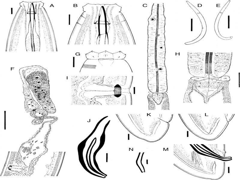
Figure 1:
Labronema montanum sp. n. from Spain. (A, B): Anterior region in lateral, median view. (C): Pharyngeal expansion. (D): Female, entire. (E): Male, entire. (F): Female, anterior genital branch. (G): Lip region in lateral, surface view. (H): Pharyngo-intestinal junction. (I): Vagina. (J): Spicule. (K, L): Female tail. (M): Male tail. (N): Lateral guiding piece. (Scale bars: A, B, G, I–M = 10 µm; C, F = 50 µm; D, E = 500 µm; H = 20 µm; N = 5 µm).

Figure 2:
Labronema montanum sp. n. from Spain (female, LM). (A): Entire. (B, C): Anterior region in lateral median view. (D): Lip region in lateral, surface view. (E, I): Vagina. (F): Anterior genital branch. (G): Pharyngeal expansion. (H): Pharyngeal enlargement. (J–M): Tail. (Scale bars: A = 500 µm; B–D, H–M = 10 µm; F, G = 50 µm.)

Figure 3:
Labronema montanum sp. n. from Spain (male, LM). (A): Entire. (B–D): Pharyngo-intestinal junction showing the three lobes. (E): Posterior body region. (F): Spicule. (G, H): Male. (I): Lateral guiding piece. (J): Sperm cells. (Scale bars: A = 500 µm; D–B = 20 µm; E = 50 µm; F–H = 10 µm; I = 5 µm; J = 10 µm).

Figure 4:
Labronema montanum sp. n. from Spain (SEM). (A): Lip region in ventral view. (B): Same in frontal view. (C): Vulva, ventral view. (D, E): Male, posterior body region showing ad-cloacal pair (D) and ventromedian series (E) of genital papillae. (F): Female tail, ventral view. (G, H): Female tail, lateral view, showing indentations of body cuticle.
Material examined
In total, 17 females and 11 males, from one location, were examined.
Morphometrics
See Table 1.
Table 1.
Morphometrics of Labronema montanum sp. n. from Spain. Measurements in μm except L in mm, and in the form: average ± sd (range).
| Holotype | Paratypes | Paratypes | |
|---|---|---|---|
| Character | ♀ | 17♀♀ | 11♂♂ |
| L | 1.74 | 1.83 ± 0.14 (1.56–2.07) | 1.87 ± 0.12 (1.68–2.08) |
| a | 17.8 | 21.3 ± 2.0 (17.8–24.4) | 22.7 ± 1.8 (20.3–25.9) |
| b | 3.7 | 3.8 ± 0.3 (3.3–4.3) | 3.7 ± 0.2 (3.3–4.1) |
| c | 62 | 68.1 ± 9.4 (56–86) | 72.2 ± 5.5 (65–84) |
| V | 58 | 58.4 ± 1.2 (56.5–60.2) | – |
| c’ | 0.6 | 0.6 ± 0.1 (0.5–0.8) | 0.6 ± 0.0 (0.6–0.7) |
| Lip region diameter | 23 | 22.0 ± 1.1 (19–23) | 22.2 ± 1.3 (19–24) |
| Odontostyle length ventral side | 27 | 25.0 ± 1.9 (21–28) | 26.0 ± 2.0 (22–29) |
| Odontostyle length dorsal side | 28 | 26.5 ± 2.1 (23–30) | 27.3 ± 2.0 (23–30) |
| Odontophore length | 43 | 42.0 ± 1.9 (37–44) | 42.7 ± 2.2(38–45) |
| Neck length | 474 | 476 ± 27 (417–514) | 500 ± 31 (450–551) |
| Pharyngeal expansion length | 238 | 242 ± 19 (205–272) | 247 ± 16 (212–264) |
| Body diam. at neck base | 92 | 80.6 ± 8.3 (65–98) | 78.9 ± 6.1 (72–91) |
| mid-body | 98 | 85.9 ± 7.6 (72–99) | 82.8 ± 8.9 (71–101) |
| anus/cloaca | 44 | 43.9 ± 3.3 (39–51) | 42.3 ± 3.7 (36–47) |
| Distance vulva – anterior end | 1015 | 1068 ± 88 (889–1232) | – |
| Prerectum length | 93 | 125 ± 23 (84–160) | 202 ± 34 (150–268) |
| Rectum/cloaca length | 61 | 60.0 ± 4.1 (52–68) | 71.6 ± 5.6 (61–80) |
| Tail length | 28 | 27.4 ± 4.5 (19–35) | 26.1 ± 2.9 (20–30) |
| Spicules length | – | – | 71.3 ± 3.6 (65–76) |
| Ventromedian supplements | – | – | (20–25) |
