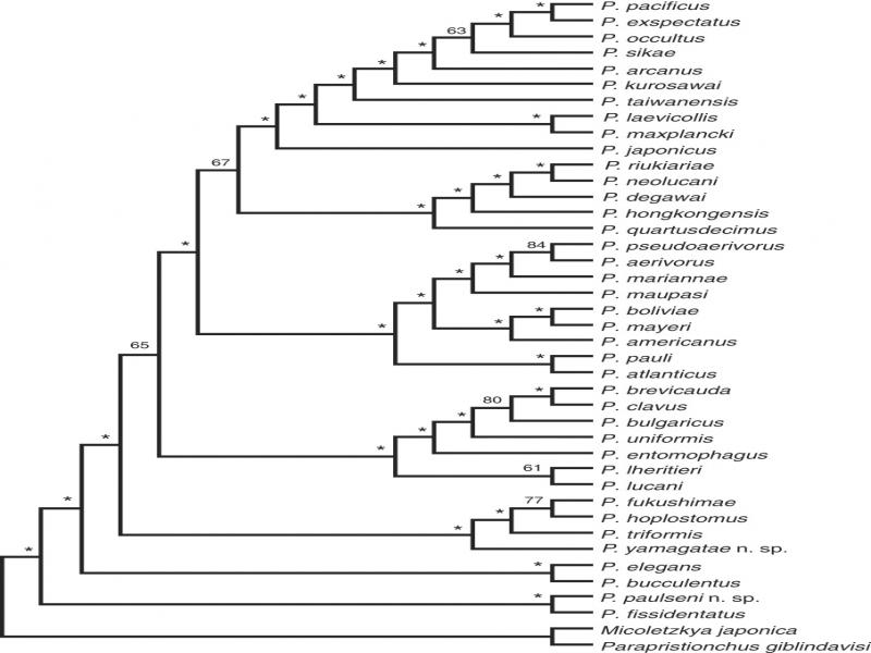
The genus Pristionchus (Kreis, 1932) with the model organism P. pacificus has emerged as an important group of nematodes to study various aspects of macroevolution, including comparative developmental biology, phenotypic plasticity, evolutionary ecology, and comparative genomics (Sommer et al., 1996; Sommer, 2015). Many of these studies critically depend on deep taxon sampling and a robust phylogenetic framework (Susoy et al., 2015). The isolation of new Pristionchus species was fostered by the original discovery that these nematodes are often found in a necromenic and entomophilic association with scarab beetles (Herrmann et al., 2006, 2007; Kanzaki et al., 2012b). Subsequent systematic samplings resulted in the characterization of more than 30 Pristionchus species with four distinct biogeographically restricted species groups (Ragsdale et al., 2015; Herrmann et al., 2016).
The three major radiations include the “maupasi species group” in America, the “pacificus species group” in Asia and the “lheritieri species group,” which contains species from Europe and Asia (Ragsdale et al., 2015). As outgroup to these radiations, the “elegans species group” found in Asia consists of only two gonochorists: P. elegans and P. bucculentus (Kanzaki et al., 2012a; Ragsdale et al., 2013, 2015). However, an additional hermaphroditic species, P. fissidentatus, is an outgroup to the “elegans species group” and is the sister group to all described beetle-associated Pristionchus species. Given that most of the seven hermaphroditic species in the genus have evolved independently of one another, they allow the detailed analysis of parallel evolution, e.g., the influence of mating-type transitions on longevity (Weadick and Sommer, 2016; Rödelsperger et al., 2018). P. fissidentatus is of particular interest, given its phylogenetic position; however, it is a hermaphroditic species that has no sister group relationship with any particular species.
Here, we describe two new species. P. yamagatae sp. n. was isolated from Holotricha sp. (Coleoptera: Melolonthinae) in Japan and belongs to the lheritieri-species-complex. P. paulseni sp. n. was isolated from Taiwan and represents the gonochoristic sister species of P. fissidentatus. This species exhibits a mouth dimorphism found in many Pristionchus species, suggesting that mouth-form plasticity is an ancestral character in the genus. Most importantly, P. paulseni sp. n. and P. fissidentatus form a new species group of the genus that represents the sister group to all described beetle-associated Pristionchus species. Thus, the gonochorist P. paulseni sp. n. has a crucial position in the phylogeny of Pristionchus. In order to characterize both species, morphology, morphometrics, mating experiments and genome-wide transcriptome sequencing are used.
Materials and methods
Nematode cultivation
One strain of P. paulseni sp. n. was isolated from an adult of Lucanus dybowski taiwanus (Miwa, 1937) (Coleoptera: Lucanidae) collected at Pilu sacred tree, Taroko National Park, Taiwan. The strain of P. yamagatae sp. n. was isolated from an adult of Holotrichia sp. (Hope, 1837) (Coleoptera: Scarabaeidae, Melolonthinae) collected at Yuza, Yamagata, Japan. Both strains have been kept in laboratory culture on NGM agar plates seeded with Escherichia coli strain OP50 under the culture code and freezing voucher numbers RS5918 (P. paulseni sp. n.) and RS5964 (P. yamagatae sp. n.).
Morphological observation and preparation of type material
Light microscopic observations for drawings and morphometrics were conducted using live nematode material, which was handpicked from culture plates (Kanzaki et al., 2013).
Molecular characterization and phylogenetic analysis
A species phylogeny of the complete Pristionchus genus was reconstructed as described in Rödelsperger et al. (2018). In the following, we give a brief summary about the generation of the Pristionchus phylogeny (please see Rödelsperger et al., (2018) for full details). For all cultivable Pristionchus species, worms were grown on NGM plates seeded with E. coli OP50 at 20°C and total RNA was isolated from 2–3 mixed-stage plates per species using standard Trizol extraction following the manufacturers’ instructions (Zymo Research, CA, USA). RNA-seq libraries were prepared using TruSeq RNA library preparation kit v2 (Illumina, Inc., CA, USA), according to the manufacturer’s instructions, from 1 µg of total RNA in each sample and sequenced on the Illumina HiSeq3000 platform, yielding a median of 14m paired reads (2 × 150 bp) per species. Raw reads were submitted to the European nucleotide archive under the study accession PRJEB20959. RNA-seq reads were assembled into transcriptomes using Trinity (version v2.2.0) (Grabherr et al., 2011). For further analysis, only the first reported isoform per gene was selected and the longest complete or partial ORF (⩾60 amino acids) was called. Orthologous clusters were generated by orthAgogue (Ekseth et al., 2014) and protein sequences were aligned using the MUSCLE software (version 3.8.31) (Edgar, 2004). 2,092 high quality alignments containing at least 14 species (without any duplication), with at least 50 amino acid positions with coverage in all represented species, were concatenated into a supermatrix spanning 350,000 amino acids. On the basis of the previous analysis of dozens of gene families (Baskaran et al., 2015), we chose the LG substitution model to reconstruct a maximum-likelihood tree using RA × ML (version 8.2.9, options –m PROTGAMMAILG –f a –N 100) (Stamatakis, 2014).
Results
Description of common characters
The majority of Pristionchus species are cryptic and typologically similar to each other, including the two novel species described here. In general, Pristionchus species are distinguished from each other by stomatal and male tail characters only (for review see Ragsdale et al., 2015). Therefore, the general morphology, e.g., body shape and gonadal structures, is described first as common characters, and then the distinctive characters are described for each species.
Adult
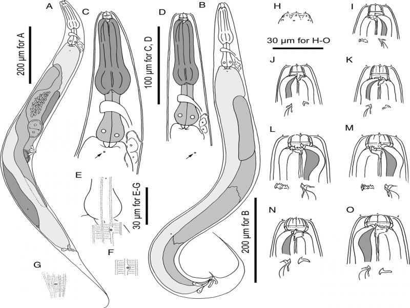
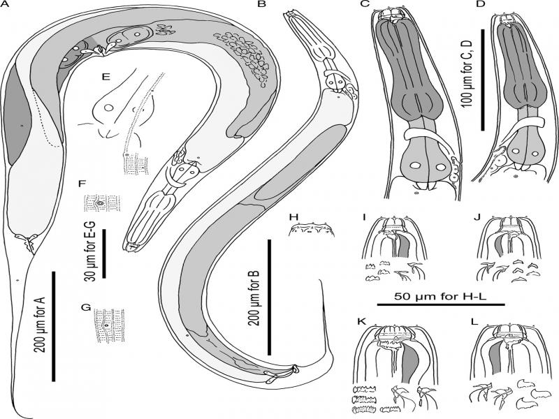
Their characteristics include a cylindrical and stout body, a thick cuticle, with fine annulation and clear longitudinal striations. Lateral field consists of two lines, weakly distinguishable from body striation with the presence of deirid. Head exists without apparent lips and with six short and papilliform labial sensillae. Four small, papilliform cephalic papillae are present in males, as typical for diplogastrid nematodes. Amphidial apertures are located on the lateral sector, slightly dorsally shifted, at level of margin of cheilo- and gymnostom. Stomatal dimorphism is present, with stenostomatous (narrow mouthed) and eurystomatous (wide mouthed) forms occurring in both males and females, but male eurystomatous form is not as common as females. Detailed stomatal morphology is described below. Dorsal pharyngeal gland is clearly observed, with a penetrating dorsal tooth to gland opening. The anterior part of pharynx (=pro and metacorpus) is 1.5 times as long as the posterior part (=isthmus and basal bulb). Procorpus is very muscular, stout, occupying half to two-thirds of the corresponding body diameter. Metacorpus is very muscular, forming a well-developed median bulb. Isthmus is narrow, not muscular. Basal bulb is glandular. Pharyngo-intestinal junction is clearly observed; it is well-developed. Nerve ring is usually surrounding the middle region of isthmus. Excretory pore is not conspicuous, ventrally located at the level of isthmus to pharyngo-intestinal junction; excretory duct extends anteriad and is reflexed back to the position of pore. Deirid is observed laterally, located from the level of pharyngo-intestinal junction to one body diameter and posterior to the junction. Hemizonid is not clearly observed. Lateral glands, small pores connected to secretory cell, are present and observed on the lateral body surface, with positions inconsistent among individuals, numbering 5 to 8 for males and 9 to 13 for females. Because the structure is very small and indistinctive, the consistency of their position and number was not confirmed by light microscopy.
Stenostomatous form
Cheilostom consists of six per- and interradial plates. Incision between plates is not easily distinguished by light microscopy. The anterior end of each plate is usually rounded and elongated to project from stomatal opening and forms a small flap. Some more details are described for each species below. The anterior end of each plate sometimes splits into two, forming two narrow flaps, i.e., number of cheilostomatal flaps varies from 6 to 12, regardless of the number of plates (=6). Gymnostom is short, with a cuticular ring-like anterior end overlapping cheilostom internally. Dorsal gymnostomatal wall is slightly thickened compared to the ventral side. Stegostom is divided into three subsections: pro-meso, meta and telostegostom. Pro-meso stegostom forms a weak cuticularized ring surrounding the anterior edge of pharynx. Metastegostom bears a conspicuous and movable triangular or flint-shaped dorsal tooth with strongly sclerotized surface, giving an appearance of an inverted V-shape in light microscopy in lateral view; a pointed left subventral ridge with three minute adventitious denticles on a plate is present; a pointed right subventral ridge exists, often with distinct distal adventitious denticle(s). Telostegostom forms a weakly sclerotized cup-like cavity connecting stoma and pharynx.
Eurystomatous form
Cheilostom is divided into six distinctive per- and interradial plates. The anterior end of each plate is usually rounded and elongated to project from stomatal opening, forming a small flap. The anterior part of each plate is sometimes split into two, forming two narrow flaps, i.e., number of cheilostomatal flaps varies from 6 to 12, regardless of the number of plates (=6). Gymnostom is present with a thick cuticle, forming a short, ring-like tube being thicker posteriorly. The anterior end of gymnostom is internally overlapping the posterior end of cheilostomatal plates. Stegostom is divided into three subsections: pro-meso, meta and telostegostom. Pro-mesostegostom forms a weak cuticularized ring surrounding the anterior edge of pharynx. Metastegostom bears large claw-like dorsal tooth, large claw-like right subventral tooth, ridge of left subventral denticles or cusps, varying in number and size. Dorsal and right subventral teeth are movable. No movement is observed for left subventral denticles. Telostegostom forms a weak sclerotized cup-like cavity connecting stoma and pharynx.
Male
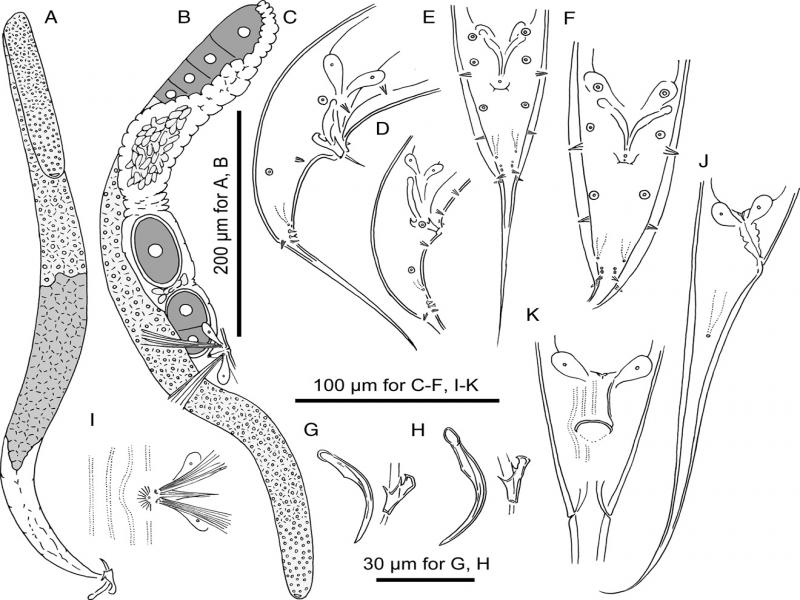
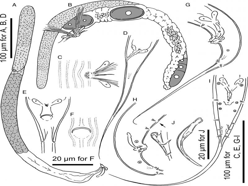
They are ventrally arcuate and become strongly ventrally curved at tail region when killed by heat. Testis is single and ventrally located; the anterior part is reflexed to the right side. Spermatogonia is arranged in three to five rows in the reflexed part; then well-developed spermatocytes are arranged in three to four rows in anterior two-thirds of the main branch; then mature amoeboid spermatids are arranged in multiple rows in remaining, proximal part of gonad. Vas deferens is not clearly separated from other parts of gonad. Three (two subventral and one dorsal) cloacal gland cells are observed at distal ends of testis and intestine. Spicules are paired and separate. Spicules are smoothly curved in ventral view, adjacent to each other for distal third of their length, smoothly tapering to pointed distal end. Spicule in lateral view is smoothly ventrally arcuate, giving spicule about 100° curvature; oval manubrium is present at the anterior end; lamina/calomus complex (blade) is clearly expanded slightly posterior to manubrium (ca 1/4 of blade length from anterior), smoothly tapering to pointed distal end. Gubernaculum conspicuous, about one-third of spicule length, is broad anteriorly such that dorsal wall is slightly recurved and that dorsal and ventral walls separate at 50 to 60° angle at the posterior end. The dorsal side of gubernaculum possesses a single, membranous, anteriorly directed process and a lateral pair of more sclerotized, anteriorly and obliquely ventrally directed processes. In lateral view, anterior half of gubernaculum is present with two serial curves separated by anteriorly and obliquely ventrally directed process, with anterior terminal curvature being highly concave and almost closed, and with deep posterior curvature being one-third of the gubernaculum length; posterior half forms a tube-like process enveloping spicules. A thick cuticle exists around tail region, falsely appearing as a narrow leptoderan bursa in ventral view. Cloacal opening (co) is slit-like in ventral view. One small, ventral, single genital papilla (vs) exists on anterior cloacal lip. Nine pairs of genital papillae (v1-v7, ad, pd) and a pair of phasmids (ph) are present, with three precloacal and six postcloacal pairs. Within those pairs, three distal ventral pairs (v5, v6, and v7) and a dorsal pair (pd) are close to each other around the posterior end of tail (just anterior to the root of spike). Anterior five pairs of papillae (v1-4 and ad) are present, almost equal in size, rather large and conspicuous; v7 and pd papillae are obviously smaller than anterior five pairs; v5 and v6 are very small, sometimes difficult to observe with light microscope. Anterior two pairs of the ventral triplet papillae (v5 and v6) papilliform are present and born from a socket-like base, v7 is simple or typical thorn-like in shape. The tip of v6 papillae is split into two small papilla-like projections. A detailed arrangement of papillae and phasmid will be described for each species below. The tail is conical, with a long spike about two to three cloacal body diameter. Bursa or bursal flap is absent.
Female
They are relaxed or slightly ventrally arcuate when killed by heat. Gonad is didelphic, amphidelphic. Each gonadal system is arranged from vulva/vagina as uterus, oviduct, and ovary. The anterior gonad is in right of intestine, with uterus and oviduct extending ventrally and anteriorly on right of intestine and with a totally reflexed (=antidromous reflexion) ovary extending dorsally on left of intestine. Oocytes are mostly arranged in three to four rows in distal two-thirds of ovary and in double or single row in rest of the ovary, with distal tips of each ovary reaching oviduct of the opposite gonad branch. The anterior end of oviduct (= junction tissue between ovary and oviduct) consists of rounded cells. The anterior part of oviduct consists of rounded cells forming simple tube. The middle part of oviduct serves as spermatheca and consists of roundish and relatively large cells. Eggs in single to multiple-cell stage or even further stages are developed at the posterior part of oviduct (= uterus), which in young female is composed of squared or angular cells, long enough to contain one well-developed oocyte. Receptaculum seminis is not observed, i.e., the organ is not independent and is a part of oviduct/uterus. Vaginal glands are present but obscure. Vagina is perpendicular to the body surface, surrounded by sclerotized tissue. Vulva is slightly protuberant in lateral view and pore-like in ventral view. Rectum is about one anal body diameter long, intestine/rectum junction is surrounded by a well-developed sphincter muscle. Three anal glands (two subventral and one dorsal) are present but not obvious. Anus is in the form of dome-shaped slit, posterior anal lip slightly protuberant. Phasmid is about one to two anal body diameter long, posterior to anus. Tail is long, smoothly tapered to distal end variable from filiform to long and conical.
Pristionchus paulseni* sp. n. (Figs 1, 2; Table 1)
Table 1
Morpometrics P. paulseni sp. n. and P. yamagatae sp. n.
| P. paulseni RS5918 | P. yamagatae RS5964 | |||
|---|---|---|---|---|
| Character | stenostomatous male | stenostomatous female | stenostomatous male | stenostomatous female |
| n | 10 | 10 | 10 | 10 |
| L | 1208±95.4 (990–1315) | 1560±230 (1222–1895) | 848±42.2 (796–916) | 1162±121.9 (937–1385) |
| L’ | 1032±92.5 (813–1138) | 1298±209.6 (978–1560) | 681±34.7 (616–724) | 908±103.7 (717–1096) |
| a | 14±1,3 (12–16) | 14±0.6 (13–15) | 14±1.4 (13–18) | 14±1.2 (12–17) |
| b | 7.2±0.8 (5.8–9.0) | 8.7±1.0 (7.3–10.1) | 5.8±0.3 (5.3–6.2) | 6.8±0.6 (5.8–7.8) |
| c | 6.9±0.8 (5.6–8.3) | 6.0±0.6 (5.0–6.9) | 5.1±0.4 (4.4–5.9) | 4.6±0.4 (4.1–5.3) |
| c’ | 4.0±0.6 (3.4–4.9) | 6.0±0.5 (5.3–6.8) | 5.1±0.6 (3.9–6) | 7.3±0.7 (6.1–8.2) |
| T or V | 58±2.9 (53–62) | 47±0.9 (45–48) | 51±3.3 (45–54) | 45±1.3 (44–48) |
| Maximum body diam. | 86±4.7 (75–91) | 114±19.3 (84–138) | 60±6.4 (47–70) | 84±9.6 (68–98) |
| Stoma length | 13.0±0.5 (12.0–13.8) | 14.4±0.4 (13.9–15.0) | 11.5±0.7 (10–12.2) | 13.3±0.5 (12.3–14) |
| Stoma diam. | 5.7±0.5 (5.0–6.8) | 6.8±0.4 (6.2–7.5) | 6±0.5 (5.4–6.7) | 7.4±0.5 (6.7–8.3) |
| Pharynx length (head to base of pharynx) | 156±13.8 (122–168) | 169±7.2 (158–178) | 135±5.4 (124–143) | 156±7.3 (146–170) |
| Anterior pharynx (pro- + metacorpus) | 96±8.3 (75–105) | 106±2.9 (102–110) | 83±3.9 (77–88) | 98±4.2 (93–106) |
| Posterior pharynx (isthmus + basal bulb) | 61±6.1 (47–68) | 64±4.8 (56–69) | 52±2.2 (47–55) | 58±3.9 (52–64) |
| Ant/total pharynx % | 61±1.3 (59–63) | 62±1.6 (60–65) | 63±4.2 (59–74) | 63±1.2 (61–65) |
| Median bulb diam. | 28±2.1 (25–31) | 34±2.8 (30–36) | 22±0.7 (21–23) | 30±3.8 (22–36) |
| Terminal bulb diam. | 26±1.8 (23–28) | 30±3.9 (25–34) | 21±1.3 (18–22) | 27±3.7 (20–33) |
| Testis length | 698±69.9 (532–787) | – | 433±37.9 (372–475) | – |
| Ant. end to vulva | – | 730±101.3 (592–860) | – | 527±48.8 (446–610) |
| Vulva to anus distance | – | 570±103.2 (424–710) | – | 380±52.7 (281–470) |
| Cloacal or anal body diam. | 44±3.4 (36–48) | 44±4.3 (37–50) | 33±3.6 (28–40) | 35±1.9 (32–38) |
| Tail length | 176±19.3 (155–220) | 262±69.9 (235–335) | 167±17.5 (140–192) | 255±27.1 (212–289) |
| Spicule length (curve) | 51±2.6 (47–54) | – | 43±1.2 (41–45) | – |
| Spicule length (chord) | 41±2.5 (36–44) | – | 36±0.8 (34–37) | – |
| Gubernaculum length | 14±1.3 (12–16) | – | 14±1.4 (13–18) | – |