
Table 1
Primers used in this study for qRT-PCR.
| Gene | Description | Forward primer (5′-3′) | Reverse primer (5′-3′) | Reference |
|---|---|---|---|---|
| SlPR1 | Lycopersicon esculentum PR1a | CCAAGACTATCTTGCGGTTCA | CGCTCTTGAGTTGGCATAGT | Li et al. (2015a) |
| SlPR2 | beta-1.3-glucanase | TCCAGGTAGAGACAGTGGTAAA | CCTAAATATGTCGCGGTTGAGA | Li et al. (2015a) |
| SlPR5 | Lycopersicon esculentum PR5 | CCCAAACACCCTAGCTGAAT | GGGCGAAAGTCATCGGTATATTA | Li et al. (2015a) |
| SlPAL | Phenylalanine ammonia-lyase | TGATGAACGGAAAGCCTGAA | CTGAGCTGCCTTGACATAAGA | Li et al. (2015a) |
| CDPK15 | Calcium-dependent protein kinase | ACGGACAATAGTGGGACA | TGCTTAACTTCAGCCTCC | Hu et al. (2016) |
| RbohB | Respiratory burst oxidase homologs | AGGGAATGATAGAGCGTCG | CATCGTCATTGGACTTGGC | Li et al. (2015b) |
| Ubi3 | Ubiquitin | GTGTGGGCTCACCTACGTTT | ACAATCCCAAGGGTTGTCAC | Bhattarai et al. (2008) |
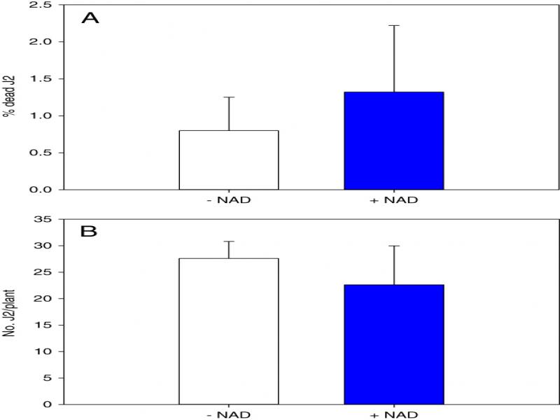
Figure 1
Effect of Nicotinamide adenine dinucleotide (NAD) treatment on Meloidogyne hapla viability and infectivity. (A) Percentage of dead J2 after incubation for 48 hr in NAD or water (control). Bars represent the average mean of 18 biological samples ± standard deviation. (B) Infectivity of NAD-incubated and water-incubated M. hapla juveniles in tomato roots 48 hr post-inoculation. Bars represent the average mean of fourteen biological samples ± standard deviation. Statistical significance of the difference was tested using Student’s t-test (*=P < 0.05).
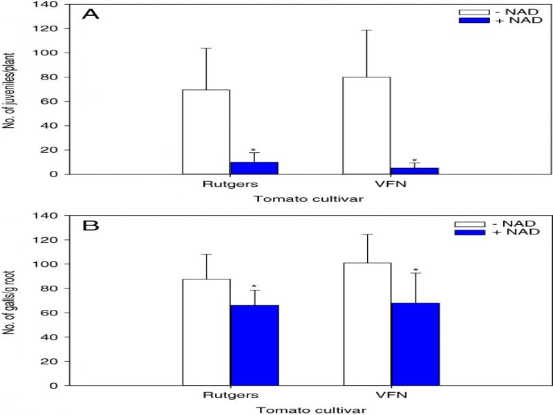
Figure 2
Effect of Nicotinamide adenine dinucleotide (NAD) on the development of Meloidogyne hapla in tomato plants. Tomato seedlings (20-d-old) were soil drenched with 5 mM NAD solution or water (−NAD) one day before M. hapla inoculation. (A) Number of J2 inside tomato cultivars; Rutgers (Mi−) and VFN (Mi+) 48 hr post-inoculation (hpi). (B) The number of galls per gram of root in tomato cultivars Rutgers and VFN 15 d post-inoculation (dpi). Bars represent the average mean of 15 biological samples ± standard deviation. Statistical significance of the difference was tested using Student’s t-test (*=P < 0.05).
Table 2
Effect of NAD application on shoot fresh weight (g) of susceptible and resistance tomato cultivars in the presence or absence of Meloidogyne hapla a.
| Inoculated | Non-inoculated | |||
|---|---|---|---|---|
| Cultivarb | ||||
| Treatmentc | Rutgers | VFN | Rutgers | VFN |
| Control | 1.12 (0.39) | 0.93 (0.23) | 0.91 (0.22) | 0.86 (0.43) |
| NAD | 1.98 (0.35)* | 2.17 (0.30)* | 1.75 (0.56)* | 1.85 (0.70)* |
1 Notes: aValues represent the mean (standard deviation in parentheses) shoot fresh weight (stem and leaves) of 15 plants. Asterisk within the same column indicates the significant difference (P<0.05) between nicotinamide adenine dinucleotide (NAD) and control; b root-knot nematode susceptible cultivar Rutgers (Mi−) and RKN resistant cultivar VFN (Mi+) were inoculated with 300 to 400 M. hapla J2 or water for the non-inoculated plants; c 21 d-old plants were soil drenched with 5 mM nicotinamide adenine dinucleotide (NAD) solution or water for control 24 hr before J2 inoculation, samples were collected 15 d after nicotinamide adenine dinucleotide (NAD) treatment.
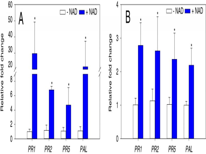
Figure 3
Relative expression of defense-related genes in response to NAD treatment. 20-d-old plants were treated with 5 mM NAD, or water (−NAD) and roots were harvested 24 hr post application. Expression levels of indicated genes; pathogenesis-related protein 1a (PR1), beta-1,3-glucanase (PR2), pathogenesis-related protein 5× (PR5), and phenylalanine ammonia-lyase (PAL) were quantified in tomato cultivars (A) Rutgers (Mi−), and (B) VFN (Mi+) using qRT-PCR. Ubiquitin gene (Ubi3) was used as an internal control. Gene expression values are presented relative to water treated control (−NAD). Bars represent the average mean of six biological samples ± standard deviation. Statistical significance of the difference was tested using Student’s t-test (*=P < 0.05).
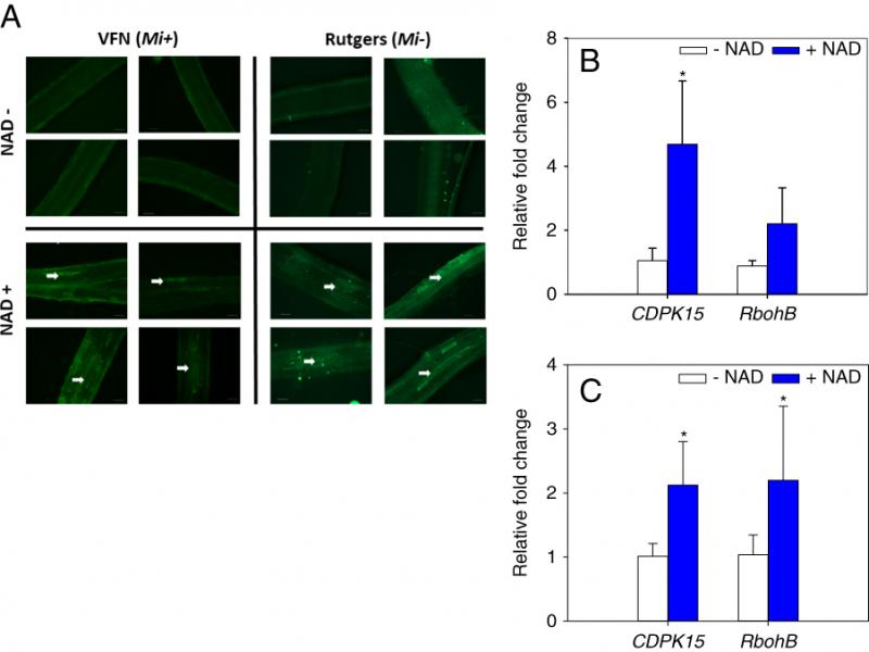
Figure 4
Nicotinamide adenine dinucleotide (NAD) treatment induces calcium signaling. (A) Microscopic observation of Ca2+ levels in roots of tomato cultivars Rutgers and VFN. Tomato seedlings were treated with 10 ml 5 mM NAD or control (−NAD) for 24 hr, then root sections (2 cm) were labeled with Ca2+ – sensitive dye, Calcium Green-1. Note Ca2+ accumulation as revealed by green fluorescence microscopy (white arrows). Scale bars = 200 μm. (B) and (C) Gene expression analysis of calcium-dependent protein kinase (CDPK15) and respiratory burst oxidase homologs (RbohB) in tomato cultivars Rutgers and VFN, respectively. Real-time PCR analysis was performed to determine the transcript levels of CDPK15 and RbohB in tomato roots pretreated with 5 mM NAD or control (−NAD) for 24 hr. Bars represent the average mean of six biological samples ± standard deviation. Statistical significance of the difference was tested using Student’s t-test (*=P < 0.05).