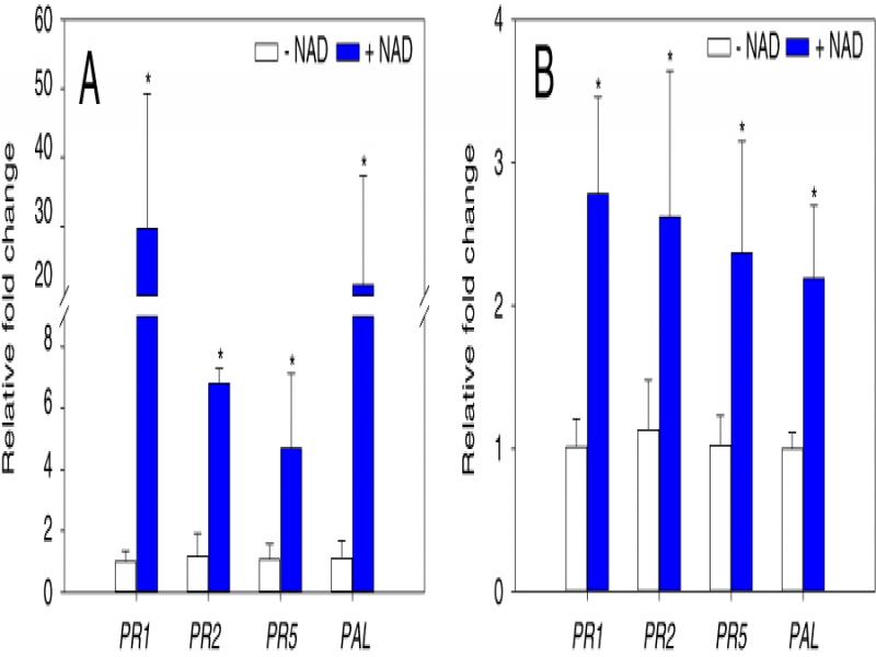
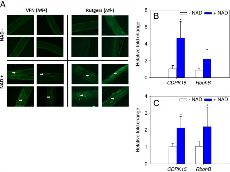
Root-knot nematodes (RKNs; Meloidogyne spp.) are sedentary endoparasitic nematodes that can infect a wide range of plant species worldwide, which results in approximately $70 billion in crop losses annually (Caboni et al., 2012). Meloidogyne spp. is ranked within the top 10 most economically devastating plant-parasitic nematodes, with Meloidogyne incognita, M. arenaria, M. hapla, and M. javanica as the four major crop-damaging species (Jones et al., 2013). In tomato, yield loss due to RKNs ranges from 25 to 100%, depending on nematode species, population densities, and tomato cultivar (Seid et al., 2015). Management practices such as chemical nematicides, cover crops, and biological control have been well documented (Monfort et al., 2007; Zasada et al., 2010; Adam et al., 2014). However, factors such as the toxic effect of chemical nematicides on the environment and humans, the wide host range of Meloidogyne spp., and the effect of soil properties limit the use of these practices (Pantelelis et al., 2006; Barbary et al., 2015). As a result of these limitations, plant resistance has become the most widely used and effective management approach to control RKN in tomato (Fuller et al., 2008).
In tomato, resistance to RKN is mediated by a single dominant R gene called Mi-1 that was introduced to cultivated tomato from a single cross with its wild relative Solanum peruvianum (formerly Lycopersicon peruvianum) (Kaloshian et al., 1998). However, the durability of Mi resistance is reduced by sustained high soil temperature (above 28°C), and the emergence of virulent nematode biotypes have been reported likely due to the intensive use of the Mi gene that applied selective pressures on nematode populations (Devran et al., 2010). In contrast to genetic resistance, induced resistance is a plant defense mechanism triggered by biological or chemical elicitros, which protects plants against a broad spectrum of biotic stresses including plant-parasitic nematodes. Following application of an elicitor, defense responses like oxidative burst, cell wall fortification, and synthesis of antimicrobial compounds such as pathogenesis-related proteins may be triggered directly or after pathogen attack (Walters et al., 2013). While the mechanisms have yet to be fully understood, elicitors such as acibenzolar-S-methyl (ASM), benzothiadiazole (BTH), DL-β-amino-n-butyric acid (BABA), and salicylic acid (SA) have been reported to induce resistance against RKN (Oka et al., 1999; Chinnasri et al., 2003; Ji et al., 2015) and are likely the result of induced plant resistance.
Plant metabolites are also known to activate defense mechanisms and induce resistance against pathogens (Rojas et al., 2014; Piasecka et al., 2015). Pyridine nucleotides, like nicotinamide adenine dinucleotide (NAD), are important redox carriers and play a crucial signaling role in response to stresses (Hashida et al., 2009). In Arabidopsis, exogenous application of NAD induces the expression of pathogenesis-related (PR) genes via the Ca2+ dependent signaling pathway, causing accumulation of SA, and enhanced disease resistance to the bacterial pathogen Pseudomonas syringae pv. tomato (Pst) (Zhang and Mou, 2009). Further characterization of NAD-dependent immune responses showed that intracellular NAD-overproducing nadC transgenic lines are more resistant to a diverse range of virulent pathogens including Pst-AvrRpm1, Dickeya dadantii, and Botrytis cinerea (Pétriacq et al., 2016). NAD can also elicit a defense-related metabolic signature detectable by mass spectrometry that is similar to those triggered by hormones and Pathogen Associated Molecular Pattern (PAMP) such as flagellin (Flg22) and fungal chitin (Pétriacq et al., 2016). The goal of this study was to evaluate NAD-induced plant resistance in a relevant crop and assess its efficacy against RKN.
Materials and Methods
Plant materials and growth conditions
Tomato (Solanum lycopersicum) lines used in this work included S. lycopersicum cv. Rutgers which does not have the Mi-1 gene, and cv. VFN (resistance to Verticillium, Fusarium, and root-knot Nematode) with the Mi-1 gene (Urban farmer LLC, Westfield, IN). Both cultivars are susceptible to M. hapla infection. Seeds were germinated in a mixture composed of sand and soil in a 1:1 (v/v) ration. Plants were grown in a growth chamber at 24°C and a 16-h light and 8-h dark regime, with daily watering. After two weeks, seedlings were washed and transferred to trays containing sand mix and kept on the bench for one week to recover before nematode inoculations.
Nematode viability
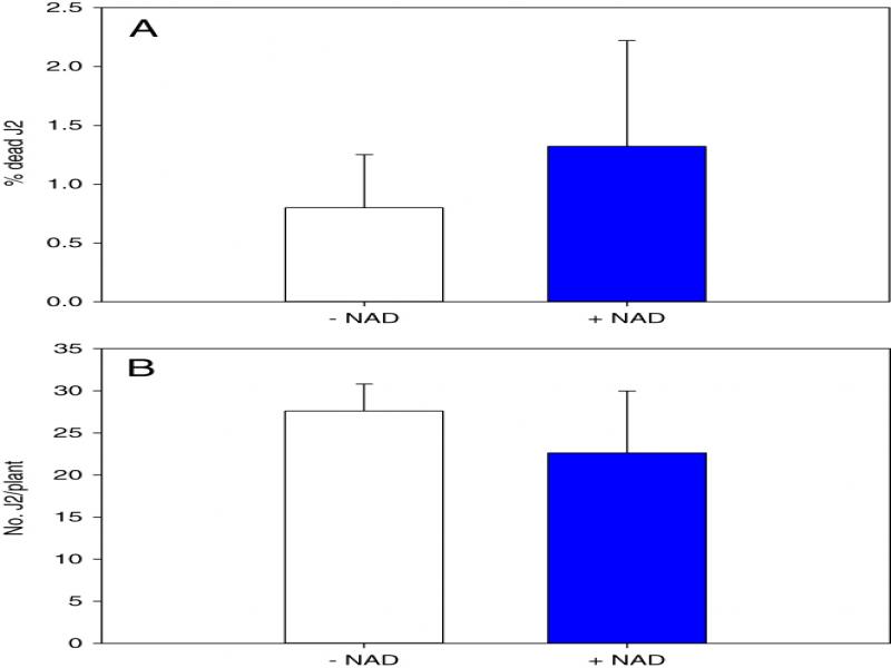
To test the negative effect of NAD on nematode mortality and infectivity, 400 J2 of M. hapla were incubated in six-well plates, each well contained 2 ml of 5 mM NAD solution or water as control under dark conditions at room temperature for 2 d. Live and dead nematodes were counted under a dissecting microscope and the percentage of dead nematodes was counted. Each treatment included six technical replicates, and the experiment was repeated three times. To investigate the effect of NAD on nematode infectivity, juveniles were collected after 2 d incubation in NAD or water, washed three times in water, and inoculated onto universal susceptible Rutgers tomato plants at100 J2 per plant.
Nematode inoculation
The root-knot nematode, M. hapla, culture was maintained on the susceptible S. lycopersicum cv. Rutgers under greenhouse temperature and light conditions. Nematode eggs were extracted from infected tomato roots using 10% commercial bleach and 40% sucrose solution (Hussey and Barker, 1973), eggs were then incubated at 25°C in hatching bowls, second-stage juveniles (J2s) were collected after 5 d. Pretreatment with NAD was performed as follows: 20 d old seedlings of cvs. Rutgers and VFN were soil drenched with 10 ml of 5 mM NAD solution (or water control), and 1 d later they were inoculated with approximately 350 to 400 J2 of M. hapla per plant by distributing the nematode suspension into three holes (2 cm deep) in the soil.
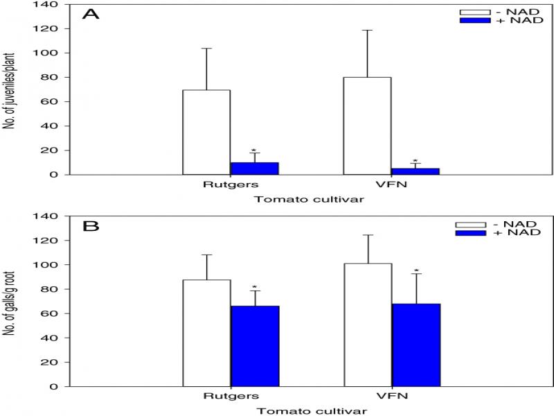
To study the effect of NAD on M. hapla development in tomato plants, two cultivars (VFN, and Rutgers), two chemical treatments (NAD and water), two sampling time points at 2, and 15 dpi (days post-inoculation), and two nematode inoculation levels (inoculated and non-inoculated) were arranged in a completely randomized design. There were five replicate pots per treatment combination and the experiment was repeated three times. Five plants were destructively sampled at 2 and 15 dpi and used to quantify J2 penetration, and number of galls, respectively. To visualize the nematodes inside the roots, tomato seedlings were washed to remove sand particles and the roots were placed in 10% commercial bleach solution for 4 min, then incubated in tap water for at least 15 min to remove excess bleach. Roots were then boiled in 3.5% acid fusion stain, after which they were washed with tap water and were boiled briefly in acidified glycerol (10:100 HCl/glycerol, vol/vol) for destaining. Number of J2 and galls were counted using stereomicroscope (LeicaMz6, Leica Microsystems, IL, USA). Fresh root and shoot weight were measured at 15 dpi on five plants per treatment, by rinsing in tap water to remove sand particles, and drying the excess water using paper towels. There were three independent biological experiments, each experiment has five biological replicates per treatment.
RNA extraction, cDNA synthesis, and real-time PCR
For RNA extraction, whole tomato roots were collected 24 h after NAD application then grounded to a fine powder in liquid nitrogen, and total RNA was extracted using Trizol reagent (Invitrogen, Carlsbad, CA, USA). DNA contamination was removed using RNase-free DNase I (Invitrogen) following the manufacturer’s instructions. RNA was quantified using a Nano-Drop 1,000 spectrophotometer (Thermo Scientific, Wilmington, DE), and integrity was verified on 1% agarose gel. First-strand cDNA was synthesized from 0.5 µg of total RNA using iScript cDNA synthesis kit (Bio-Rad, Hercules, CA, USA).
Quantitative PCR was performed using iTaq universal SYBR Green Supermix (Bio-Rad) and Applied Biosystems Step One Plus detection system. The reaction mix consisted of 5 µl master mix, 0.5 µl of reverse and forward primers (500 nM final concentration), 2 µl of diluted cDNA (10 ng final concentration), and the final volume was adjusted to 10 µl with RNase DNase free water (Invitrogen). The primers sequences used for real-time PCR are listed in Table 1. The thermal cycling protocol was 2 min at 95°C, 40 cycles of 3 s at 95°C, 30 s at 60°C, followed by melting curve data collection to check for nonspecific amplification and primer dimers. Relative gene expression was calculated using the 2−ΔΔct method (Livak et al., 2001), in which the transcription levels of the target genes in control seedlings (J2 inoculated and water treated) were used as reference for expression analysis, and Ubiquitin was used as the internal control gene expression.
Table 1
Primers used in this study for qRT-PCR.
| Gene | Description | Forward primer (5′-3′) | Reverse primer (5′-3′) | Reference |
|---|---|---|---|---|
| SlPR1 | Lycopersicon esculentum PR1a | CCAAGACTATCTTGCGGTTCA | CGCTCTTGAGTTGGCATAGT | Li et al. (2015a) |
| SlPR2 | beta-1.3-glucanase | TCCAGGTAGAGACAGTGGTAAA | CCTAAATATGTCGCGGTTGAGA | Li et al. (2015a) |
| SlPR5 | Lycopersicon esculentum PR5 | CCCAAACACCCTAGCTGAAT | GGGCGAAAGTCATCGGTATATTA | Li et al. (2015a) |
| SlPAL | Phenylalanine ammonia-lyase | TGATGAACGGAAAGCCTGAA | CTGAGCTGCCTTGACATAAGA | Li et al. (2015a) |
| CDPK15 | Calcium-dependent protein kinase | ACGGACAATAGTGGGACA | TGCTTAACTTCAGCCTCC | Hu et al. (2016) |
| RbohB | Respiratory burst oxidase homologs | AGGGAATGATAGAGCGTCG | CATCGTCATTGGACTTGGC | Li et al. (2015b) |
| Ubi3 | Ubiquitin | GTGTGGGCTCACCTACGTTT | ACAATCCCAAGGGTTGTCAC | Bhattarai et al. (2008) |
| Inoculated | Non-inoculated | |||
|---|---|---|---|---|
| Cultivarb | ||||
| Treatmentc | Rutgers | VFN | Rutgers | VFN |
| Control | 1.12 (0.39) | 0.93 (0.23) | 0.91 (0.22) | 0.86 (0.43) |
| NAD | 1.98 (0.35)* | 2.17 (0.30)* | 1.75 (0.56)* | 1.85 (0.70)* |