The family Tylenchidae (Örley, 1880) is a cosmopolitan group of nematodes in the suborder Tylenchina (Chitwood, 1950) and currently contains over 400 species (Siddiqi, 2000; Geraert, 2008). Members of the family have high ecological diversity and abundance, and are associated with algae, mosses, lichens, fungi, and plant roots. Some of the species are considered as weak parasites of plants (Siddiqi, 2000) and typically show low interspecific and high intraspecific morphological variability (Qing and Bert, 2018). Many species are poorly described because of an overreliance on morphological characters using light microscopy (LM) and a limited number of specimens which has led to taxonomic confusion (Qing and Bert, 2017, 2018). Recently, molecular data have been used to improve taxonomic studies of the family members (Palomares-Rius et al., 2009; Bert et al., 2010; Atighi et al., 2013; Panahandeh et al., 2014, 2015a, 2015b, 2016, 2018; Soleymanzadeh et al., 2016; Pereira and Baldwin, 2016; Pereira et al., 2016; Qing et al., 2016, 2017; Pedram et al., 2018; Qing and Bert, 2018). Unfortunately, molecular data are often not available for all genera and species, and some genera are only rarely recovered after their original descriptions.
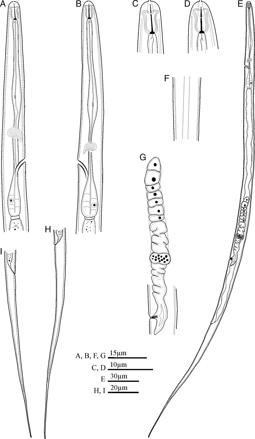
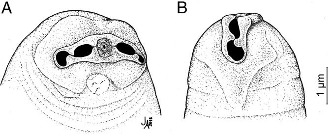
According to a comprehensive review of the family Tylenchidae by Geraert (2008), the family includes 42 valid genera. Recently, two other monotypic genera were added, i.e., Discopersicus (Yaghoubi et al., 2016) from Iran, and Labrys (Qing and Bert, 2018) from China with L. chinensis (Qing and Bert, 2018) as its type species (Qing and Bert, 2018). The latter genus is mainly characterized by having a unique offset labial plate, tapering toward both tips and detached from the adjacent cuticle in scanning electron microscopy (SEM) images, protruding lips under LM, laterally elongate amphidial apertures, two incisures in lateral field, delicate stylet, its shaft two times longer than the cone, elongate fusiform weakly developed median bulb having a distinct weakly sclerotized valvular apparatus, wide excretory pore with heavily sclerotized duct, a round spacious postvulval uterine sac (PUS) and spicules with sharp protrusion (Qing and Bert, 2018). Immediately following the description of L. chinensis, it was reported from Iran and confirmed to be identical following morphological and morphometric analyses (Konani et al., 2018).
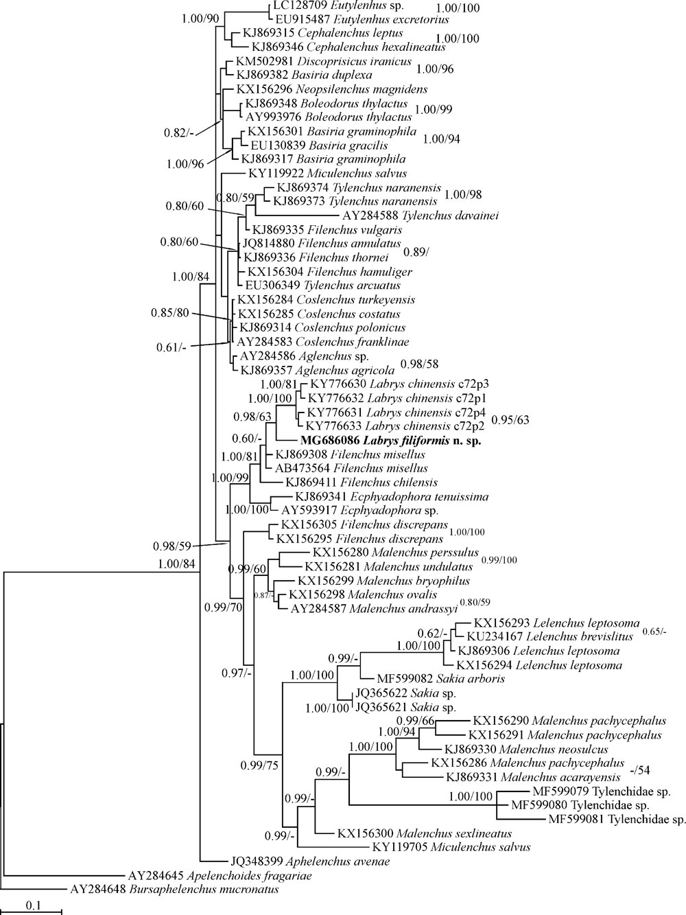
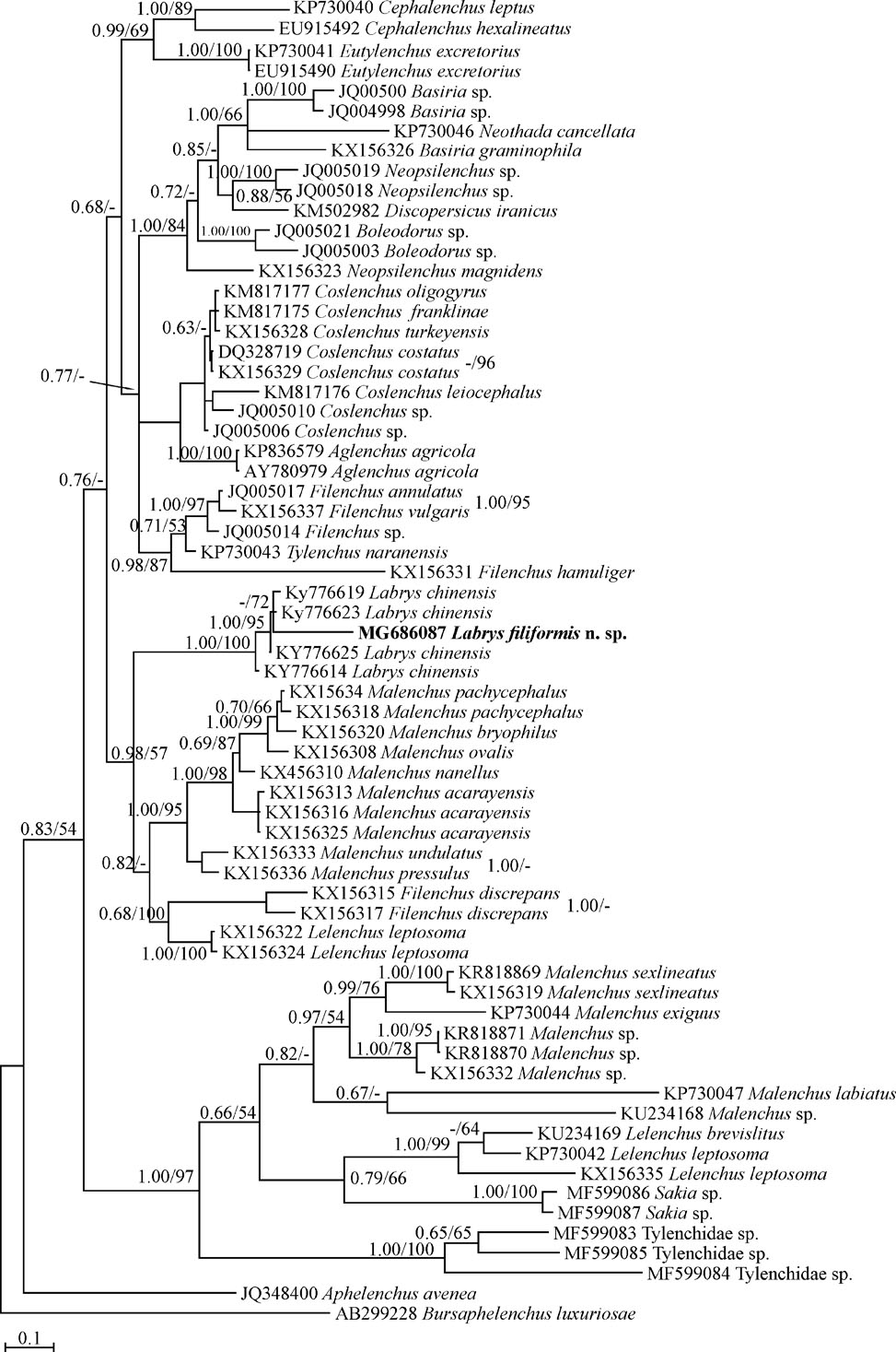
During our extensive study of tylenchids in northern Iran (Panahandeh et al., 2014, 2015a, 2015b, 2016; Soleymanzadeh et al., 2016; Mobasseri et al., 2017; Konani et al., 2018; Pedram et al., 2018), one population of the genus Labrys representing the second species of the genus was recovered from a soil sample of the natural forests in Gilan province. It is characterized using molecular and morphological characters and described herein as L. filiformis n. sp.
Material and methods
Sampling, extraction and morphological study
The tray method (Whitehead and Hemming, 1965) was used to extract nematodes from several soil samples collected from undisturbed forests of northern Iran. The specimens were collected and concentrated using a 500 mesh sieve (equal to 25 μm openings). Nematodes of interest were hand-picked under a Nikon SMZ1000 stereomicroscope and were heat killed by adding boiling 4% formaldehyde solution, and processed to anhydrous glycerin according to De Grisse (1969) for preparation of permanent slide mounts.
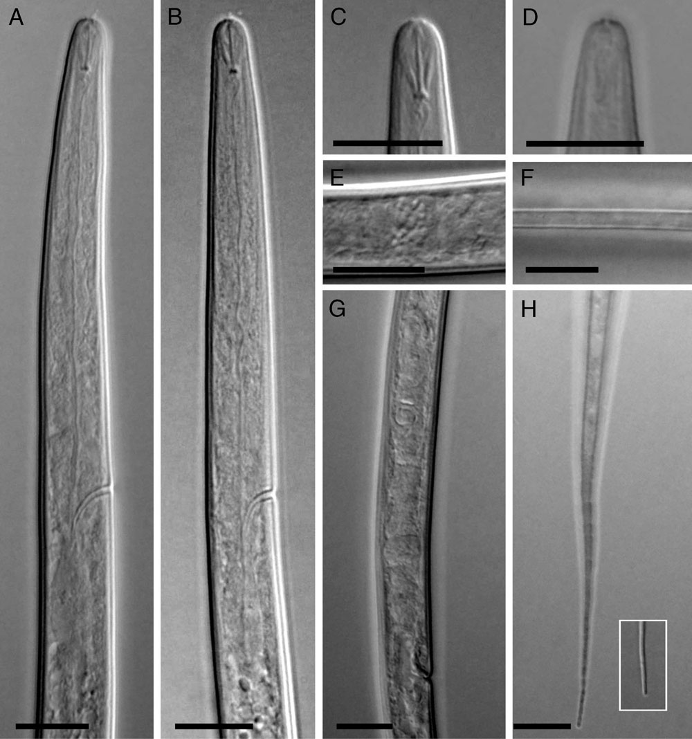
Study and drawings of morphological and morphometric characters were done with a Nikon E600 light microscope equipped with a drawing tube. The hand-drawn sketches were redrawn in CorelDRAW® software version 17. Photographic images of specimens were taken with an Olympus DP72 digital camera attached to an Olympus BX51 microscope equipped with differential interference contrast.
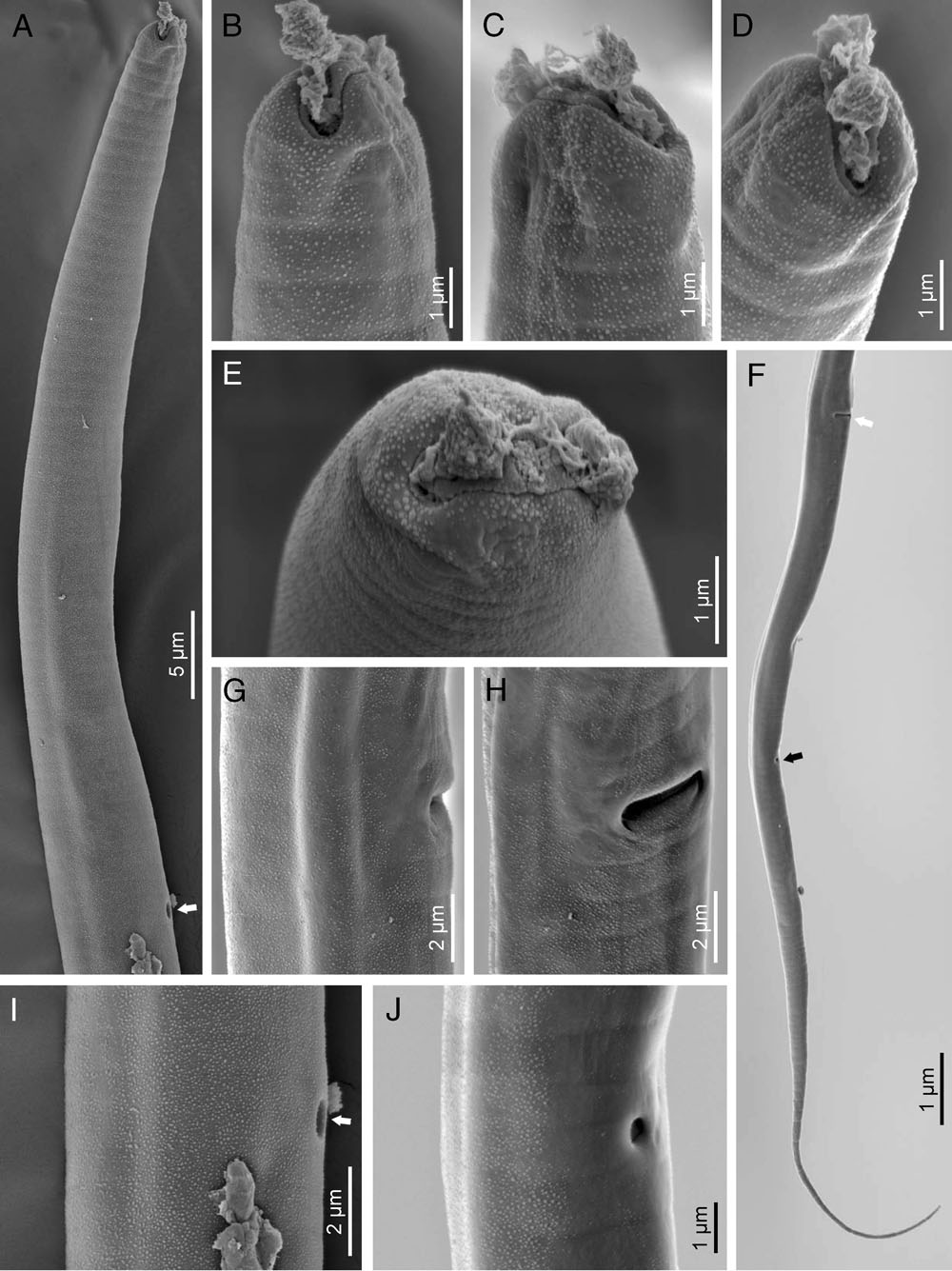
Specimens preserved in glycerine were selected for observation under SEM according to Abolafia (2015). The nematodes were hydrated in distilled water, dehydrated in a graded ethanol-acetone series, critical point dried, coated with gold, and observed with a Zeiss Merlin microscope (5 kV) (Zeiss, Oberkochen, Germany).
DNA extraction, PCR and sequencing
For genomic DNA extraction, a single live nematode specimen was picked out and transferred to a small drop of TE buffer (10 mM Tris-Cl, 0.5 mM EDTA; pH 9.0, QIAGEN Inc., Valencia CA, USA) on a clean slide, studied mainly for its lip region structure, stylet, metacorpus and excretory duct nature, and squashed using a clean slide cover. The suspension was collected by adding 30 µl TE buffer. The DNA sample was stored at ‒20°C until used as PCR templates. To amplify the near-full length fragment of the 18S rDNA and D2-D3 domains of the 28S rDNA, three sets of primers were used in the PCR reactions. The near-full length fragment of the 18S rDNA was amplified with forward primer 1096 F (5′-GGTAATTCTGGAGCTAATAC-3′) and reverse primer 1912R (5′-TTTACGGTCAGAACTAGGG-3′), forward primer 1813F (5′-CTGCGTGAGAGGTGAAAT-3′) and reverse primer 1573R (5′-TACAAAGGGCAGGGACGTAAT-3′) (Holterman et al., 2006; Mullin et al., 2005). Primers for amplification of the D2-D3 domains of the 28S rDNA were forward primer D2A (5′-ACAAGTACCGTGAGGGAAAGT-3′) and reverse primer D3B (5′ TGCGAAGGAACCAGCTACTA-3′) (Nunn, 1992). The PCR amplification and sequencing were done as described by Panahandeh et al. (2016). The amplicon sizes were verified in 1.2% agarose gel and visualized by staining with DNA Green Viewer™ (0.05 μl/ml). The PCR products were sequenced in both directions using the same primers with an ABI 3730XL sequencer (Bioneer Corporation, South Korea). Newly obtained sequences of the studied species were deposited into the GenBank database under accession numbers: MG686086 for near full-length 18S, and MG686087 for partial 28S rDNA D2–D3.
Phylogenetic analyses
The recently obtained sequences were manually checked, edited, and assembled using CodonCode Aligner v. 6.0.2 (CodonCode Corporation, MA, USA, www.codoncode.com). Using the BLAST homology search program in the GenBank database, these sequences were compared with other relevant available sequences. Several representatives of the family Tylenchidae were selected for both dataset analyses. Multiple alignments of the selected DNA sequences with newly obtained sequences were conducted using MUSCLE (Edgar, 2004) in MEGA6 (Tamura et al., 2013). The ambiguously aligned parts and divergent regions were eliminated using the online version of Gblocks 0.91b (Castresana, 2000) with all three less stringent parameters (http://molevol.cmima.csic.es/castresana/Gblocks_server.html). The best-fitting substitution model for both datasets was selected using PAUP*/MrModeltest.2 (Nylander, 2004). Bayesian analyses were carried out on MrBayes 3.1.2 (Rounquist and Huelsenbeck, 2003) under the Akaike-supported model, a general time reversible model, including among-site rate heterogeneity and estimates of invariant sites (GTR+G+I), for both genes, with five independent runs and 107 generations. The Markov chains were sampled every 100 generations for estimating the posterior probabilities of the phylogenetic trees (Larget and Simon, 1999) using the 50% majority rule. Twenty-five percent of the converged runs were regarded as burnin. The Tracer v1.5 software (Rambaut and Drummond, 2009) was used to visualize the results of each run in order to check the effective sample size of each parameter. The maximum likelihood (ML) analysis was performed with 103 bootstraps (BS) replicates for both datasets under the same nucleotide substitution model as in the Bayesian inference (BI) using RaxmlGUI 1.1 (Silvestro and Michalak, 2012). The output files of the phylogenetic programs were visualized using Dendroscope v.3.2.8 (Huson and Scornavacca, 2012). The Bayesian posterior probability (BPP) and ML BS values exceeding 0.60 and 50%, respectively, were plotted on Bayesian 50% majority-rule consensus trees after redrawing in CorelDRAW® software version 17.
Results
Labrys filiformis n. sp.*
Measurements: See Table 1.
Table 1
Morphometrics of Labrys filiformis n. sp. All measurements are in µm and in the form: mean ± standard deviation (range).
| Character | Holotype | Paratype |
|---|---|---|
| n | female | 8 females |
| L | 427 | 440 ±17 (425-463) |
| a | 38.8 | 41.6 ± 2.3 (38.7-46.2) |
| b | 4.9 | 5.1 ± 0.2 (4.9-5.4) |
| c | 3.9 | 3.8 ± 0.2 (3.5-4.0) |
| c' | 13.8 | 14.6 ± 1.1 (13.2-15.9) |
| V | 60.4 | 59.9 ± 1.21 (57.7-61.5) |
| V' | 81.4 | 81.5 ± 1.3 (79.9-84.1) |
| Stylet length | 6 | 6.2 ± 0.4 (6-7) |
| Median bulb valve – anterior end | 44 | 43.7 ± 1.8 (40-46) |
| Excretory pore – anterior end | 69 | 65.5 ± 2.6 (62-69) |
| Neck length (stoma + pharynx) | 87 | 86.1 ± 2.3 (82-90) |
| Lip region to vulva | 258 | 263 ± 8 (256-277) |
| Body width | 11 | 10.6 ± 8.3 (10-11) |
| Anal body width | 8 | 8.0 ± 0.5 (7-9) |
| Anterior genital branch length | 63 | 62.5 ± 5.7 (56-72) |
| Postvulval uterine sac | 6 | 6.4 ± 1.1 (5-8) |
| Vulva – anterior end | 258 | 263 ± 8 (256-277) |
| Vulva to anus distance | 59 | 60.1 ± 5.2 (49-66) |
| Tail length | 110 | 116.6 ± 8.6 (106-127) |