
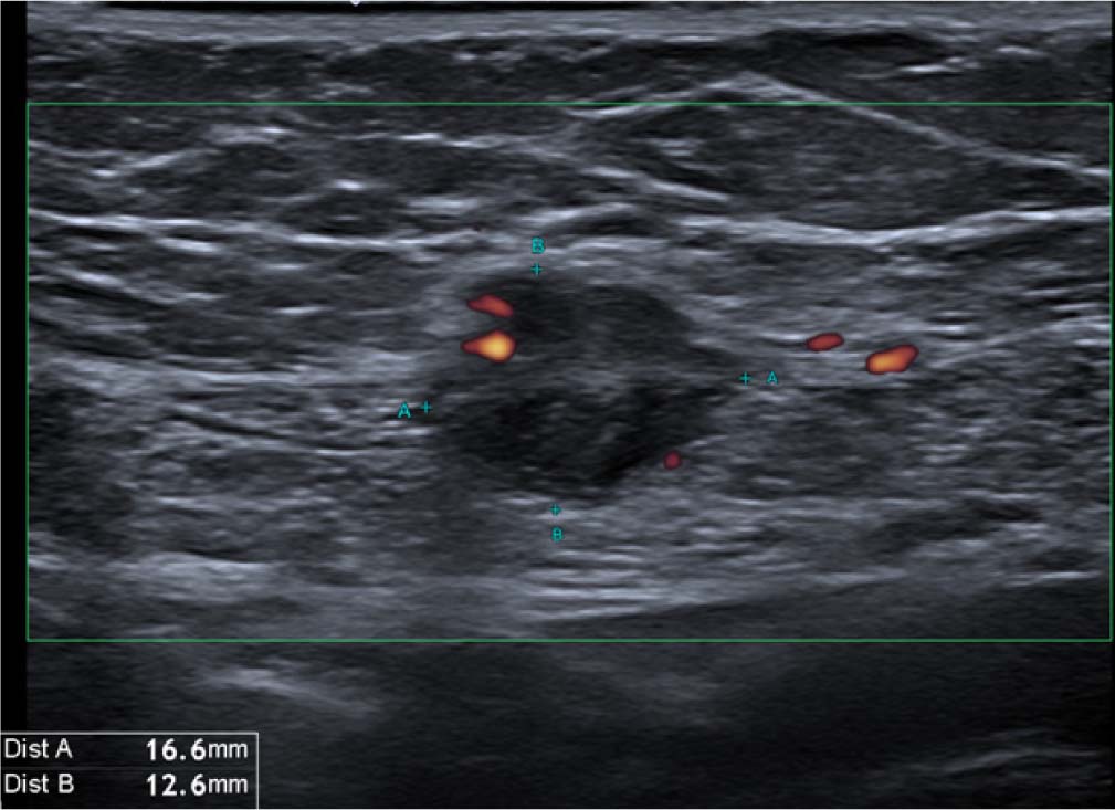
Fig. 1.
A 36-year-old female with a small lump in the anterior abdominal wall adjacent to a caesarean-section scar. The patient reported cyclical changes and pain in the lump related to her menstrual cycle. Biopsy confirmed endometriosis.
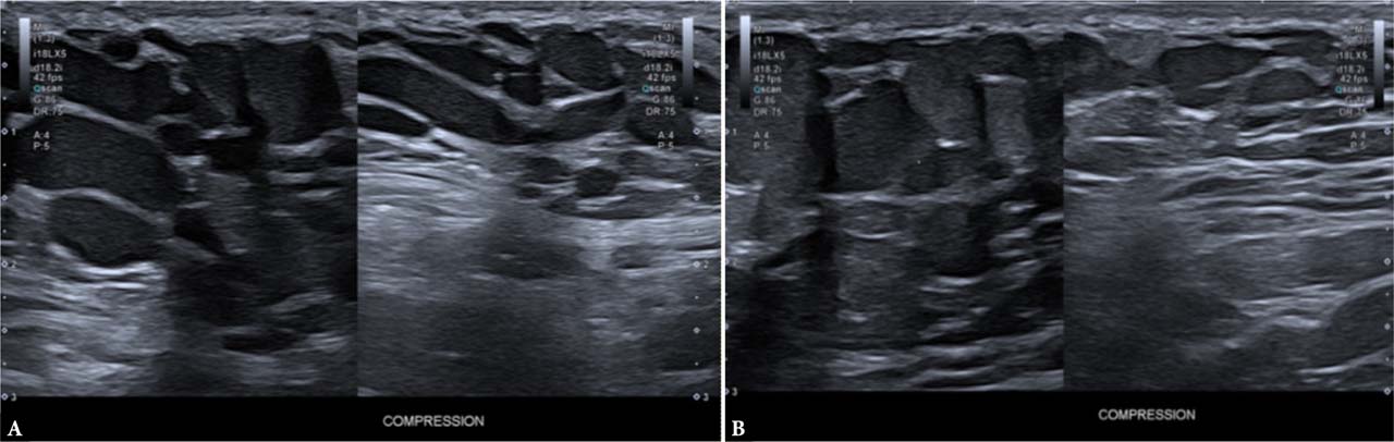
Fig. 2.
A. Right axilla. B. Left axilla. Bilateral axillary soft tissue masses in a lactating female. First noticed during pregnancy, increased in size after delivery. Findings consistent with bilateral ectopic breast tissue. Note the dilated but compressible lactiferous ducts.
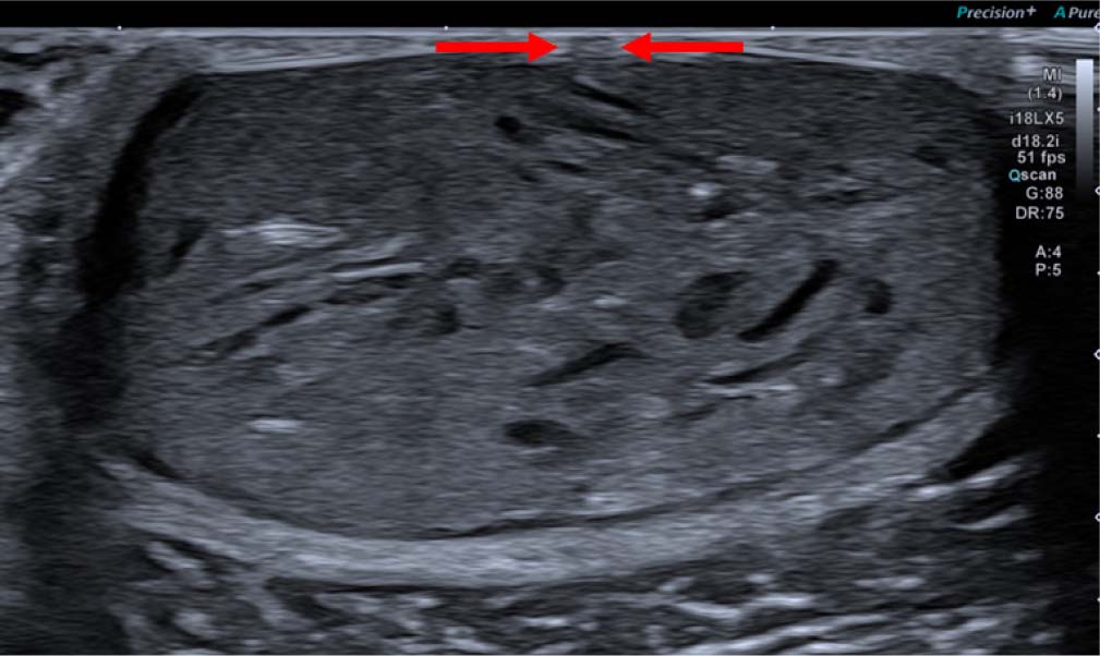
Fig. 3.
Ultrasound image of a subcutaneous soft tissue lesion demonstrating heterogenous echogenicity with internal hyperechogenic foci (keratin flecks), suggestive of an epidermoid cyst. Note the punctum (arrows).
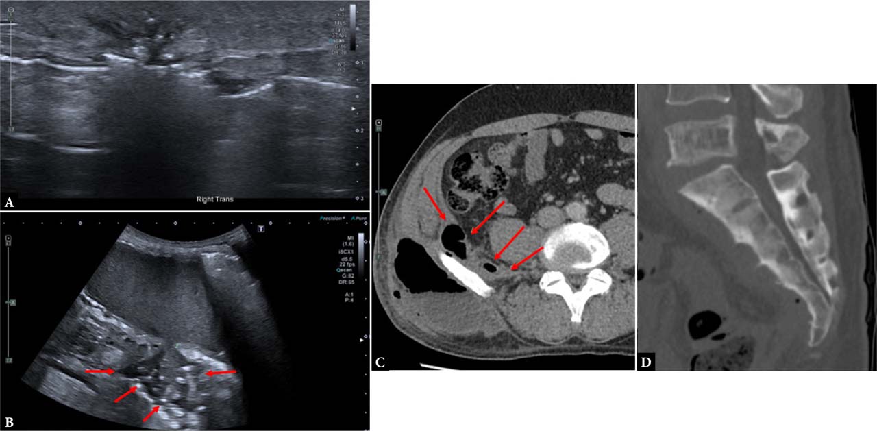
Fig. 4.
A. Ultrasound image obtained using a high-frequency linear probe. B. Ultrasound image obtained with a curvilinear probe. C. Axial CT image of the right lumbar region on soft tissue window. D. Sagittal CT image of the sacrum. Patient referred for evaluation of a lump in the right lumbar region. Note that initial examination with a high-frequency linear probe demonstrated only trace fluid and ill-defined superficial echogenicities with posterior acoustic shadowing indicating air. The operator then switched to a curvilinear probe with reduced frequency to allow for deeper penetration, which demonstrated a significantly larger and deeper collection (image B). CT scan performed immediately afterward demonstrated a large gas-containing subcutaneous lumbar collection with deep extension into the right iliac fossa and spine. Sagittal images of the sacrum demonstrated diffuse sclerotic changes consistent with chronic osteomyelitis. Interestingly, apart from the lump and some pain, the patient was systemically well.
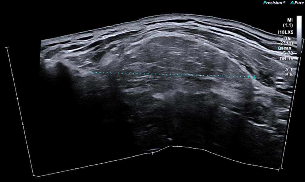
Fig. 5.
Ultrasound image with ultrawide view.
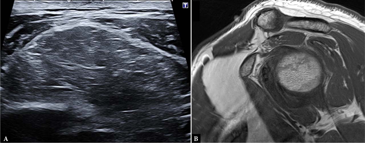
Fig. 6.
A. Ultrasound image on a soft tissue lesion in the left infraclavicular fossa. Note the hypoechogenic lesion of relatively similar echogenicity to adjacent muscles and with echogenic linear striations, which may make differentiation from muscle difficult. However, the striations are not parallel to muscle fibers, and there is an ovoid configuration to the lesion. B. Sagittal T1-weighted MRI image confirming an intermuscular lipoma.
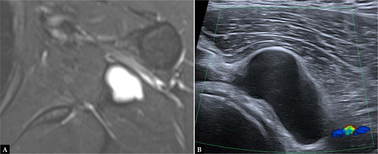
Fig. 7.
A. Axial STIR MRI demonstrating a hyperintense lesion with subtle heterogeneity adjacent to the glenohumeral joint. B. Ultrasound image of an anechoic lesion adjacent to the glenohumeral joint. No abnormal vascularity on Doppler ultrasound suggestive of a paralabral cyst. Occasionally, differentiation from a myxoid lesion such as an intramuscular myxoma may be difficult, and biopsy may be warranted.
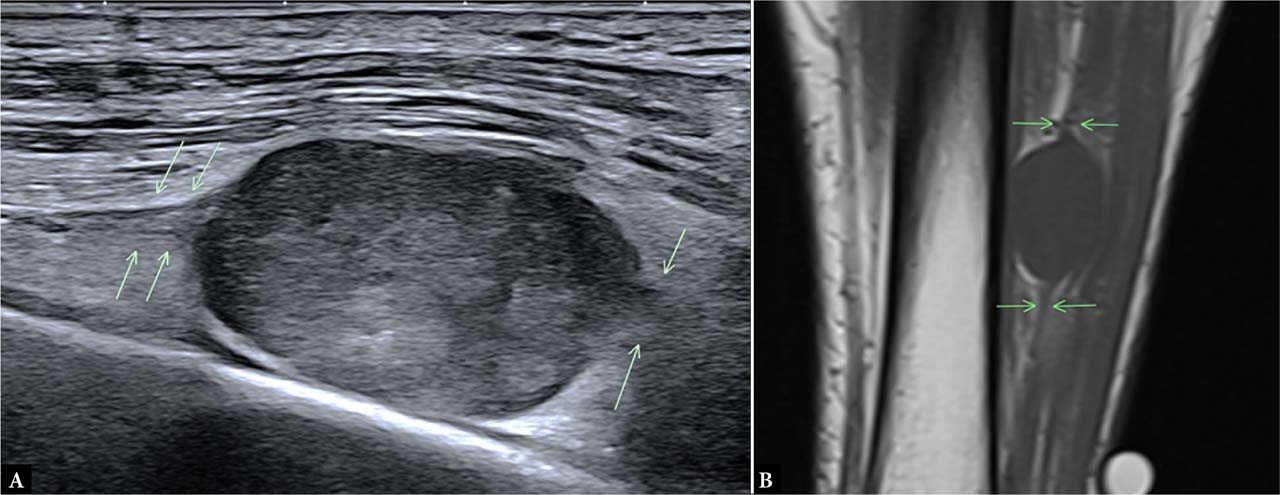
Fig. 8.
A. Long-axis ultrasound image of an oval soft tissue lesion in the leg. Note the tubular structures entering and exiting the lesion (arrows). B. Coronal T1-weighted MRI image confirming the same (arrows). Findings are suggestive of a nerve sheath tumor, confirmed on biopsy.
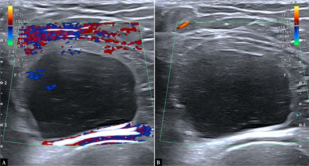
Fig. 9.
Color Doppler ultrasound image with dual view. Note the foci of color in image A, which may be mistaken for vascularity. However, in image B, the previously identified foci are no longer visible, indicating that they were artefactual. In image B, the scale has been increased and motion was eliminated by stabilizing the examiner’s hand.
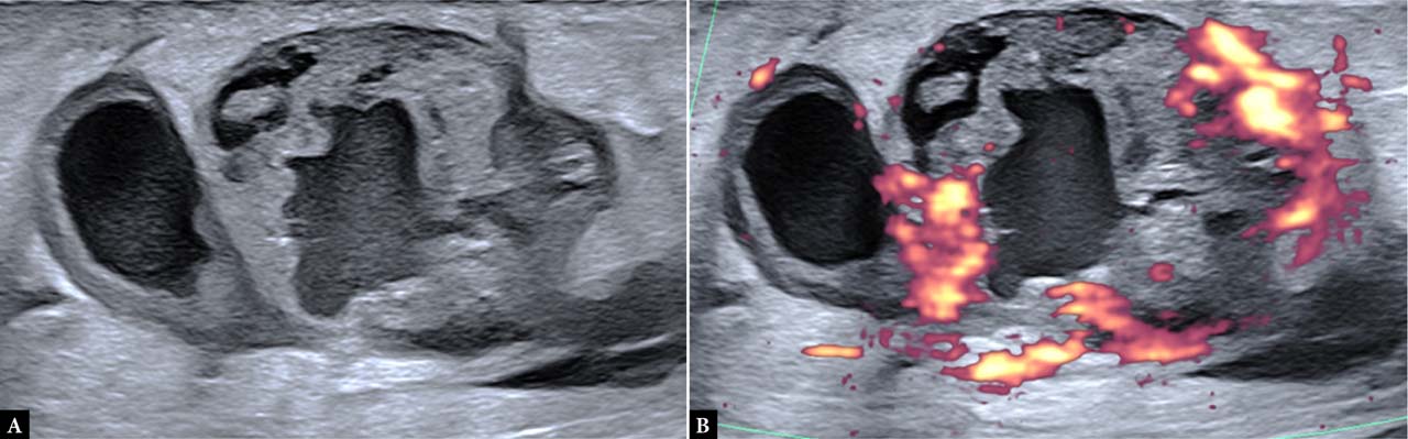
Fig. 10.
The patient noted a lump in her calf after trauma with subsequent skin bruising. The lump initially decreased in size but demonstrated regrowth. Ultrasound images demonstrated a lobulated lesion with anechoic foci and a heterogenous solid component demonstrating marked vascularity. Biopsy confirmed metastatic melanoma.