
Introduction
Assessing soft tissue lumps is a major part of the routine daily practice of musculoskeletal radiologists. Most of these lumps are non-neoplastic and are usually traumatic, inflammatory, or post-traumatic in origin. Of the small number of neoplastic lesions that are encountered, the majority are benign, while malignant lesions account for only a tiny percentage, with a reported benign-to-malignant ratio of 150:1(1). These soft tissue lesions are usually clinically palpable and may even be visible. Ultrasound is usually the first imaging modality used to assess these lesions.
Ultrasound is readily available, inexpensive, and safe for all patients, including children and pregnant women. It avoids the potential risks associated with ionizing radiation from radiography, CT, and nuclear medicine imaging, as well as potential MRI-related risks during pregnancy or the risks associated with contrast medium administration in CT and MRI. It is particularly useful in distinguishing cysts from solid lesions(2). Ultrasound clinics provide radiologists with a unique opportunity to meet patients, obtain a more relevant and detailed clinical history, and physically examine patients(3). The major limitations of ultrasound are operator dependence, the need for appropriate experience, and reduced reliability in the assessment of deep soft tissue masses.
This article aims to provide practical tips and pointers to help residents and junior consultants in musculoskeletal radiology build on their existing experience and refine their skills, allowing them to maximize the potential of this readily available modality.
How we do it
The first opportunity to assess the patient is, in fact, well before we meet them. It begins when the referral is received from the clinician requesting an ultrasound examination of a specific soft tissue lump. In musculoskeletal tumors, whether soft tissue or bone, there are three essential parameters that can significantly narrow and refine the differential diagnosis: the patient’s age, sex, and the location of the lesion. These details are usually provided in the referral. The clinical information will usually further include the duration of the lesion, any change in size, pain, and whether there is a relevant history of trauma, travel, intervention, or a personal or family history of malignancy.
It is good practice to review previous relevant imaging, as it is possible that the lesion was there before but was only recently noticed by the patient, their family, or their physician. This is usually the case when patients sustain an injury to a specific location, which brings their attention to a lesion in the vicinity, or when there is a recent history of weight loss (intentional or unintentional), which may make soft tissue lesions more perceptible or highlight normal prominent anatomical structures, such as costochondral junctions or sternoclavicular joints.
With all this information available, the radiologist should be able to justify and approve the request.
The clinical encounter
Physically meeting the patient is where ultrasound has an advantage over other imaging modalities. It should be considered equivalent to an outpatient clinic visit by the referring clinician. This encounter is a valuable opportunity to obtain further relevant clinical information.
Observe the patient as they enter the room. An unwell patient with pyrexia and diaphoresis referred for assessment of a new soft tissue lump should raise suspicion of an abscess. Surgical dressing over the location of the lesion may suggest recent trauma, ulceration, or intervention.
After conducting formal introductions and confirming the patient’s identity, it is good practice to establish the patient’s understanding of their current condition and their expectations from the examination. This may help reduce anxiety, especially if biopsy is being considered. Ask the patient to point out the lesion, even if it appears obvious; one does not want to scan the wrong lump. It is not uncommon for patients to struggle to locate the lesion on the day of examination, especially if it is small, too deep, or mobile. Remember that patients may be anxious; do not rush them and give them sufficient time to identify the lesion. Sometimes, they are only able to locate it in certain positions. Always try to reassure them and explain what you are doing as you go along.
History is the most important part of the puzzle. Confirm the duration of the lesion and how the patient first noticed it. A common scenario is that it was noticed after “knocking” the area or after losing weight; determine whether the weight loss was intentional. Weight loss may make normal anatomical structures, such as the costochondral junctions or the sternoclavicular joints, more prominent or may make pre-existing soft tissue lesions more noticeable. Ask whether the patient has noticed any change in size, consistency, or associated symptoms. A sudden increase in size of a previously stable lesion that has been present for years, a change in consistency, or new pain in a previously painless lesion are alarming features. In females of reproductive age, ask whether the patient has noticed any cyclical changes in the lesion. A long-standing soft tissue lesion in a woman of reproductive age, exhibiting cyclical changes related to the menstrual cycle, should raise suspicion of endometriosis (Fig. 1). Bilateral axillary soft tissue lesions in a lactating female that increase in size after delivery should raise the possibility of ectopic breast tissue (Fig. 2). A history of trauma may suggest a hematoma or infection. Gardening may suggest a foreign body, such as a thorn or splinter, or a foreign body reaction. Insect bites or animal scratches may suggest an inflammatory reaction or infection. Always ask about personal and family medical history. A history of rheumatoid arthritis combined with a soft tissue lump on the extensor surfaces should raise the possibility of a rheumatoid nodule. A previous history of malignancy should always prompt consideration of metastasis in the differential diagnosis.
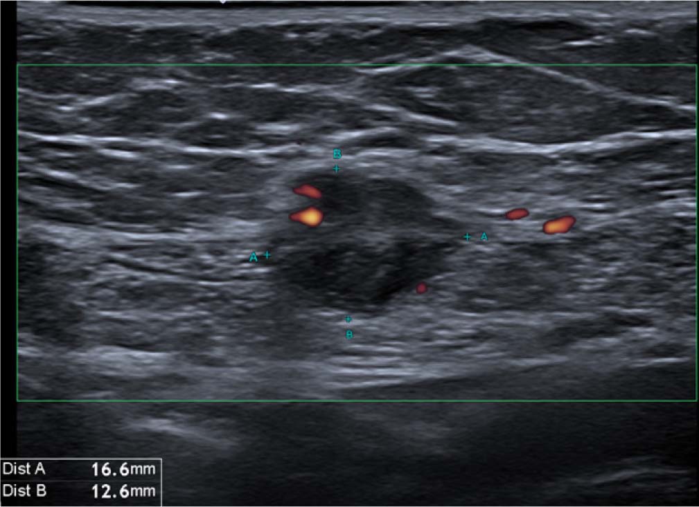
Fig. 1.
A 36-year-old female with a small lump in the anterior abdominal wall adjacent to a caesarean-section scar. The patient reported cyclical changes and pain in the lump related to her menstrual cycle. Biopsy confirmed endometriosis.
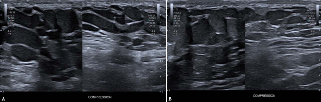
Fig. 2.
A. Right axilla. B. Left axilla. Bilateral axillary soft tissue masses in a lactating female. First noticed during pregnancy, increased in size after delivery. Findings consistent with bilateral ectopic breast tissue. Note the dilated but compressible lactiferous ducts.
Always take the opportunity to inspect the lesion if it is visible, including the appearance of the overlying skin. Skin discoloration may indicate a vascular malformation. Red, shiny skin may indicate infection or inflammation. The presence of a punctum may point toward an epidermoid cyst (Fig. 3). A surgical scar may suggest that the lump is due to scar tissue/fibrosis, a stitch granuloma, prominent underlying metalwork, or, in the context of malignancy, recurrence or metastasis.
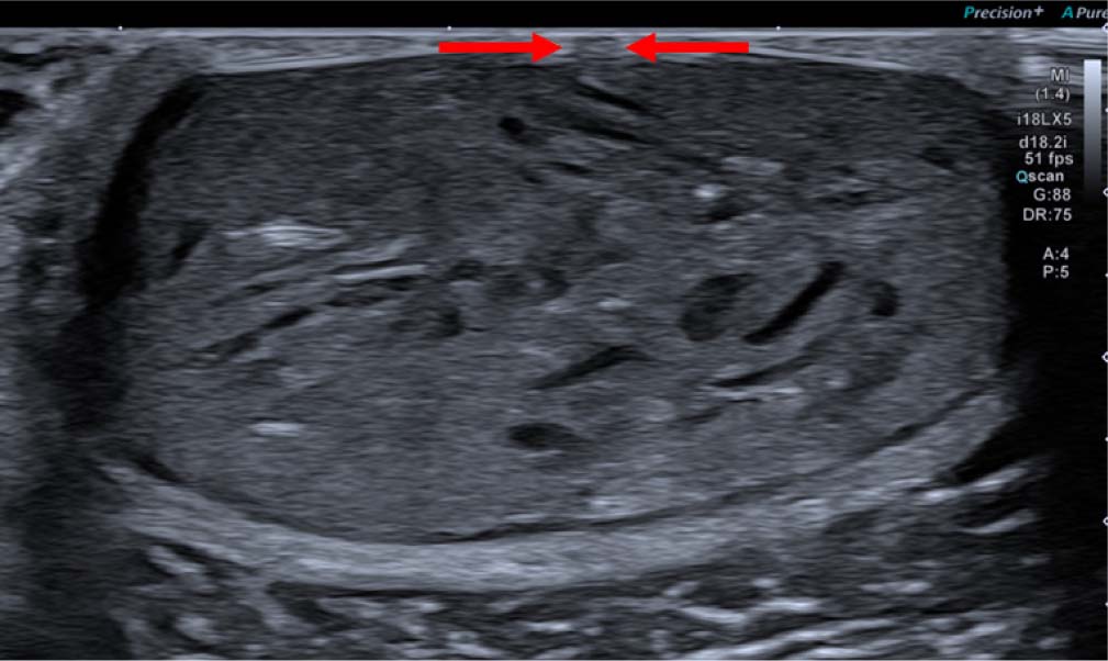
Fig. 3.
Ultrasound image of a subcutaneous soft tissue lesion demonstrating heterogenous echogenicity with internal hyperechogenic foci (keratin flecks), suggestive of an epidermoid cyst. Note the punctum (arrows).
Always confirm whether the lesion is painful before palpation. Assess its consistency and mobility.
Sonographic examination
Ensure optimal positioning that is comfortable to the patient for the duration of the examination while also providing the person performing the scan with adequate access to the lesion and sufficient exposure, while maintaining the patient’s dignity. The presence of a chaperone can provide reassurance to both the patient and the person performing the scan, especially when the lesion is located in an intimate anatomical region.
Usually, a high-resolution 12–18 MHz linear transducer is optimal for most musculoskeletal soft tissue lesions(4). Use a generous amount of gel and avoid excessive pressure, as this may efface slow vascular flow and small vessels or even compress subtle soft lesions. Adjust the gain as needed and consider reducing the transducer frequency for deeper lesions. Be prepared to use a curvilinear abdominal probe to explore the possibility of deep extension of the lesion, particularly in the context of abscesses and postoperative collections (Fig. 4).
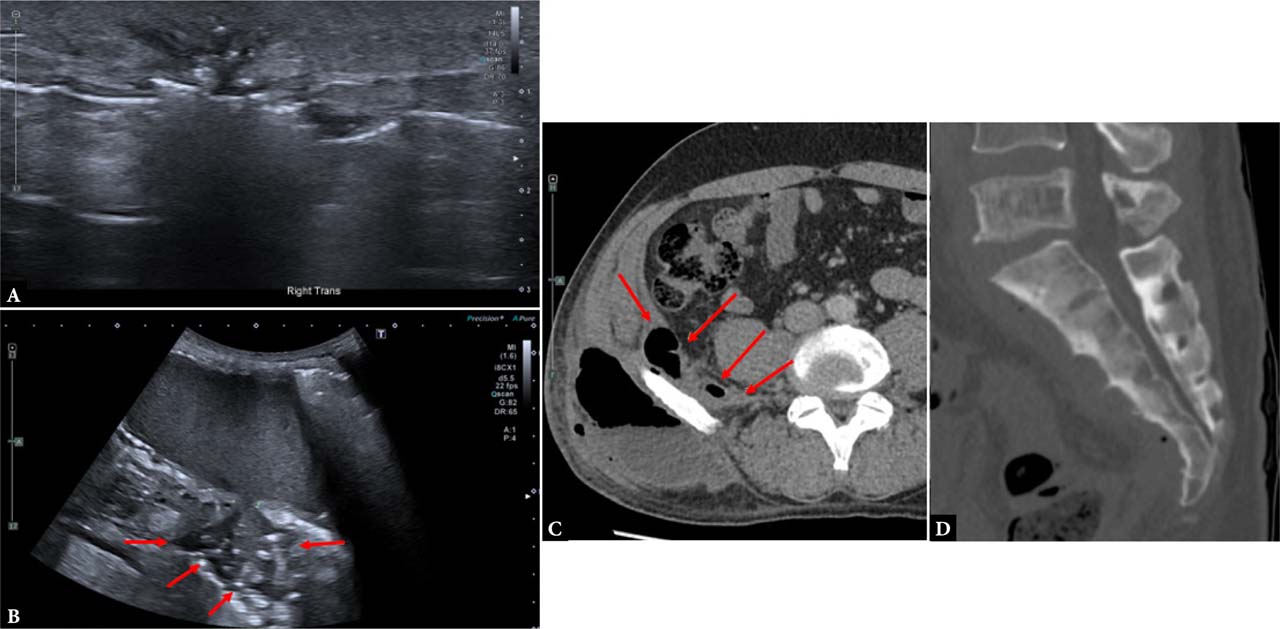
Fig. 4.
A. Ultrasound image obtained using a high-frequency linear probe. B. Ultrasound image obtained with a curvilinear probe. C. Axial CT image of the right lumbar region on soft tissue window. D. Sagittal CT image of the sacrum. Patient referred for evaluation of a lump in the right lumbar region. Note that initial examination with a high-frequency linear probe demonstrated only trace fluid and ill-defined superficial echogenicities with posterior acoustic shadowing indicating air. The operator then switched to a curvilinear probe with reduced frequency to allow for deeper penetration, which demonstrated a significantly larger and deeper collection (image B). CT scan performed immediately afterward demonstrated a large gas-containing subcutaneous lumbar collection with deep extension into the right iliac fossa and spine. Sagittal images of the sacrum demonstrated diffuse sclerotic changes consistent with chronic osteomyelitis. Interestingly, apart from the lump and some pain, the patient was systemically well.
The first few sweeps across the length and width of the lesion will provide an initial assessment of its size, location (whether superficial or deep to the fascia, or intramuscular), and internal architecture. Always measure the lesion in two orthogonal plains. Save images without measurements for future assessment and comparison. For large lesions, use an extended field-of-view technique to demonstrate the full extent of the lesion (Fig. 5). Multiple small, painful, echogenic subcutaneous lesions suggest angiolipomas in Dercum disease. Lipomas may demonstrate echogenicity similar to the surrounding fat or may appear hyperechogenic or hypoechogenic. Occasionally, lipomas, especially intermuscular lipomas, contain internal striations that make them difficult to differentiate from adjacent muscles. Assessing the orientation of these striations is a helpful discriminator, as muscle fibers run parallel on the long-axis view (Fig. 6). Anechoic foci within a solid lesion indicate cystic degeneration or necrosis and should be avoided during biopsy. Diffusely hypoechogenic lesions with posterior acoustic shadowing may indicate fibromatosis or calcification. Radiographs can confirm the presence of the latter.
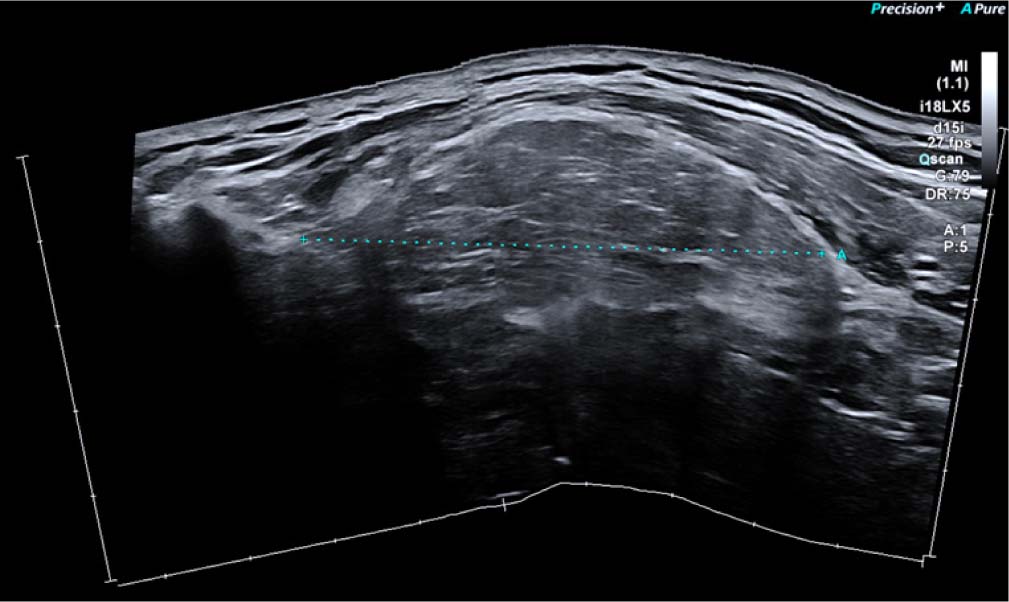
Fig. 5.
Ultrasound image with ultrawide view.
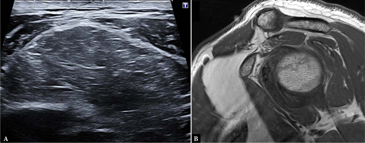
Fig. 6.
A. Ultrasound image on a soft tissue lesion in the left infraclavicular fossa. Note the hypoechogenic lesion of relatively similar echogenicity to adjacent muscles and with echogenic linear striations, which may make differentiation from muscle difficult. However, the striations are not parallel to muscle fibers, and there is an ovoid configuration to the lesion. B. Sagittal T1-weighted MRI image confirming an intermuscular lipoma.
Assess the lesion’s relationship to the surrounding structures. Anechoic lesions that communicate with the adjacent joint are the hallmarks of ganglion cysts. Anechoic lesions overlying bony prominences are consistent with bursae. A compressible anechoic lesion intimately associated with a vein is likely to represent a varix. However, lack of compressibility may be explained by the presence of an internal thrombus or a solid soft tissue component; therefore, vascular leiomyoma or leiomyosarcoma should be excluded, and biopsy would be warranted. Anechoic or hypoechoic lesions adjacent to the glenoid or acetabular labrum or to the menisci suggest a paralabral or parameniscal cyst (Fig. 7). However, increasing complexity warrants biopsy to rule out malignant pathology, such as synovial sarcoma. Demonstrating a tubular structure entering and exiting the lesion is pathognomonic of a nerve sheath lesion (Fig. 8). Solid intra-articular nodular soft tissue lesions may represent either a tenosynovial giant cell tumor or focal synovitis. A solid soft tissue lesion intimately associated with a tendon should raise the possibility of a tenosynovial giant cell tumor of the tendon sheath.
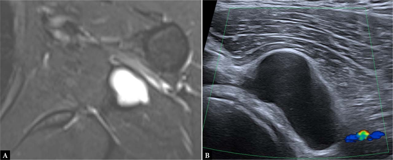
Fig. 7.
A. Axial STIR MRI demonstrating a hyperintense lesion with subtle heterogeneity adjacent to the glenohumeral joint. B. Ultrasound image of an anechoic lesion adjacent to the glenohumeral joint. No abnormal vascularity on Doppler ultrasound suggestive of a paralabral cyst. Occasionally, differentiation from a myxoid lesion such as an intramuscular myxoma may be difficult, and biopsy may be warranted.
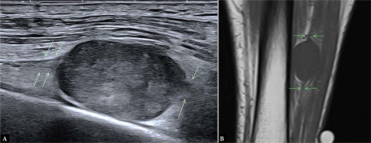
Fig. 8.
A. Long-axis ultrasound image of an oval soft tissue lesion in the leg. Note the tubular structures entering and exiting the lesion (arrows). B. Coronal T1-weighted MRI image confirming the same (arrows). Findings are suggestive of a nerve sheath tumor, confirmed on biopsy.
Dynamic assessment may be useful in assessing the lesion’s relationship to adjacent structures, such as muscles and tendons, and its impact on function, including joint mobility or nerve impingement.
Vascularity can be assessed with color Doppler, which is sensitive to high arterial flow. Be mindful of noise and motion artifacts. Adjust the scale to a level where noise is minimized and ensure the transducer is kept steady during Doppler assessment by stabilizing the scanning hand on the patient. However, avoid applying firm pressure to the lesion to avoid compressing the internal vessels (Fig. 9). Power Doppler is more sensitive for detecting low- and slow-flow vascularity. Most modern ultrasound machines offer microvascular assessment algorithms for small vessels and slow flow(4). Avascular or minimally vascular lesions are often associated with low biological activity, while hypervascular lesions are more concerning for malignancy, due to the chaotic rather than uniform distribution of the vessels(5,6). Identifying a feeding vascular pedicle is useful for surgical and interventional planning. Spectral waveform analysis is not routinely used in our practice.
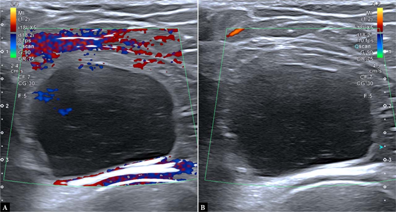
Fig. 9.
Color Doppler ultrasound image with dual view. Note the foci of color in image A, which may be mistaken for vascularity. However, in image B, the previously identified foci are no longer visible, indicating that they were artefactual. In image B, the scale has been increased and motion was eliminated by stabilizing the examiner’s hand.
Elastography
Sonoelastography has been used in clinical practice for the assessment of breast, liver, thyroid, prostate, and musculoskeletal pathology, including soft tissue tumors. The underlying principle is that tissues respond to applied stress or pressure through longitudinal deformation, such as compression or stretching, and shear deformation, such as twisting. This tissue response is termed “strain”(7). In clinical practice, elastography is performed by applying mechanical stress to tissues and measuring tissue displacement before and immediately after stress as an estimate of strain(8). Two main methods are used clinically: compression/strain elastography and shear wave elastography. Despite the potential of this technique and evidence suggesting that it may help differentiate benign from malignant tumors, its use remains limited. A study by Pass et al. showed that there is no added role for shear wave elastography in soft tissue tumors(9). It certainly does not eliminate the need for biopsy.
Contrast-enhanced ultrasound (CEUS)
Ultrasound contrast agents have the potential to identify small vessels through application of ultrasound “microbubbles”. CEUS may be used as an additional tool for characterizing tumor perfusion patterns and identifying viable tissue to target for biopsy(10,11). However, CEUS use in soft tissue tumors imaging remains limited and does not replace the need for biopsy. It also increases costs and requires additional examination time and operator experience for accurate interpretation.
By the end of a thorough clinical and sonographic assessment, the lesion can usually be categorized into one of three categories: clearly benign, clearly malignant, or indeterminate. Experienced radiologists can correctly identify the type of tumor in approximately three-quarters of cases based on ultrasound features alone, with a negative predictive value approaching 99%(12,13). Patients with a confident diagnosis of a benign lesion can be safely discharged. Patients with clearly malignant lesions require further workup, including biopsy. The literature remains divided on whether MRI is required prior to biopsy. At our institution, MRI is routinely obtained before biopsy as a baseline examination to assess the extent of the lesion, define its anatomy, and evaluate its relationship to adjacent structures, including neurovascular bundles, to aid surgical planning.
Indeterminate lesions require further evaluation with MRI. CT and radiography may also be used to assess for calcification and evaluate the adjacent bones for scalloping, periosteal reaction, or destruction. If the lesion remains indeterminate following further imaging, then one might consider biopsy or imaging follow-up in 3–6 months. Hematomas are a good example of lesions in which sometimes it is not possible to rule out that the hematoma is secondary to bleeding in a pre-existing soft tissue lesion. Hematomas classically appear as heterogenous lesions with both echogenic and anechoic foci. It is not uncommon to find solid nodular areas or even vascularity within these hematomas, which may represent entrapped vessels or vascular ingrowth(14). These features can make the diagnosis challenging. Follow-up is appropriate to confirm a reduction in size or complete resolution of the hematoma without a residual soft tissue component. However, biopsy may occasionally be required to establish a definitive diagnosis (Fig. 10).
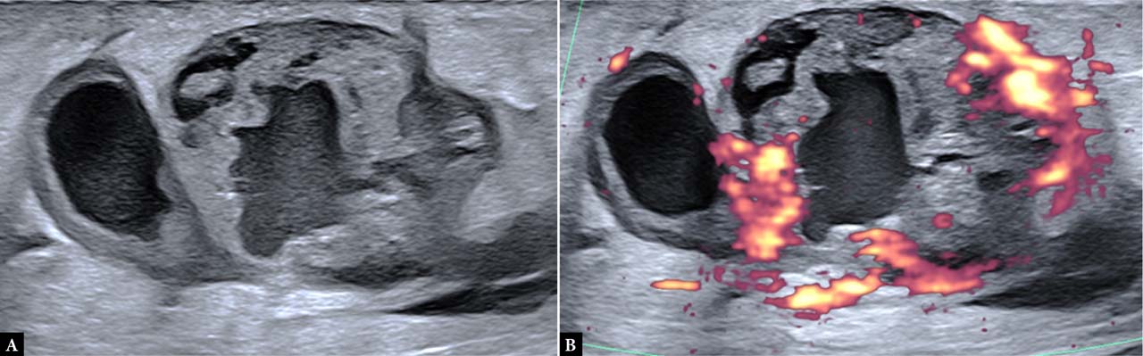
Fig. 10.
The patient noted a lump in her calf after trauma with subsequent skin bruising. The lump initially decreased in size but demonstrated regrowth. Ultrasound images demonstrated a lobulated lesion with anechoic foci and a heterogenous solid component demonstrating marked vascularity. Biopsy confirmed metastatic melanoma.
Conclusion
Soft tissue masses are common, and the vast majority are benign. Ultrasound is usually the first-line imaging modality used for the assessment of these lesions. It is highly reliable, readily available, inexpensive, and safe, making it a powerful diagnostic modality. Following a systematic approach to the assessment of such lesions, combined with deploying all the available tools and opportunities that it offers, will maximize its strength in achieving a confident diagnosis.