
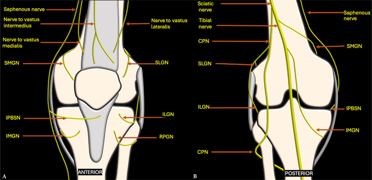
Fig. 1.
Schematic diagram depicting the sensory innervation of the knee, with genicular nerves arising from various nerves around the joint as seen from the A. anterior and B. posterior aspects. CPN – common peroneal nerve; ILGN – inferolateral genicular nerve; IMGN – inferomedial genicular nerve; IPBSN – infrapatellar branch of the saphenous nerve; RPGN – recurrent peroneal genicular nerves; SLGN – superolateral genicular nerve; SMGN – superomedial genicular nerve
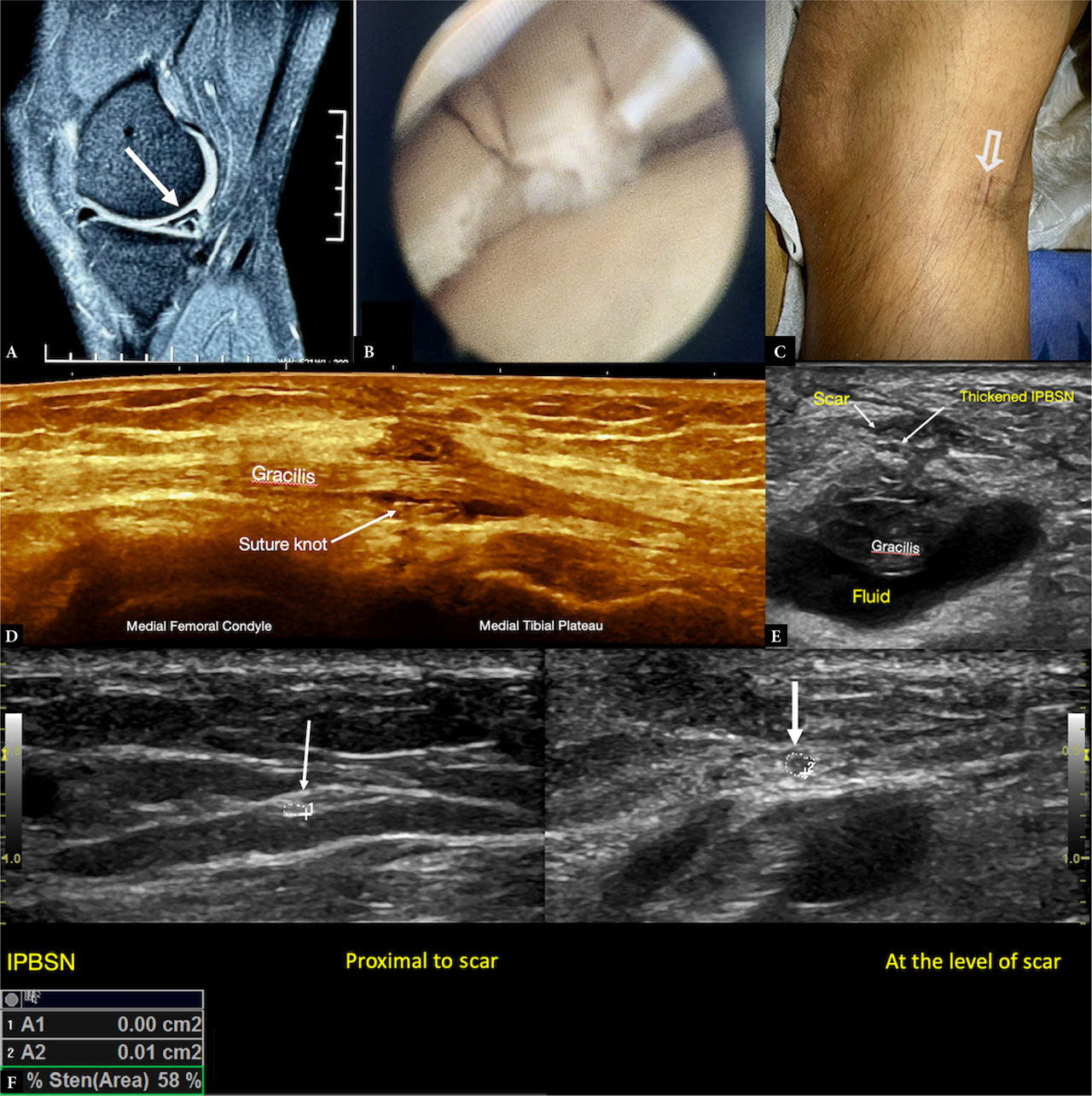
Fig. 2.
Infrapatellar branch of the saphenous nerve (IPBSN) injury following medial meniscal repair surgery. A. Proton-density fat-suppressed sagittal MRI image showing an oblique tear in the posterior horn of the medial meniscus B. Arthroscopic image of the torn meniscus C. Scar site corresponding to the site of the ultrasonographic Tinel sign D. Longitudinal view at the scar site revealing a suture knot around the gracilis muscle E. Transverse view across the scar site showing a thickened IPBSN adjacent to the scar, overlying the gracilis muscle. F. Cross-sectional area of the nerve (arrow) measured proximal and at the scar site shows focal enlargement with increased hypoechogenicity of the nerve at the scar site consistent with neuroma formation. The IPBSN was traced for its origin from the saphenous nerve; in this patient, it followed a more posterior course than usual. IPBSN – infrapatellar branch of the saphenous nerve
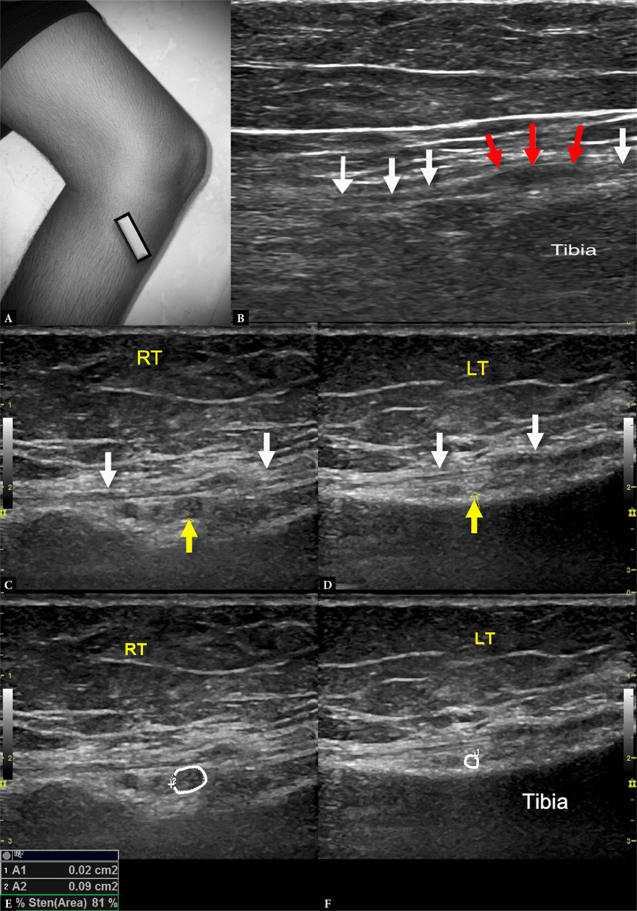
Fig. 3.
Post-traumatic inferomedial genicular nerve (IMGN) neuroma in a 53-year-old woman with chronic inferomedial right knee pain with history of trauma A. Probe positioned at the proximal tibial shaft B. Long-axis view of the IMGN showing focal thickening of the nerve (red arrows) at the site of the scar – neuroma in continuity, with normal caliber of the nerve proximal and distal (white arrows). Short-axis view of the nerve (yellow arrow) deep to the MCL (white arrows) and just superficial to the tibial cortex on the C. affected (right) side showing a bulky hypoechoic nerve and D. on the normal contralateral (left) side for comparison. Cross-sectional area comparison on the E. right and F. left side shows a marked difference in caliber; the abnormal nerve caliber may be overlooked if seen in isolation due to the inherently small size of the nerve
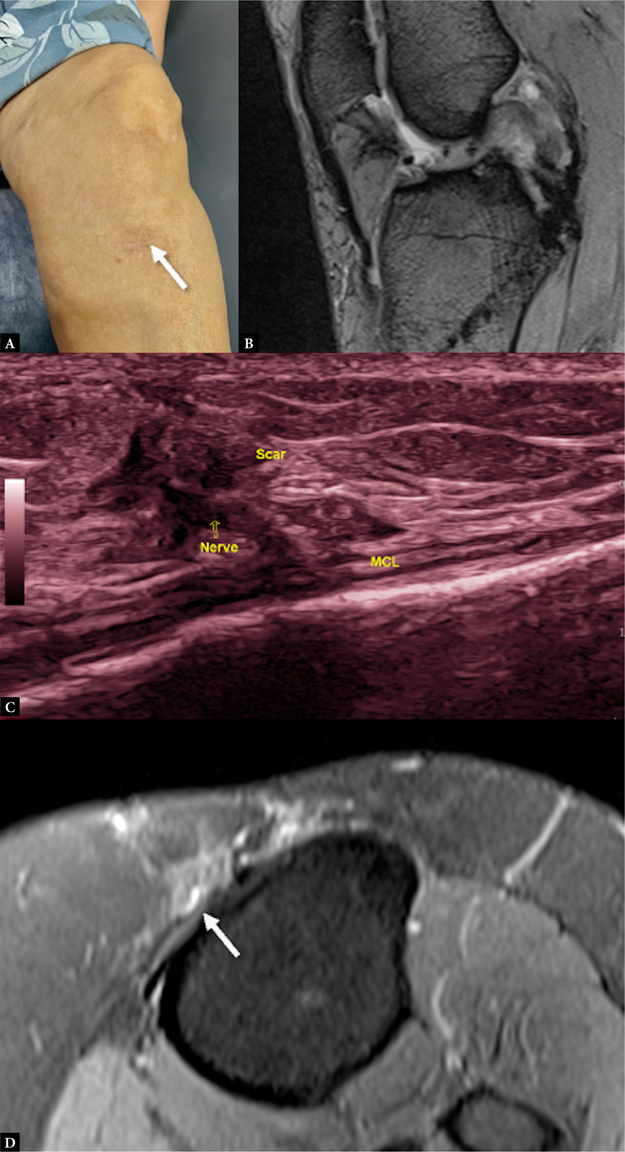
Fig. 4.
Inferomedial genicular nerve scar entrapment following posterior cruciate ligament (PCL) repair A. Clinical image showing the scar site B. Fat-suppressed proton density sagittal weighted MRI showing postoperative changes in PCL; no obvious abnormality was appreciated on MRI explaining the patient’s symptoms C. Ultrasound image with the probe placed along the distal tibial insertion site of the medial collateral ligament (MCL) showing nerve entrapped within scar tissue, corresponding precisely to the site of maximum tenderness. D. Retrospective review of the axial MRI images reveals the entrapped nerve as a focal hyperintense structure (arrow)
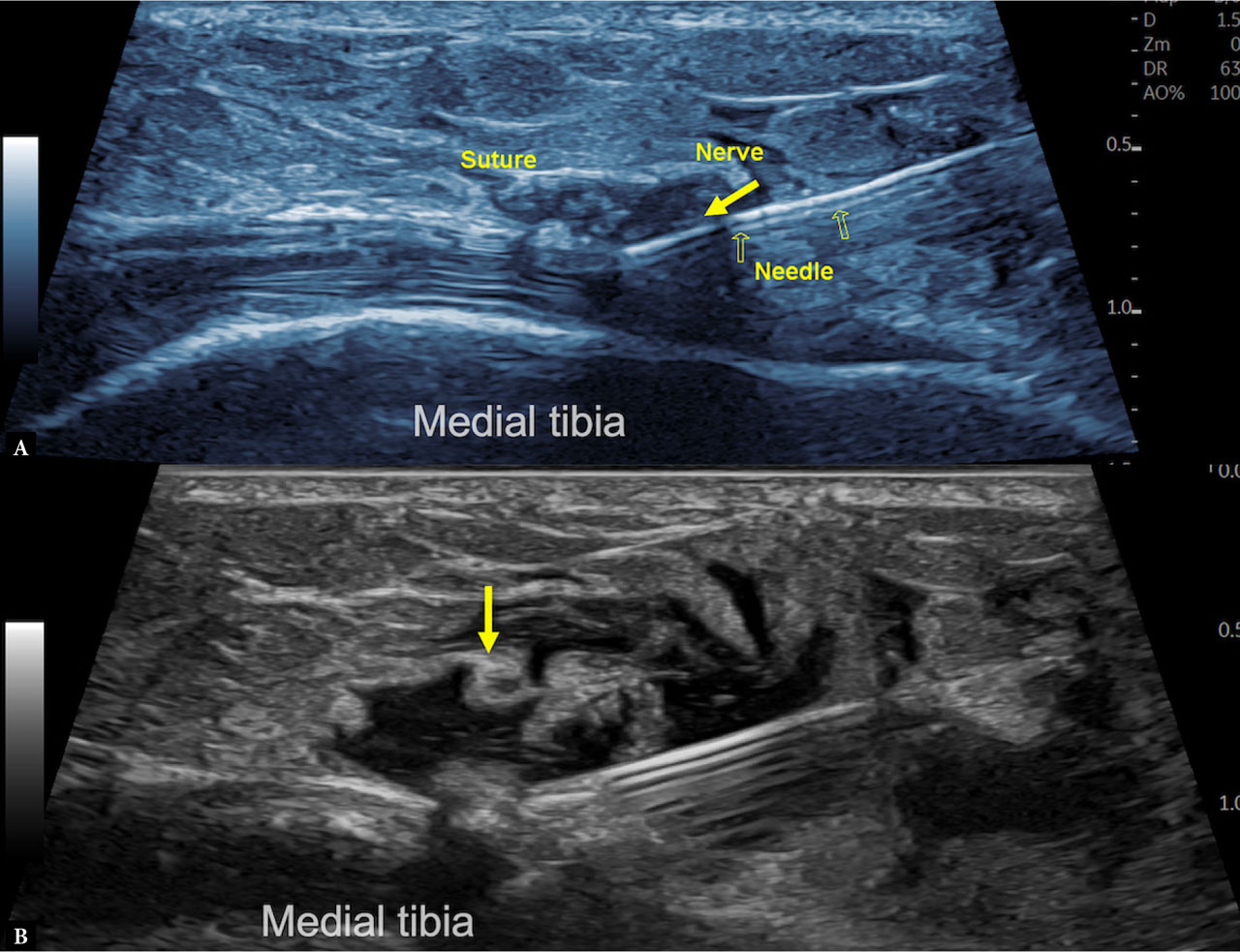
Fig. 5.
US-guided therapeutic hydrodissection of the inferomedial genicular nerve in the patient described in Fig. 4. A. Longitudinal view along the medial proximal tibial shaft near the medial collateral ligament attachment site corresponding to the scar site, showing the entrapped nerve within the scar; the needle is advanced to the target site using ‘in-plane approach’ with the tip directed deep to the nerve. B. Approximately 4–5 mL of injectate (3 mL 5% dextrose + 1 mL steroid + 1 mL local anesthetic) was delivered circumferentially to release the nerve from the scar
Tab. 1.
Relevant anatomy of the six genicular nerves
| Genicular Nerve | Origin | Accompanying vessel | Relevant anatomical landmark | Area of innervation | Special point |
|---|---|---|---|---|---|
| Superomedial genicular nerve | Femoral nerve branch (nerve to vastus medialis) Tibial nerve | Superomedial genicular arteries | Bony: adductor tubercle Soft tissue: femoral attachment of MCL, vastus medialis and adductor magnus | Medial patellar retinaculum; medial knee joint capsule | Largest and the most consistent genicular nerve |
| Superolateral genicular nerve | Femoral nerve branch Sciatic nerve | Superolateral genicular artery | Bony: bony ridge separating the lateral and posterior cortex Soft tissue: femoral attachment of LCL, vastus lateralis and biceps femoris muscle | Superolateral knee capsule | |
| Inferomedial genicular nerve | Tibial nerve Sciatic nerve | Inferomedial genicular artery | Bony: tibial tuberosity and medial tibial condyle Soft tissue: tibial insertion of MCL | Medial anterior and inferior knee capsule | Susceptible to iatrogenic injury |
| Inferolateral genicular nerve | CPN | Inferolateral genicular arteries | Bony: lateral tibial plateau Soft tissue: LCL | Anterolateral knee capsule and proximal tibiofibular joint | Close to CPN motor branches, but CPN is inferior to fibular neck |
| RPGN | CPN proximal DPN | Recurrent anterior tibial artery – branch of proximal ATA | Bony: Gerdy’s tubercle Soft tissue: tibialis anterior muscle | Inferolateral and anterior knee, tibiofibular joint | Close to CPN and anterior tibial vessels |
| Infrapatellar branch of the saphenous nerve | Saphenous nerve | Descending genicular artery in proximal course | Bony: patellar tip and tibial tuberosity Soft tissue: MCL | Medial knee joint | Highly susceptible to iatrogenic injury Subcutaneous course |
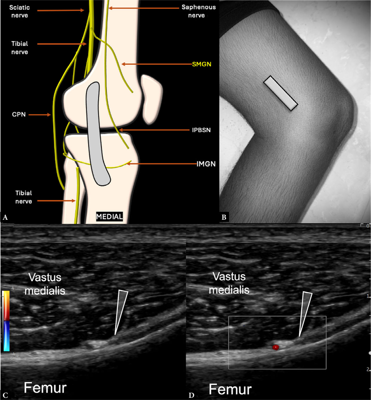
Fig. 6.
Imaging and anatomy of the superomedial genicular nerve (SMGN): A. Schematic showing the location of the SMGN cranial to the femoral insertion of the medial collateral ligament. B. Transducer positioned in a coronal plane along the metaphyseal–diaphyseal junction of the distal medial femur. Short-axis sonographic image of the SMGN (arrowhead) deep to the vastus medialis C. on grayscale and D. adjacent to the superomedial genicular arteries demonstrated with color Doppler. CPN – common peroneal nerve; IMGN – inferomedial genicular nerve; IPBSN – infrapatellar branch of the saphenous nerve
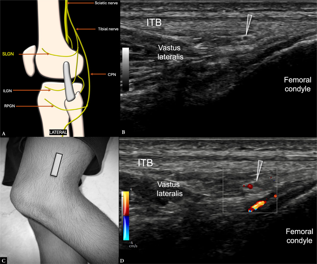
Fig. 7.
Imaging and anatomy of the superolateral genicular nerve (SLGN): A. Schematic showing the location of the SLGN cranial to the lateral collateral ligament. B. Short-axis sonographic image of the SLGN (arrowhead) deep to the iliotibial band (ITB) and vastus lateralis myotendinous junction on grayscale and D. adjacent to the superolateral genicular artery demonstrated with color Doppler. C. Transducer positioned in a coronal plane along the metaphyseal–diaphyseal junction of the femur. CPN – common peroneal nerve; ILGN – inferolateral genicular nerve; RPGN – recurrent peroneal genicular nerves
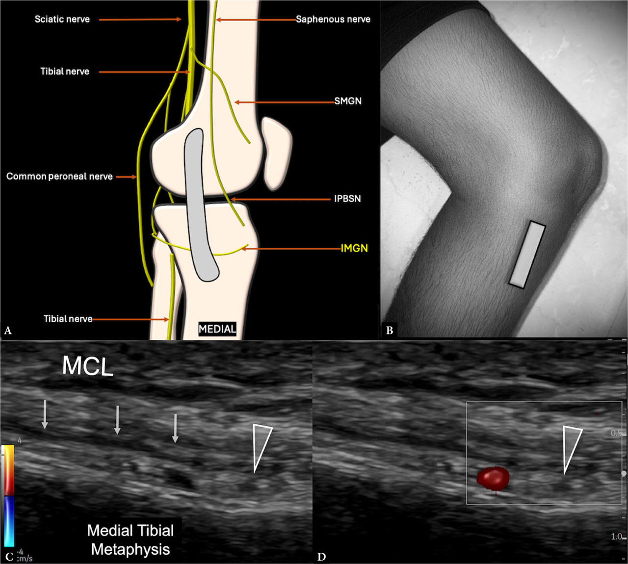
Fig. 8.
Imaging and anatomy of the inferomedial genicular nerve (IMGN): A. Schematic showing the location of the IMGN deep to the medial collateral ligament (MCL). B. Transducer positioned in a coronal plane along the metaphyseal–diaphyseal junction of the tibia. Short-axis sonographic image of the IMGN (arrowhead) deep to the distal MCL C. on grayscale and D. adjacent to the inferomedial genicular artery (IMGA) demonstrated with color Doppler. IPBSN – infrapatellar branch of the saphenous nerve; SMGN – superomedial genicular nerve
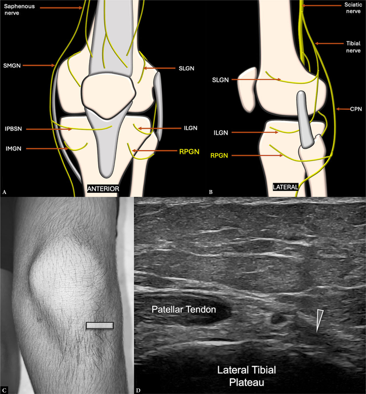
Fig. 9.
Imaging and anatomy of the recurrent peroneal genicular nerve (RPGN): A. and B. Schematic showing the location of the RPGN in the anterolateral knee, lateral to the patellar tendon. C. Transducer positioned in an axial plane at the lateral tibial plateau. D. Short-axis sonographic image of the RPGN (arrowhead) in its distal course, cranial to the tibialis anterior muscle and adjacent to the recurrent anterior tibial artery. CPN – common peroneal nerve; ILGN – inferolateral genicular nerve; IMGN – inferomedial genicular nerve; IPBSN – infrapatellar branch of the saphenous nerve; SLGN – superolateral genicular nerve; SMGN – superomedial genicular nerve
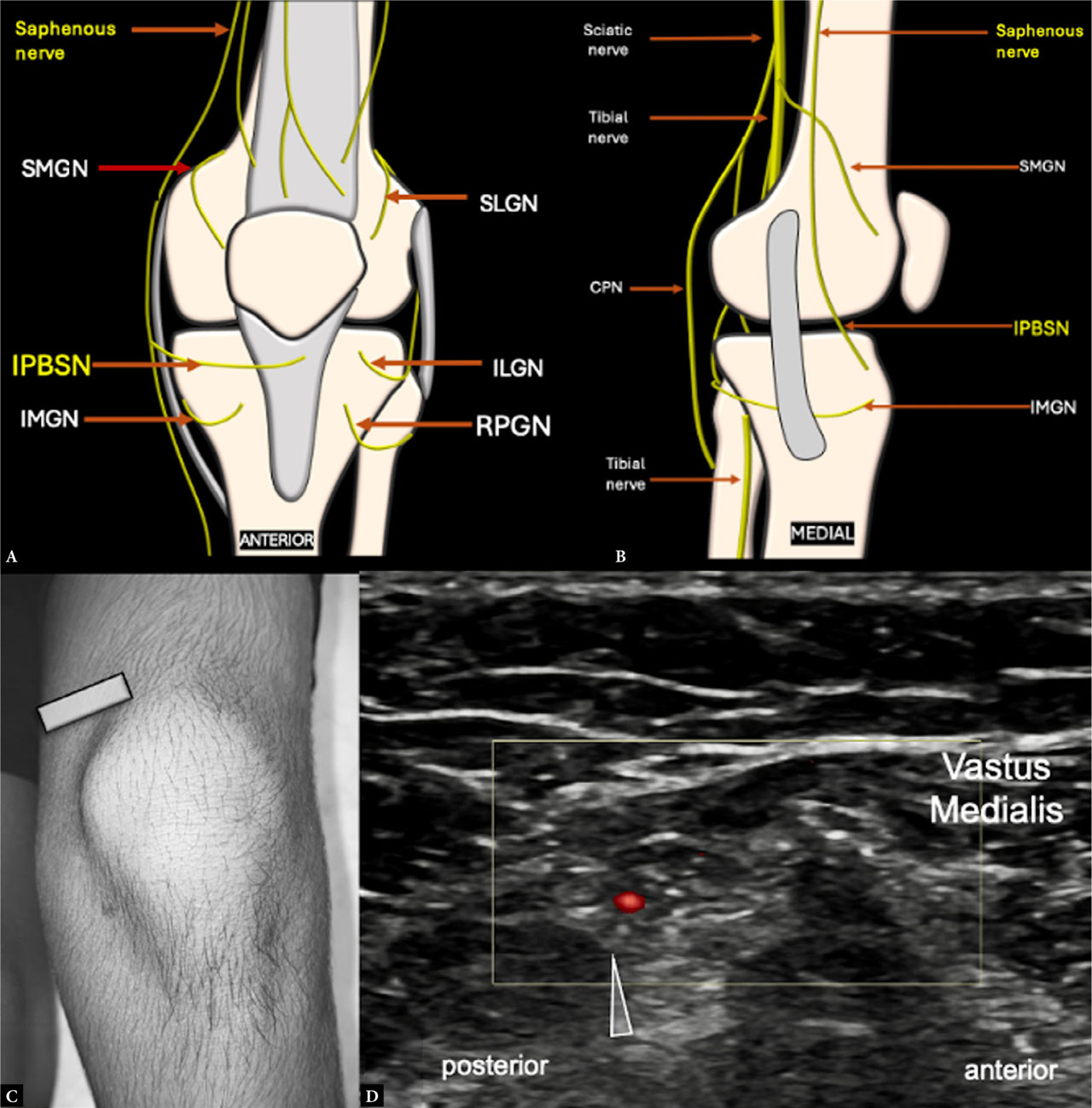
Fig. 10.
Imaging and anatomy of the infrapatellar branch of the saphenous nerve (IPBSN): A. and B. Schematic showing the location of the IPBSN in the medial aspect of the knee in the superficial plane. C. Transducer positioned in an axial plane along the medial femoral condyle. D. Short-axis sonographic image of the IPBSN (arrowhead) posteromedial to the vastus medialis and superficial to the MCL, usually accompanied by a small artery demonstrated with color Doppler. CPN – common peroneal nerve; ILGN – inferolateral genicular nerve; IMGN – inferomedial genicular nerve; RPGN – recurrent peroneal genicular nerves; SLGN – superolateral genicular nerve; SMGN – superomedial genicular nerve
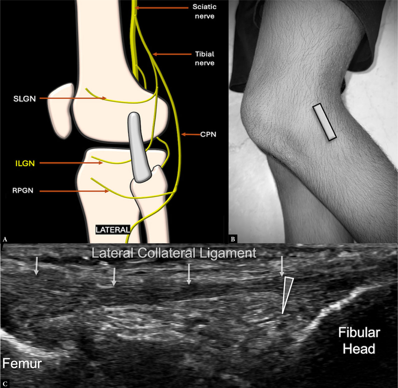
Fig. 11.
Imaging and anatomy of the inferolateral genicular nerve (ILGN): A. Schematic showing the location of the ILGN deep to the lateral collateral ligament (LCL) and cranial to the fibular head. B. Transducer positioned in a coronal plane along the lateral tibial plateau. C. Short-axis grayscale sonographic image of the ILGN (arrowhead) deep to the LCL and cranial to the fibular head. CPN – common peroneal nerve; RPGN – recurrent peroneal genicular nerves; SLGN – superolateral genicular nerve