
Introduction
The term genicular nerves, first introduced by Choi et al., refers to a network of sensory nerves that innervate the knee joint capsule, ligaments, and surrounding structures, playing a crucial role in transmitting pain signals(1). A detailed understanding of their anatomy, particularly when visualized with ultrasound, is essential for accurate diagnosis and effective management of knee pain. The genicular nerves, which are often difficult to visualize because of their small size, travel alongside the genicular arteries, making the arteries valuable sonographic landmarks when attempting to locate the nerves(2). Given that knee pain is a prevalent and often debilitating condition affecting a significant portion of the population, precise targeting of the genicular nerves has become increasingly important(3). While traditional approaches to managing chronic knee pain include pharmacology, physical therapy, and surgery, a subset of patients continue to experience persistent symptoms, necessitating alternative strategies. Genicular nerve blocks and radiofrequency ablation (RFA) have emerged as minimally invasive techniques offering promising results in alleviating chronic knee pain by selectively targeting the sensory innervation of the joint(4). This article provides a comprehensive overview of genicular nerve anatomy, ultrasound scanning techniques, and interventional approaches, equipping clinicians with a detailed guide to effectively utilize ultrasound-guided procedures for the diagnosis and management of knee pain.
Sensory innervation of knee
The genicular nerves form a complex network of sensory fibers responsible for innervating the knee joint capsule, ligaments, and surrounding structures. These nerves play a crucial role in transmitting pain signals from the knee to the central nervous system. However, variability persists in the reported number and origin of the nerve branches supplying the anterior knee capsule(5). It is well established that significant contributions arise from the femoral, sciatic, obturator, common peroneal, and saphenous nerves. These nerves are both motor and sensory in nature; however, the terminal or distal-most branches supplying the muscles that cross the knee joint are purely sensory and primarily innervate the joint itself(6,7) (Fig. 1).
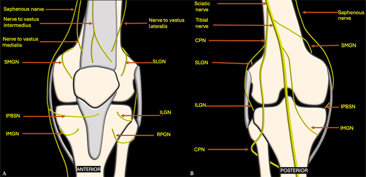
Fig. 1.
Schematic diagram depicting the sensory innervation of the knee, with genicular nerves arising from various nerves around the joint as seen from the A. anterior and B. posterior aspects. CPN – common peroneal nerve; ILGN – inferolateral genicular nerve; IMGN – inferomedial genicular nerve; IPBSN – infrapatellar branch of the saphenous nerve; RPGN – recurrent peroneal genicular nerves; SLGN – superolateral genicular nerve; SMGN – superomedial genicular nerve
According to Tran et al., up to eleven nerves and their terminal branches may supply the anterior knee capsule(8). Multiple cadaveric studies have identified six consistent articular branches – namely, the superomedial, superolateral, inferomedial, inferolateral, recurrent peroneal genicular nerves (RPGN), and the infrapatellar branch of the saphenous nerve (IPBSN) – with relatively predictable anatomical locations. Although the proximal trajectories of these nerve branches may vary, they tend to have consistent distal contact points on the femur and tibia, making them suitable targets for nerve blocks or ablative procedures(6,9).
Each genicular nerve supplies a distinct region of the knee joint, allowing for precise, targeted interventions based on the specific location of pain(5). The superomedial genicular nerve (SMGN) innervates the medial patellar retinaculum and the medial aspect of the knee joint capsule. The IPBSN contributes to sensory innervation of the medial knee joint, along with the SMGN. The superolateral genicular nerve (SLGN) supplies the superolateral portion of the tibiofemoral joint capsule. The inferomedial genicular nerve (IMGN) innervates the medial, anterior, and inferior regions of the knee joint capsule. The inferolateral genicular nerve (ILGN) is responsible for innervation of the anterolateral tibiofemoral joint and the proximal tibiofibular joint. Lastly, the RPGN supplies the inferolateral and anterior aspects of the knee joint(10).
The genicular nerves, often difficult to visualize on ultrasound because of their small size, travel alongside the genicular arteries. This relationship makes the arteries valuable sonographic landmarks when attempting to locate the nerves. Similar to the nerves, the arteries demonstrate variability in their origins but have consistent distal courses that correspond to the trajectories of the genicular nerves(11). The knee joint is supplied by a network of six genicular arteries. The descending genicular artery originates from the femoral artery, while the popliteal artery gives rise to the superomedial genicular arteries (SMGA), superolateral genicular arteries (SLGA), inferomedial genicular arteries (IMGA), inferolateral genicular arteries (ILGA), and the middle genicular arteries. The recurrent anterior tibial artery (RATA) branches off from the anterior tibial artery(2).
Understanding these specific nerve territories enables clinicians to tailor interventions more effectively, targeting the precise areas of joint pain. For effective pain management, it is essential to target these sensory branches while sparing the motor fibers to avoid unwanted weakness or motor deficits. Therefore, performing distal nerve blocks or ablations that focus specifically on the sensory components – known as motor-sparing nerve blocks – is crucial. This approach ensures adequate pain relief or neuromodulation while minimizing the risk of motor impairment and associated complications(9).
Ultrasound scanning technique for genicular nerves
Ultrasound imaging is an invaluable tool for visualizing genicular nerves and guiding interventional procedures. High-resolution ultrasound facilitates direct visualization of nerves as well as the surrounding soft-tissue and bony landmarks.
For optimal imaging, a very high frequency (12–15 MHz or higher) transducer with a small footprint, such as a hockey-stick probe, is recommended for superficial areas, whereas a medium-frequency (6–15 MHz) linear transducer with a larger footprint is suitable for deeper regions or patients with larger body habitus. The high-frequency transducer provides the best resolution for the small-caliber genicular nerves (0.5–1.5 mm in diameter), which can be challenging to visualize directly. A larger-footprint probe may initially be used to locate the genicular vessels, after which a high-frequency probe is advisable for identifying the nerves. Utilizing color Doppler or power Doppler is essential for identifying accompanying genicular vessels, which serve as crucial landmarks for nerve localization(10).
The patient is typically positioned supine with the knee slightly flexed and rotated as needed to access the specific nerve being examined. Nerves should be visualized in both short- and long-axis views. The ultrasonographic appearance of normal peripheral nerves reflects their macroscopic anatomy. In the short-axis view, nerves exhibit a “honeycomb” pattern, with hypoechoic fascicles surrounded by hyperechoic perineurium and encased in echogenic epineurium. In the longitudinal view, nerves resemble “train tracks,” with fascicles appearing as linear hypoechoic structures outlined by hyperechoic borders. As nerves progress distally, their cross-sectional area (CSA) typically decreases as branches are given off(12). Since genicular nerves are terminal branches, they are generally monofascicular, possessing a small CSA and appearing as small, round hypoechoic structures in short-axis views. They may resemble adjacent genicular arteries on grayscale imaging; therefore, color Doppler is employed to differentiate between them. A low pulserepetition frequency, typically between 3 and 4 cm/s, is generally recommended to enhance visualization of low-velocity flow within genicular vessels(10).
Clinical implications
Understanding the anatomy of the genicular nerves and the corresponding ultrasound scanning protocols has several important clinical implications:
Perioperative pain management
Genicular nerve blocks can be incorporated into multimodal analgesia regimens for patients undergoing knee surgery, such as total knee arthroplasty. Targeting specific genicular nerves helps reduce postoperative pain, decrease opioid consumption, and improve functional outcomes(13).
Chronic pain palliation
RFA of the genicular nerves presents an effective treatment option for chronic knee pain arising from osteoarthritis or other conditions, particularly after total knee arthroplasty. RFA works by heating the nerves to disrupt their ability to transmit pain signals(14,15). RFA can be performed at temperatures ranging from 60 to 80 degrees for 60 to 120 seconds, depending on manufacturer specifications and operator preference. Procedures may be performed under ultrasound or fluoroscopic guidance with local anesthesia or sedation.
Post-intervention nerve injury diagnosis and treatment
A thorough understanding of genicular nerve anatomy is essential for diagnosing nerve injuries following knee surgery, trauma, or other interventions(16). Ultrasound imaging can reveal nerve damage, entrapment, or neuroma formation (Fig. 2). Applying focal compression with the transducer at a site of ultrasonographic abnormality may elicit pain or a tingling sensation – a response known as the ultrasonographic Tinel sign (Fig. 3). This sign helps pinpoint the exact site of neuropathy, thereby enhancing diagnostic accuracy and guiding management(17) (Fig. 4, Fig. 5).
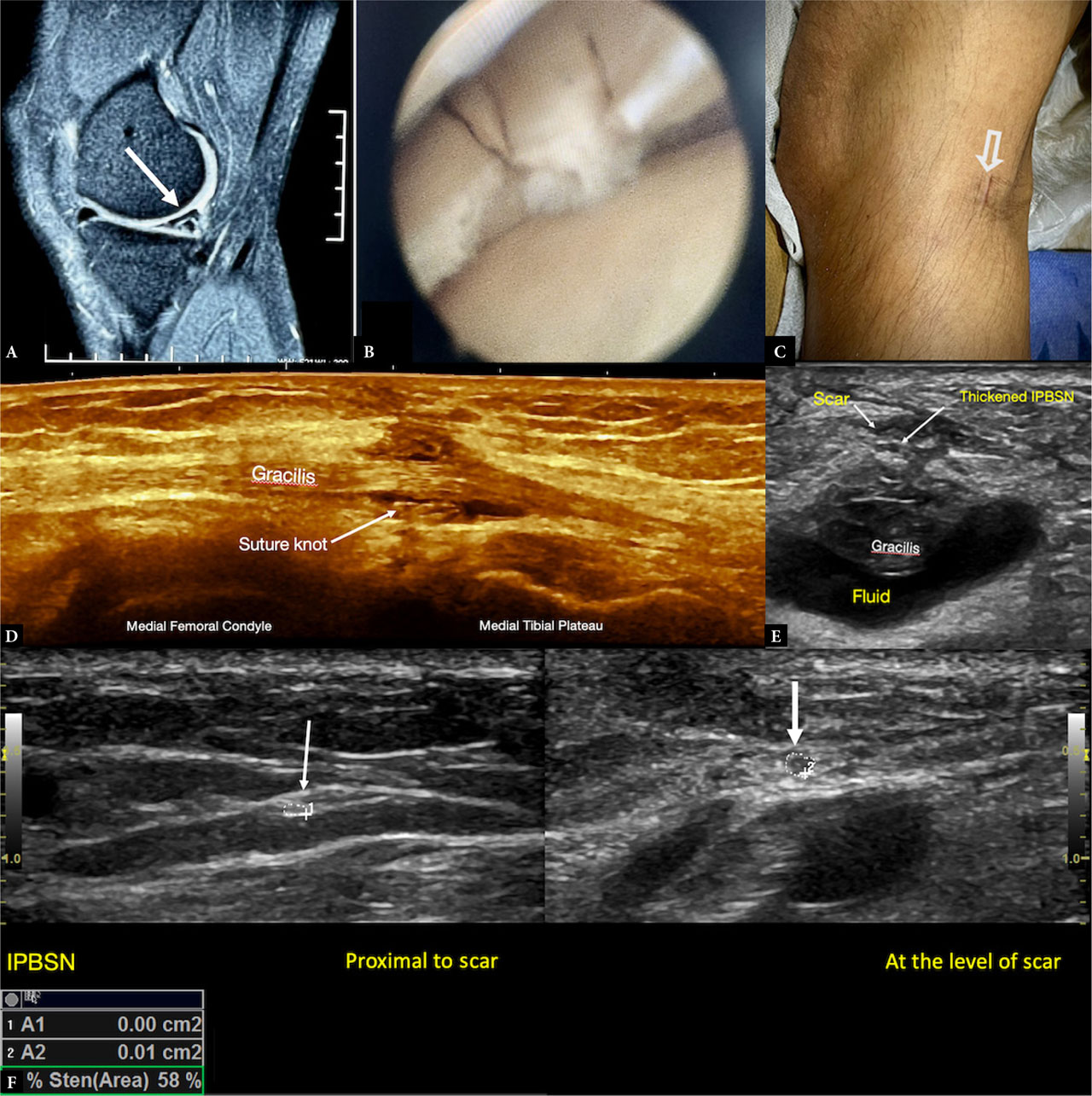
Fig. 2.
Infrapatellar branch of the saphenous nerve (IPBSN) injury following medial meniscal repair surgery. A. Proton-density fat-suppressed sagittal MRI image showing an oblique tear in the posterior horn of the medial meniscus B. Arthroscopic image of the torn meniscus C. Scar site corresponding to the site of the ultrasonographic Tinel sign D. Longitudinal view at the scar site revealing a suture knot around the gracilis muscle E. Transverse view across the scar site showing a thickened IPBSN adjacent to the scar, overlying the gracilis muscle. F. Cross-sectional area of the nerve (arrow) measured proximal and at the scar site shows focal enlargement with increased hypoechogenicity of the nerve at the scar site consistent with neuroma formation. The IPBSN was traced for its origin from the saphenous nerve; in this patient, it followed a more posterior course than usual. IPBSN – infrapatellar branch of the saphenous nerve
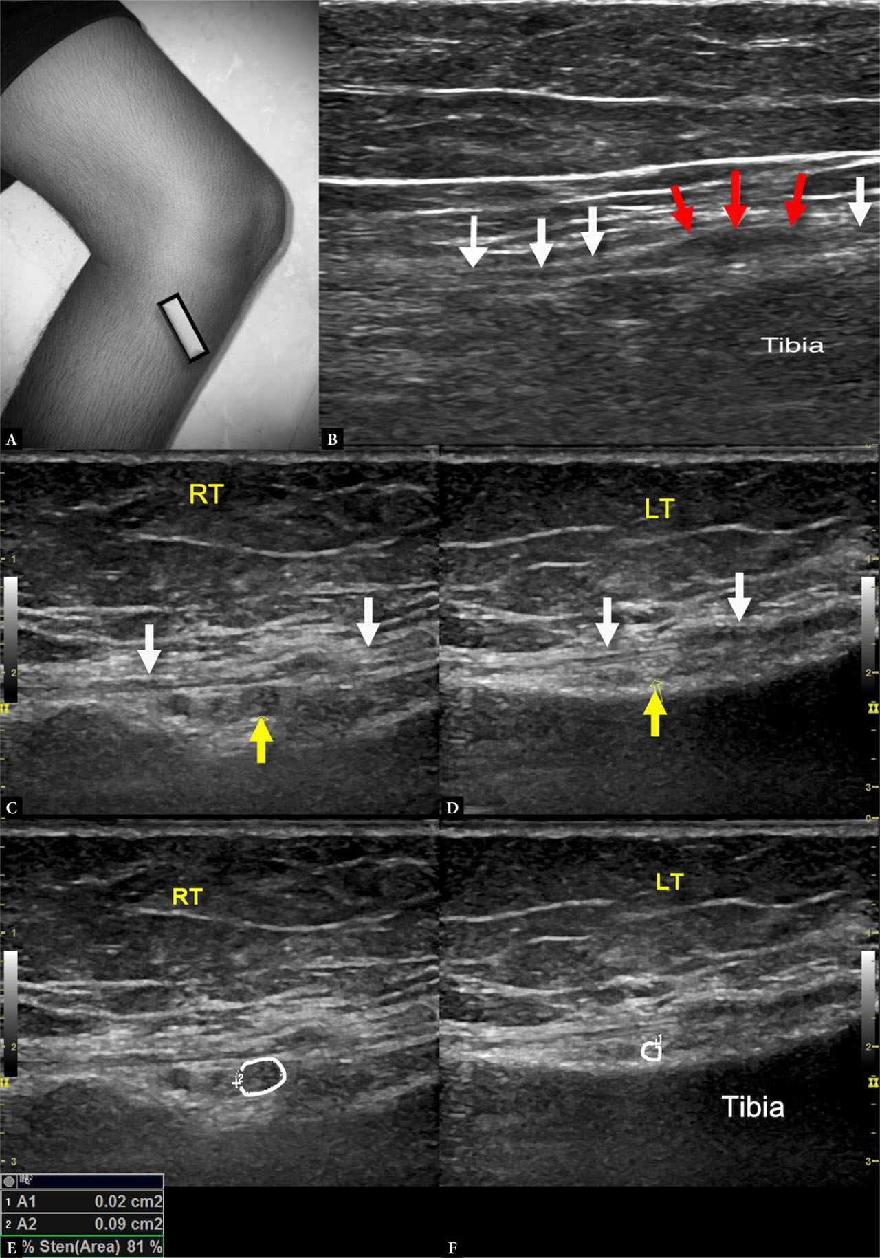
Fig. 3.
Post-traumatic inferomedial genicular nerve (IMGN) neuroma in a 53-year-old woman with chronic inferomedial right knee pain with history of trauma A. Probe positioned at the proximal tibial shaft B. Long-axis view of the IMGN showing focal thickening of the nerve (red arrows) at the site of the scar – neuroma in continuity, with normal caliber of the nerve proximal and distal (white arrows). Short-axis view of the nerve (yellow arrow) deep to the MCL (white arrows) and just superficial to the tibial cortex on the C. affected (right) side showing a bulky hypoechoic nerve and D. on the normal contralateral (left) side for comparison. Cross-sectional area comparison on the E. right and F. left side shows a marked difference in caliber; the abnormal nerve caliber may be overlooked if seen in isolation due to the inherently small size of the nerve
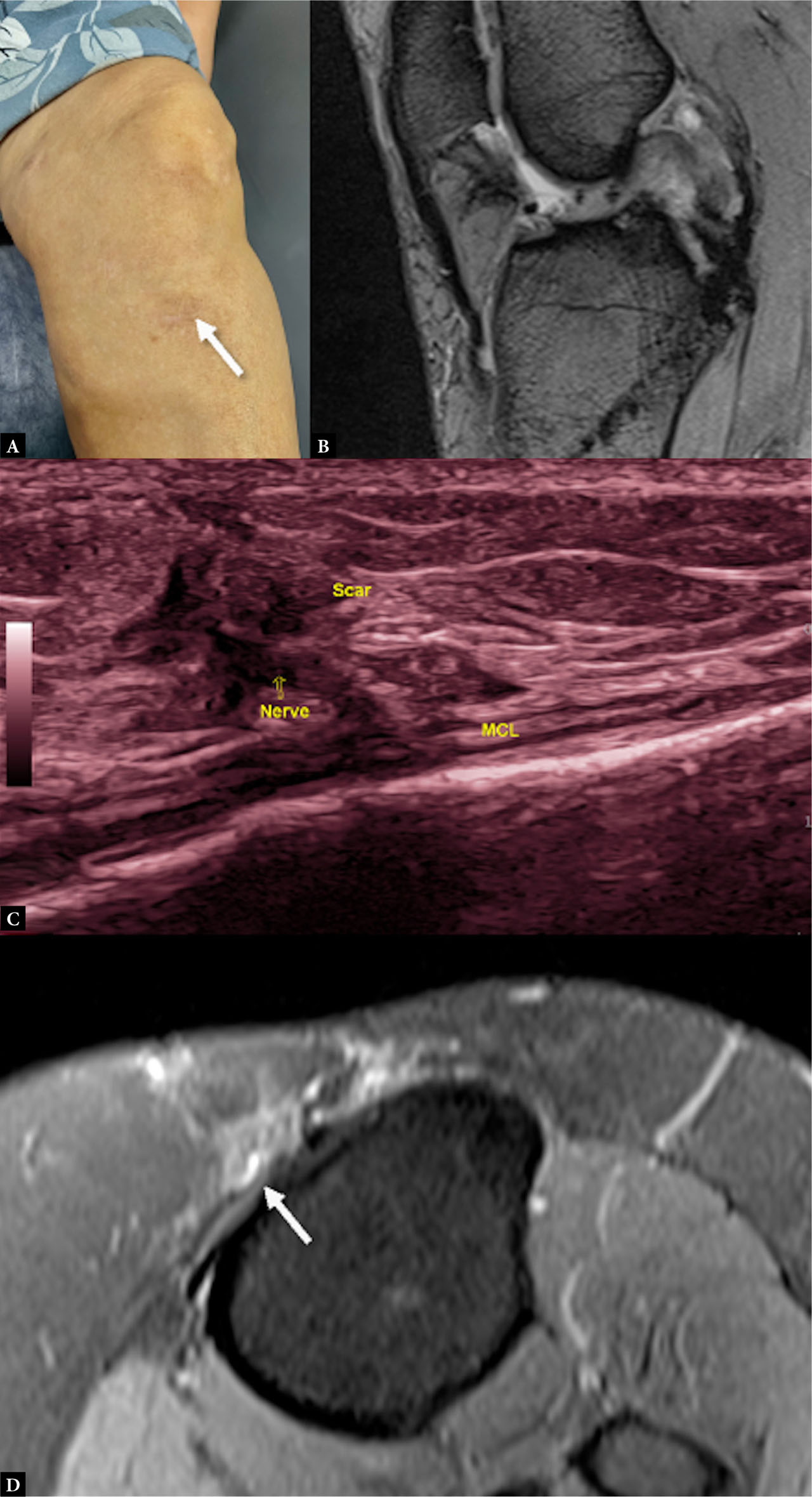
Fig. 4.
Inferomedial genicular nerve scar entrapment following posterior cruciate ligament (PCL) repair A. Clinical image showing the scar site B. Fat-suppressed proton density sagittal weighted MRI showing postoperative changes in PCL; no obvious abnormality was appreciated on MRI explaining the patient’s symptoms C. Ultrasound image with the probe placed along the distal tibial insertion site of the medial collateral ligament (MCL) showing nerve entrapped within scar tissue, corresponding precisely to the site of maximum tenderness. D. Retrospective review of the axial MRI images reveals the entrapped nerve as a focal hyperintense structure (arrow)
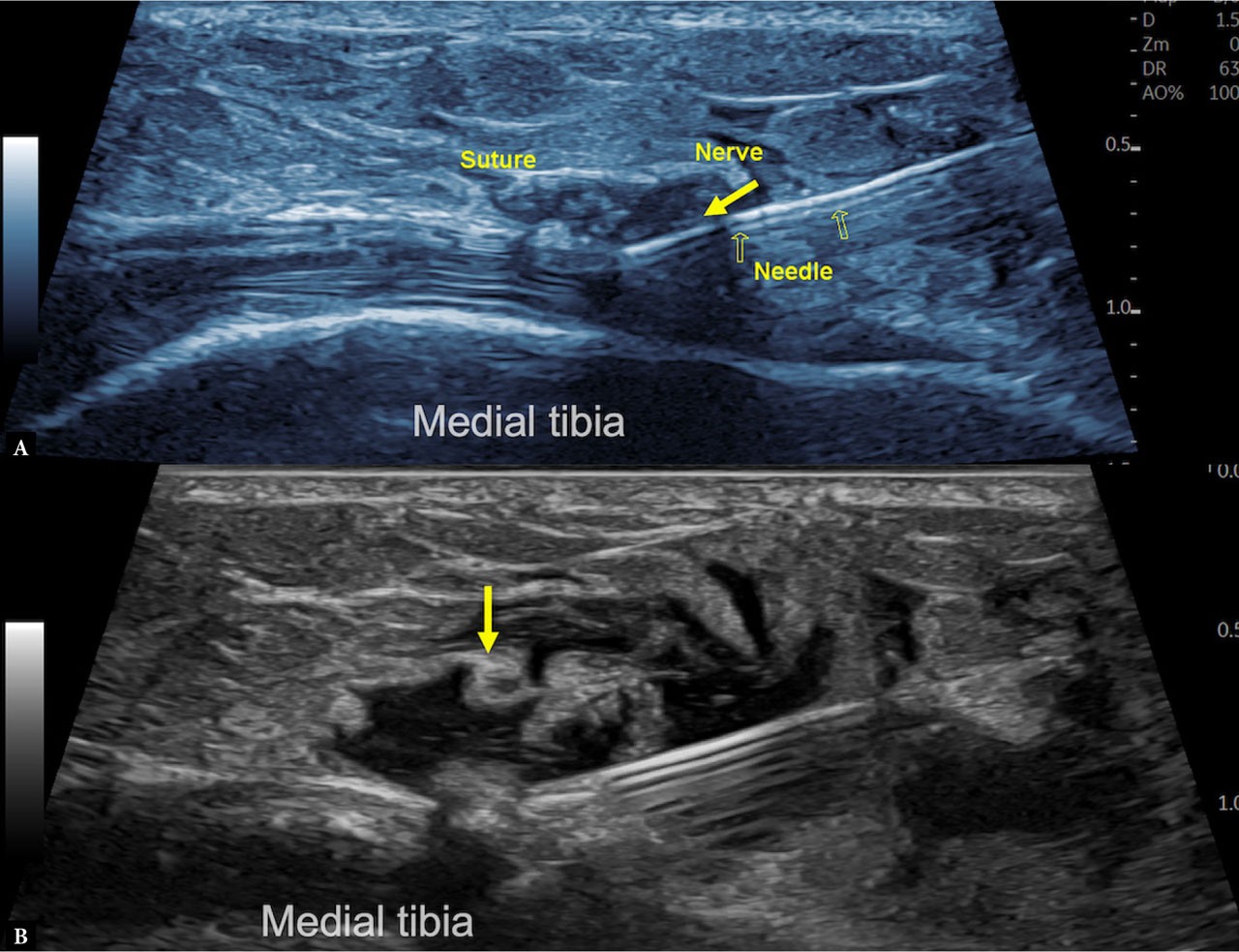
Fig. 5.
US-guided therapeutic hydrodissection of the inferomedial genicular nerve in the patient described in Fig. 4. A. Longitudinal view along the medial proximal tibial shaft near the medial collateral ligament attachment site corresponding to the scar site, showing the entrapped nerve within the scar; the needle is advanced to the target site using ‘in-plane approach’ with the tip directed deep to the nerve. B. Approximately 4–5 mL of injectate (3 mL 5% dextrose + 1 mL steroid + 1 mL local anesthetic) was delivered circumferentially to release the nerve from the scar
The role of genicular nerve blocks and radiofrequency ablation
Genicular nerve blocks
Genicular nerve blocks involve injecting a local anesthetic, often combined with a corticosteroid, near the genicular nerves to temporarily inhibit nerve conduction. This procedure provides immediate pain relief and also serves as a diagnostic tool: a significant reduction in pain following the block suggests that the targeted nerves are contributing to the patient’s symptoms(18).
Radiofrequency ablation (RFA)
RFA may be employed as a longer-lasting solution, particularly for patients who experience substantial pain relief from a genicular nerve block. RFA uses thermal energy to create a lesion on the nerve, disrupting its ability to transmit pain signals and thereby providing sustained pain relief(18).
While the traditional method for genicular nerve pain reduction, as described by Choi et al. with fluoroscopic guidance and later adapted to ultrasound using the genicular arteries, targeted three nerves (SMGN, SLGN, and IMGN), current approaches increasingly utilize a five-nerve target (SMGN, SLGN, IMGN, IPBSN, and RPGN) based on recent cadaveric and clinical studies incorporating modified landmarks for potentially better results(1,19,20). Possible complications of genicular nerve block or ablation include injury to muscles or blood vessels, unintentional puncture of nearby motor nerves, and, rarely, complex regional pain syndrome following radiofrequency ablation. When targeting the RPGN, there is a risk of foot drop due to common peroneal nerve (CPN) block. The ILGN is usually avoided as a target because of the potential risk of injury to the adjacent CPN(21).
The detailed anatomy, ultrasound scanning protocols, and approaches for targeting the six genicular nerves for both nerve blocks and RFA are described below, providing clinicians with a comprehensive guide to optimizing pain management interventions (Tab. 1).
Tab. 1.
Relevant anatomy of the six genicular nerves
| Genicular Nerve | Origin | Accompanying vessel | Relevant anatomical landmark | Area of innervation | Special point |
|---|---|---|---|---|---|
| Superomedial genicular nerve | Femoral nerve branch (nerve to vastus medialis) Tibial nerve | Superomedial genicular arteries | Bony: adductor tubercle Soft tissue: femoral attachment of MCL, vastus medialis and adductor magnus | Medial patellar retinaculum; medial knee joint capsule | Largest and the most consistent genicular nerve |
| Superolateral genicular nerve | Femoral nerve branch Sciatic nerve | Superolateral genicular artery | Bony: bony ridge separating the lateral and posterior cortex Soft tissue: femoral attachment of LCL, vastus lateralis and biceps femoris muscle | Superolateral knee capsule | |
| Inferomedial genicular nerve | Tibial nerve Sciatic nerve | Inferomedial genicular artery | Bony: tibial tuberosity and medial tibial condyle Soft tissue: tibial insertion of MCL | Medial anterior and inferior knee capsule | Susceptible to iatrogenic injury |
| Inferolateral genicular nerve | CPN | Inferolateral genicular arteries | Bony: lateral tibial plateau Soft tissue: LCL | Anterolateral knee capsule and proximal tibiofibular joint | Close to CPN motor branches, but CPN is inferior to fibular neck |
| RPGN | CPN proximal DPN | Recurrent anterior tibial artery – branch of proximal ATA | Bony: Gerdy’s tubercle Soft tissue: tibialis anterior muscle | Inferolateral and anterior knee, tibiofibular joint | Close to CPN and anterior tibial vessels |
| Infrapatellar branch of the saphenous nerve | Saphenous nerve | Descending genicular artery in proximal course | Bony: patellar tip and tibial tuberosity Soft tissue: MCL | Medial knee joint | Highly susceptible to iatrogenic injury Subcutaneous course |
Superomedial genicular nerve (SMGN)
The SMGN was previously described as a branch originating from the tibial nerve. However, recent studies have identified it as a terminal branch of the nerve to the vastus medialis, arising from the femoral nerve. The SMGN is the largest and most consistent of the genicular nerves. It originates at the junction of the middle and distal thirds of the thigh(22).
After its origin, it descends deep to the adductor magnus tendon and eventually makes contact with the bone approximately 1 cm anterior to the most prominent part of the adductor tubercle. The nerve then courses anteriorly alongside the SMGA, remaining close to the bone and deep to the vastus medialis muscle. It is located cranial to the proximal origin of the medial collateral ligament (MCL) before giving branches to the knee capsule(6).
Ultrasound scanning protocol
The patient is positioned supine with the knee flexed approximately 30–45 degrees and the leg slightly externally rotated. The ultrasound transducer is aligned in a coronal plane over the medial femorotibial joint line, then moved cephalad and posteriorly to identify the adductor tubercle and the insertion of the adductor magnus tendon. Color Doppler helps identify the adjacent SMGA, located deep to the vastus medialis and directly over the bony cortex of the distal femur. Typically, the SMGN is found immediately adjacent to the SMGA. In the coronal plane, the nerve is visualized in short axis. Once identified, the transducer is rotated 90 degrees to obtain a long-axis image. Key landmarks for identifying the SMGN include the adductor tubercle, the femoral origin of the MCL, and the SMGA. To target the nerve for RFA, after localizing it in the short-axis view, the transducer is rotated to acquire a long-axis image. The RF cannula is then advanced in-plane from anterior to posterior until it reaches the posterior cortex of the femur, positioned either at or just above the adductor tubercle(19) (Fig. 6).
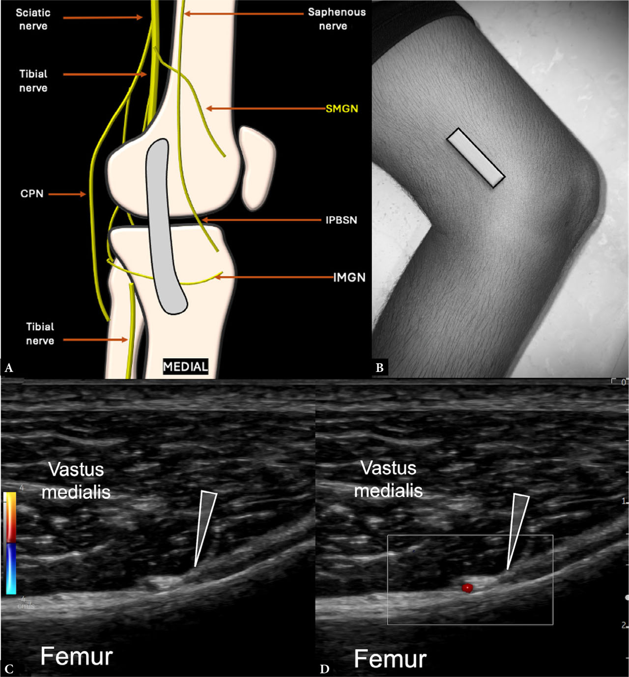
Fig. 6.
Imaging and anatomy of the superomedial genicular nerve (SMGN): A. Schematic showing the location of the SMGN cranial to the femoral insertion of the medial collateral ligament. B. Transducer positioned in a coronal plane along the metaphyseal–diaphyseal junction of the distal medial femur. Short-axis sonographic image of the SMGN (arrowhead) deep to the vastus medialis C. on grayscale and D. adjacent to the superomedial genicular arteries demonstrated with color Doppler. CPN – common peroneal nerve; IMGN – inferomedial genicular nerve; IPBSN – infrapatellar branch of the saphenous nerve
Superolateral genicular nerve (SLGN)
The SLGN exhibits variable anatomical origins. Traditionally, it has been described as arising from the nerve to the vastus lateralis, a branch of the femoral nerve(5). However, other anatomical studies have reported its origin from the sciatic nerve, and rarely from the CPN. Despite this variability, the distal course of the SLGN remains relatively consistent. It typically accompanies the SLGA, traveling deep to the distal biceps femoris tendon and the vastus lateralis muscle, over the junction of the femoral shaft and the lateral femoral epicondyle(9).
Ultrasound scanning protocol
The patient is positioned with the knee flexed approximately 30 degrees and the leg slightly internally rotated. The ultrasound transducer is aligned in a coronal plane over the lateral femorotibial joint line and moved cephalad to locate the junction between the lateral femoral condyle and the femoral shaft. Color Doppler assists in identifying the adjacent SLGA, which lies deep to the myotendinous junction of the vastus lateralis and directly over the bony cortex of the distal femur. In the coronal view, the nerve is visualized in short axis. Once identified, the transducer is rotated 90 degrees to obtain a long-axis view(19).
For RFA targeting, traditional techniques involve positioning the needle at the bony cortex near the artery, around the junction of the femoral shaft and the lateral femoral epicondyle. The RF cannula is advanced in-plane from anterior to posterior, with the tip positioned midway through the femoral depth(1). More recently, alternative approaches have been described, such as targeting the nerve in the superolateral popliteal fossa, medial to the biceps femoris tendon. In this method, the RF cannula is inserted obliquely at approximately 45 degrees to the coronal plane and advanced in-plane until the tip reaches the posterior edge of the lateral femoral cortex. This cortical crest – the bony ridge separating the lateral and posterior cortex – is easily identifiable and serves as a useful landmark for accurate placement(20,23) (Fig. 7).
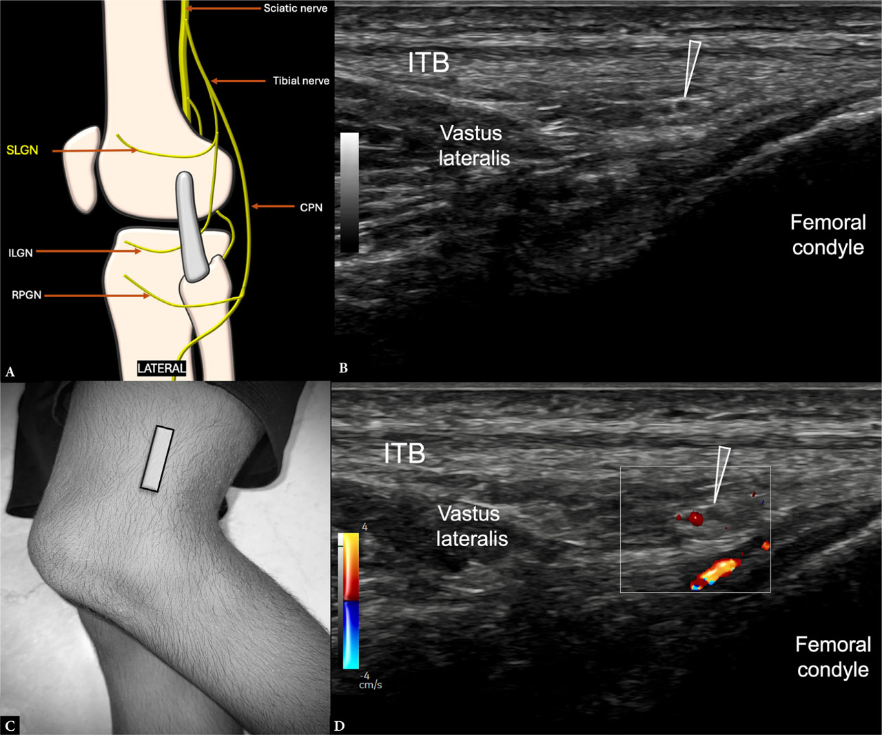
Fig. 7.
Imaging and anatomy of the superolateral genicular nerve (SLGN): A. Schematic showing the location of the SLGN cranial to the lateral collateral ligament. B. Short-axis sonographic image of the SLGN (arrowhead) deep to the iliotibial band (ITB) and vastus lateralis myotendinous junction on grayscale and D. adjacent to the superolateral genicular artery demonstrated with color Doppler. C. Transducer positioned in a coronal plane along the metaphyseal–diaphyseal junction of the femur. CPN – common peroneal nerve; ILGN – inferolateral genicular nerve; RPGN – recurrent peroneal genicular nerves
Inferomedial genicular nerve (IMGN)
The IMGN has historically been described as originating from the tibial nerve within the popliteal fossa. However, other studies have indicated that it may also arise from the sciatic nerve. After its origin, the nerve curves around the tibia – distal to the tibial plateau – and emerges anteriorly, passing deep to the MCL. It typically lies just proximal to the tibial insertion of the MCL, alongside the IMGA(7). The IMGN, along with the IPBSN, is particularly vulnerable to accidental injury during surgical procedures, including open or arthroscopic surgeries such as total knee replacement, tibial nailing, and anterior cruciate ligament (ACL) reconstruction at the medial tibia(16).
Ultrasound scanning protocol
For ultrasound visualization, the patient is positioned with the knee flexed between 30 and 45 degrees, with the leg slightly externally rotated. The transducer is initially aligned in a coronal plane over the medial femorotibial joint line and then moved distally to identify the metaphyseal–diaphyseal junction of the tibia and the distal insertion of the MCL. The nerve appears in short axis as a small hypoechoic structure along the tibial bone surface adjacent to the inferomedial genicular vessels. Rotating the transducer 90 degrees provides a long-axis view of the nerve(24).
For RFA targeting, the traditional method involves positioning the needle at the bony cortex near the IMGA, around the proximal tibial metaphyseal–diaphyseal junction. The RF cannula is advanced inplane from anterior to posterior, with the tip situated at the mid-point of the tibial width, close to the periosteum(1). Alternatively, the midpoint between the most prominent part of the medial tibial condyle and the tibial insertion of the MCL can be used as a landmark. In this approach, the needle is inserted approximately 1 cm medial to the tibial tuberosity and directed toward the target, with the tip positioned at the medial edge of the medial cortex. This landmark-based technique may be particularly useful when Doppler signals from the arteries are weak, ensuring accurate nerve targeting(23,25) (Fig. 8).
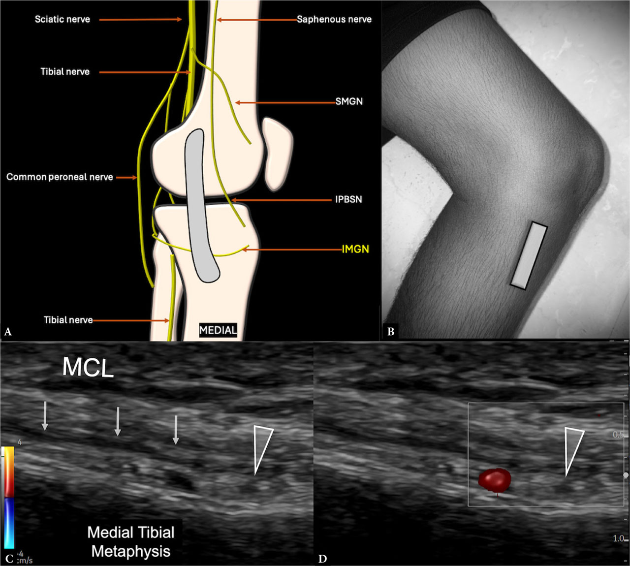
Fig. 8.
Imaging and anatomy of the inferomedial genicular nerve (IMGN): A. Schematic showing the location of the IMGN deep to the medial collateral ligament (MCL). B. Transducer positioned in a coronal plane along the metaphyseal–diaphyseal junction of the tibia. Short-axis sonographic image of the IMGN (arrowhead) deep to the distal MCL C. on grayscale and D. adjacent to the inferomedial genicular artery (IMGA) demonstrated with color Doppler. IPBSN – infrapatellar branch of the saphenous nerve; SMGN – superomedial genicular nerve
Recurrent peroneal genicular nerve (RPGN)
The origin of the RPGN is variably described, with some reports indicating that it arises either from the CPN before its bifurcation or from the deep peroneal nerve. At the level of the fibular neck, the RPGN courses proximally, ascending alongside the RATA, which originates from the proximal anterior tibial artery (ATA). The nerve and artery run together along the anterolateral surface of the tibia, deep to the tibialis anterior muscle, heading superiorly toward the distal patellar tendon. The RPGN is considered one of the most consistent branches responsible for articular innervation. Although it is not routinely targeted during conventional genicular nerve RFA procedures – primarily because of its proximity to the CPN and the associated risk of foot drop – it holds clinical significance as a major contributor to inferior anterolateral knee pain, as well as pain in the tibiofibular joint(26).
Ultrasound scanning protocol
To visualize the RPGN with ultrasound, the patient is positioned with the knee slightly flexed and the leg rotated slightly inward. The transducer is aligned in a coronal plane over the lateral femorotibial joint line, then moved distally to locate the insertion of the iliotibial tract at Gerdy’s tubercle. The transducer is subsequently rotated 90 degrees and shifted about 1–2 cm distally to visualize the tibialis anterior muscle. In short axis, the nerve appears as a hypoechoic, oval-shaped structure within the subcutaneous tissue, just above the periosteum of the anterior tibia, coursing alongside the RATA. The artery can be confirmed with Doppler imaging(10).
For RF treatment, the target point is approximately 2 cm distal to Gerdy’s tubercle, where the nerve runs anterior to the tibial periosteum and deep to the tibialis anterior muscle, aligned with the RATA, and located near the midpoint of the tibial width. The transducer is positioned in an axial plane, and the RF cannula is inserted just lateral to the tibial tuberosity, advancing toward the bone while maintaining an in-plane approach(23) (Fig. 9).
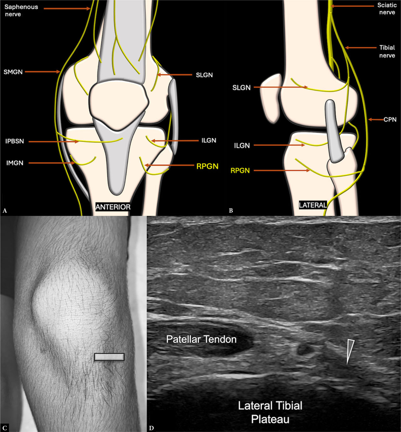
Fig. 9.
Imaging and anatomy of the recurrent peroneal genicular nerve (RPGN): A. and B. Schematic showing the location of the RPGN in the anterolateral knee, lateral to the patellar tendon. C. Transducer positioned in an axial plane at the lateral tibial plateau. D. Short-axis sonographic image of the RPGN (arrowhead) in its distal course, cranial to the tibialis anterior muscle and adjacent to the recurrent anterior tibial artery. CPN – common peroneal nerve; ILGN – inferolateral genicular nerve; IMGN – inferomedial genicular nerve; IPBSN – infrapatellar branch of the saphenous nerve; SLGN – superolateral genicular nerve; SMGN – superomedial genicular nerve
Infrapatellar branch of the saphenous nerve (IPBSN)
The IPBSN originates from the saphenous nerve at the level of the adductor canal. It transitions from a deep position within the canal to a superficial course between the vastus medialis and sartorius muscles in the distal thigh. Along with the SMGN, it contributes to sensory innervation of the medial knee joint. Its pathway generally parallels that of the descending genicular artery but can be distinguished by its superficial trajectory over the proximal MCL, in contrast to the deeper location of the SMGN near the femoral bone surface. Surgical interventions on the anteromedial knee (such as total knee arthroplasty, arthroscopic surgery, anterior cruciate ligament reconstruction, and meniscectomy) carry a risk of iatrogenic injury to the IPBSN(27).
Ultrasound scanning protocol
Fonkoue et al. described the “treatment line” for the IPBSN as a longitudinal path approximately 4 cm medial to the patellar apex, connecting transverse lines passing through the patellar tip and the top of the tibial tuberosity. To visualize the nerve at the knee, the probe is aligned in a coronal plane along this line, identifying the nerve as a hypoechoic, ovoid structure within the subcutaneous tissue overlying the MCL, often accompanied by a small artery. If the nerve is not visible, the probe is shifted posteromedially and cephalad (retrograde exploration) to search for it. Alternatively, the saphenous nerve may be followed from its emergence at mid-femoral level (anterograde exploration) toward the knee(23) (Fig. 10).
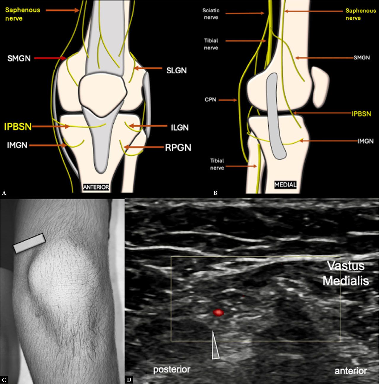
Fig. 10.
Imaging and anatomy of the infrapatellar branch of the saphenous nerve (IPBSN): A. and B. Schematic showing the location of the IPBSN in the medial aspect of the knee in the superficial plane. C. Transducer positioned in an axial plane along the medial femoral condyle. D. Short-axis sonographic image of the IPBSN (arrowhead) posteromedial to the vastus medialis and superficial to the MCL, usually accompanied by a small artery demonstrated with color Doppler. CPN – common peroneal nerve; ILGN – inferolateral genicular nerve; IMGN – inferomedial genicular nerve; RPGN – recurrent peroneal genicular nerves; SLGN – superolateral genicular nerve; SMGN – superomedial genicular nerve
For RFA, if direct visualization is unsuccessful, an indirect targeting approach involves placing the transducer in the coronal plane along the treatment line. The RF cannula is then inserted near the superior edge of the line and advanced distally, deep into the subcutaneous tissue just above the MCL. The cannula should be positioned as deeply as possible within the subcutaneous tissue to minimize the risk of skin burns caused by the heat generated during RFA(20).
Inferolateral genicular nerve (ILGN)
The ILGN typically originates from the CPN within the popliteal fossa. It courses inferolaterally toward the lateral joint line, positioned deep to the biceps femoris muscle, the lateral collateral ligament (LCL), and the iliotibial band(7). The nerve travels alongside the inferolateral genicular vessels, just above the lateral tibial plateau cortex and cranial to the fibular head, before branching to innervate the joint capsule. It supplies sensory innervation to the anterolateral aspect of the tibiofemoral joint and the proximal tibiofibular joint(5). Differentiating the ILGN from the CPN and the RPGN is essential: the ILGN closely follows the periosteum of the lateral tibia, whereas the CPN is associated with the fibular neck and does not accompany genicular vessels(28).
Ultrasound scanning protocol
For ultrasound visualization, the patient is placed supine with the knee slightly flexed and the leg mildly internally rotated. The transducer is aligned in a coronal plane over the lateral femorotibial joint line and subsequently moved distally to identify the LCL insertion at the fibular head. The nerve appears in its short-axis view as a small hypoechoic structure along the surface of the lateral tibial plateau, just above the fibular head and beneath the LCL. On color Doppler, it can be identified coursing with the inferolateral genicular vessels. Rotating the transducer 90 degrees provides a long-axis view. Because of the ILGN’s proximity to the CPN, there is a potential risk of accidental CPN blockade, which may result in iatrogenic foot drop if not carefully avoided(10,29) (Fig. 11).
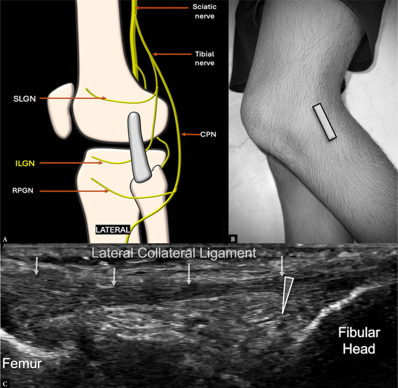
Fig. 11.
Imaging and anatomy of the inferolateral genicular nerve (ILGN): A. Schematic showing the location of the ILGN deep to the lateral collateral ligament (LCL) and cranial to the fibular head. B. Transducer positioned in a coronal plane along the lateral tibial plateau. C. Short-axis grayscale sonographic image of the ILGN (arrowhead) deep to the LCL and cranial to the fibular head. CPN – common peroneal nerve; RPGN – recurrent peroneal genicular nerves; SLGN – superolateral genicular nerve
USG versus fluoroscopy guidance
Ultrasound guidance for genicular nerve block and RFA offers several advantages over traditional fluoroscopy. While fluoroscopy-guided procedures rely primarily on bony landmarks to approximate the position of the targeted nerves, ultrasound incorporates both bony and soft-tissue landmarks – including ligaments, muscles, and vessels – for more precise localization(22). Since the vascular patterns accompanying each genicular nerve are well established, arterial blood flow visualized with Doppler can further aid in the accurate identification of the nerves. An additional benefit of real-time ultrasound imaging is that it facilitates dynamic assessment, enabling clinicians to observe needle advancement relative to surrounding structures. As a result, needles can be inserted and advanced without applying direct pressure to the periosteum, reducing patient discomfort and pain during the procedure. Importantly, ultrasound does not expose patients or clinicians to radiation, making it a safer and more desirable option(12,30).
However, when comparing nerve blocks with RFA, it is important to recognize that RFA requires greater precision because the lesion produced is typically confined to a small, well-defined area. In contrast, for nerve blocks, the injectate tends to diffuse over a broader region, making precise anatomical landmarks less critical. Ultimately, the success of both genicular nerve block and RFA depends on accurately targeting the articular branches innervating the knee joint capsule, highlighting the importance of precise visualization techniques. The growing number of publications on ultrasound-guided genicular nerve blocks and RFA for chronic knee pain reflects increasing clinical interest in these techniques(18).
Conclusion
Ultrasound-guided genicular nerve blocks and RFA are valuable tools in the multimodal management of chronic knee pain, offering a targeted and minimally invasive approach to pain relief.
A thorough understanding of genicular nerve anatomy, precise ultrasound scanning techniques, and careful patient selection are critical for optimizing outcomes and minimizing complications. Although further research is needed to fully elucidate long-term efficacy and comparative effectiveness of these interventions, current evidence supports their role in reducing pain, improving function, and decreasing opioid consumption in carefully selected patients. With continued adoption and ongoing refinement of techniques, ultrasound-guided genicular nerve interventions have the potential to significantly improve the management of chronic knee pain, enhancing quality of life for many individuals.