
Introduction
Migraine is currently the second most disabling condition worldwide, affecting an estimated 12% of the population(1). Managing this condition is crucial given its profound global and personal burden. Within the migraine spectrum, high-frequency episodic migraine (EM) and chronic migraine (CM) particularly benefit from prophylactic therapies aimed at reducing the frequency, duration, and severity of headache attacks(2).
Currently, migraine is recognized as a complex disorder involving altered connectivity across various brain regions, alongside functional changes in sensory and pain-processing circuits. The trigeminovascular system plays a central role in this pathophysiological cascade, with neuropeptides acting as key mediators; among these, calcitonin gene-related peptide (CGRP) has been established as a primary therapeutic target(3).
Monoclonal antibodies (mAbs) targeting CGRP (eptinezumab, fremanezumab, and galcanezumab) or its receptor (erenumab), hereafter referred to as “anti-CGRP mAbs”, have significantly improved the management of EM and CM. They provide a substantial reduction in the number of migraine attacks and related disability with minimal side effects(4). However, CGRP is also a potent vasodilator that plays a protective role against excessive vasoconstriction, particularly in the peripheral microvasculature and cerebral circulation(5). This has raised concerns regarding potential cardiac and cerebrovascular adverse events associated with anti-CGRP therapies due to altered vascular flow dynamics(6,7). Preclinical studies have suggested an increased risk of acute vascular events, such as stroke, in animal models treated with CGRP-targeted agents(8). Moreover, a recent study observed subtle alterations in cerebral hemodynamics in clinical responders to mAb prophylactic treatment, including reduced cerebral blood flow velocity (CBFv)(9). To date, randomized clinical trials (RCTs) of anti-CGRP mAbs in migraine have not demonstrated a significant increase in cerebrovascular adverse events. Nonetheless, patients with cardiovascular or cerebrovascular comorbidities were generally excluded, and follow-up periods were relatively short and primarily focused on migraine-related outcomes, limiting the power to detect rare or long-term vascular complications(8).
Cerebral vasomotor reactivity (CVR) – the capacity of cerebral arterioles to dilate or constrict in response to vasoactive stimuli – is considered a robust indicator of cerebrovascular health and a potential predictor of stroke risk(10). Among the available methods for evaluating CVR, the breath-holding index (BHI), derived from the breath-holding test (BHT) combined with transcranial Doppler (TCD) ultrasonography, has emerged as a non-invasive and reliable tool(11).
As anti-CGRP monoclonal antibodies represent emerging treatment options that are likely to become first-line therapies for migraine prevention(8), further investigation into their long-term cerebrovascular safety is essential. The aim of this study was to evaluate whether migraine patients treated with anti-CGRP mAbs exhibit an abnormal cerebrovascular response to hypercapnia, a potential indicator of increased cerebrovascular risk, using transcranial color-coded Doppler (TCCD) ultrasonography for the assessment of BHI.
Materials and methods
Study design
This single-center, cross-sectional, case-control study was conducted at the Headache Outpatient Clinic of the Neurology Unit at Ferrara University Hospital, between February 2023 and March 2024. The study protocol was approved by the Ethics Committee of Area Vasta Emilia Centro (ID: 696/2023/Oss/AOUFe). All procedures performed in this study were in accordance with the 1964 Helsinki Declaration and its later amendments. The privacy rights of human subjects were observed, and written informed consent was obtained from all participants before study initiation.
Patient recruitment
Migraine patients treated at the Headache Clinic and receiving anti-CGRP monoclonal antibody therapy according to current guidelines(12) were considered for recruitment. They were classified as having EM or CM, as defined by the International Classification of Headache Disorders (ICHD)(13), based on their clinical status at the time of the hemodynamic assessment. Inclusion criteria for migraine patients were: 1) a confirmed diagnosis of migraine without aura (MwA) according to the ICHD(13); 2) age over 18 years; 3) initiation of anti-CGRP mAb therapy and regular follow-up at our center; 4) completion of a minimum pharmacokinetic period sufficient to ensure that steady-state drug levels had been reached. Exclusion criteria included: 1) diagnosis of migraine with aura, as per ICHD criteria(13); 2) history of vascular accidents, established cerebrovascular or cardiovascular disease, uncontrolled vascular risk factors (e.g., diabetes), malignancies, or other neurological disorders; 3) ongoing vasoactive therapy (e.g., calcium channel blockers, beta-blockers) or concurrent migraine prophylactic treatments; 4) inability to complete a 30-second BHT; 5) lack of informed consent. The sample size was estimated based on a presumed effect size of 0.6, consistent with previous paired-sample studies evaluating the effect of migraine prophylaxis on hypercapnic cerebral hemodynamic response(14,15), and was adjusted accordingly for the study design. Assuming a two-sided alpha of 0.05 and a power of 80%, a minimum of 36 participants per group was determined sufficient; to increase statistical power and improve the reliability of our estimates, we slightly expanded the sample size in both the case and control groups.
Outcome measures
The primary objective of the study was to evaluate the hemodynamic response to hypercapnia in MwA patients treated with anti-CGRP mAbs (erenumab, fremanezumab, galcanezumab) and to determine whether their BHI values are comparable to those observed in individuals at increased cerebrovascular risk. Exploratory outcomes included assessing associations between TCCD-derived parameters, including BHI, and clinical response to therapy, as well as reporting any treatment-related adverse events.
Methods
All subjects underwent ultrasound examination in a temperature-controlled room (22–24°C) under quiet conditions, using an Esaote MyLab Alpha portable Doppler machine equipped with a 7–12 MHz probe for carotid vessels and a 2–5 MHz probe for intracranial vessels. Patients were examined in the supine position through the temporal bone window. To minimize the effect of circadian variations, the instrumental examination was performed for all participants between 2:00 and 3:00 PM. Patients were advised to avoid triptans for at least three days prior to the scheduled assessments. All measurements were obtained when patients had been free from headache and analgesic use for at least 24 hours. Data were collected by a specialist experienced in TCCD. Peak systolic velocity (PSV), enddiastolic velocity (EDV), and mean flow velocity (MFV) of the right and left middle cerebral arteries (MCAs) were angle-corrected and measured at a depth of insonation ranging from 52 to 64 mm via the temporal bone window. The same depth was used for each patient. The Gosling Pulsatility Index (PI) was automatically calculated as (systolic flow velocity – diastolic flow velocity) / MFV and was assessed to estimate MCA resistance. Evaluation was performed at rest and after 30 seconds of apnea. The BHI was calculated offline as the percent increase in the MFV of the MCA recorded after apnea, divided by the seconds of breath-holding after a normal inspiration: ((BH MFV – rest MFV) / rest MFV) × 100 / seconds of apnea. The BHIs from three consecutive breath-holding periods, each separated by five minutes of normal breathing, were averaged to calculate the mean BHI for both sides, as there were no clinically or statistically significant differences among the three measurements. The mean BHI was then selected from the side with the lowest value and calculated the average of each parameter corresponding to that same side. The mean BHI in healthy subjects was estimated to be approximately 1.5%/sec(16), while a BHI value below 0.69%/sec was considered highly predictive of cerebrovascular complications(11). The timing of the evaluation was selected based on the time required to reach a steady-state concentration for each drug: at least 12 weeks from the start of treatment for erenumab, 1 month for galcanezumab, and 6 months for fremanezumab; therefore, the TCCD assessment was conducted two weeks after the last injection, taking into account the estimated half-life of each anti-CGRP mAb(17). Color Doppler ultrasonography of the bilateral internal carotid artery was performed to rule out steno-occlusive pathology. Clinical improvement was assessed by comparing the Migraine Disability Assessment Score (MIDAS) before treatment with the score obtained after three months of completed therapy. Both MIDAS scores were collected retrospectively from routine clinical documentation corresponding to the pre-treatment evaluation and the three-month follow-up visit. A reduction of at least 50% in the total MIDAS score was considered indicative of therapeutic success. Adverse events related to treatment were reported during periodic medical examinations. Physical examination, collection of anamnestic data, and administration of the MIDAS questionnaire were conducted by a neurologist specializing in headache disorders.
Statistical analysis
Data are presented as absolute numbers, percentages, mean ± standard deviation (SD) for normally distributed variables, or median and interquartile range (IQR) for non-normally distributed variables. Dichotomous variables were compared using the chi-square test or Fisher's exact test, while continuous variables were compared by Student's t-test or the Mann-Whitney U test, as appropriate, based on data distribution. ANOVA or the Kruskal-Wallis test was applied to compare more than two independent samples. Normality was assessed using the Shapiro–Wilk test. Statistical significance was defined as p <0.05. Statistical analyses were performed with SPSS Statistics (Version 29).
Results
Forty patients with MwA were considered eligible for the study and recruited as cases. Forty healthy subjects matched for age, sex, smoking habits, and common controlled vascular risk factors agreed to participate as controls. Selected and clinically stable comorbidities, including treated hypothyroidism or hyperthyroidism, anxiety-depressive disorders, and fibromyalgia, were similarly distributed across groups. Forty percent of patients had CM, while 60% had EM, with a mean disease duration of 36.3 (± 11.4) years. A total of 27.5% of patients were treated with fremanezumab 225 mg monthly, while 10% received fremanezumab 675 mg every three months; 32.5% were treated with galcanezumab 120 mg monthly, and 30% received erenumab 140 mg monthly. Anti-CGRP mAbs were self-administered at home according to the prescribed schedule. No signs of steno-occlusive disease were detected upon examination of the bilateral internal carotid arteries. Descriptive characteristics of the study population are reported in Tab. 1 and Tab. 2.
Tab. 1.
Characteristics of the studied population
| Cases (N = 40) | |
|---|---|
| Chronic migraine (n) | 16.0 (40.0) |
| Episodic migraine (n) | 24.0 (60.0) |
| Years of illness (years) | 36.3 (11.4) |
| Therapy with anti-CGRP mAbs (months)* | 16.0 (6.5–31.0) |
| Triptans | 8.0 (20.0) |
| NSAIDs | 13.0 (32.5) |
| Paracetamol | 1.0 (2.5) |
| Opioids | 3.0 (7.5) |
| Triptans + NSAIDs | 14.0 (35.0) |
| NSAIDs + Paracetamol | 1.0 (2.5) |
| MIDAS T0* | 81.5 (65.3–114.8) |
| MIDAS T1* | 25.5 (17.5–33.8) |
| Fremanezumab 225 mg monthly | 11.0 (27.5) |
| Fremanezumab 675 mg every 3 months | 4.0 (10.0) |
| Galcanezumab 120 mg monthly | 13.0 (32.5) |
| Erenumab 140 mg monthly | 12.0 (30.0) |
[i] Data are presented as mean ± standard deviation (* median and IQR) or number and percentage. N – number; CGRP mAB – monoclonal antibodies targeting calcitonin gene-related peptide; mg – milligrams; MIDAS – Migraine Disability Assessment Test; T0 – prior to initiation of anti-CGRP mAb therapy; T1 – after 3 months of completed anti-CGRP mAb therapy
Tab. 2.
Characteristics of the population evaluated with TCCD
| Cases (N = 40) | Controls (N = 40) | P value | |
|---|---|---|---|
| Age (years) | 52.0 (±10.9) | 50.0 (±7.5) | 0.268 |
| Female (n) | 31.0 (77.5) | 30.0 (75.0) | 0.541 |
| Essential hypertension (n) | 8.0 (20.0) | 4.0 (10.0) | 0.210 |
| Dyslipidemia (n) | 1.0 (2.5) | 2.0 (5.0) | 0.556 |
| Anxious-depressive disorder (n) | 12.0 (30.0) | 10.0 (25.0) | 0.616 |
| Hyperthyroidism (n) | 4.0 (10.0) | 1.0 (2.5) | 0.165 |
| Hypothyroidism (n) | 11.0 (27.5) | 8.0 (20.0) | 0.430 |
| Fibromyalgia (n) | 3.0 (7.5) | 0 | 0.074 |
| Smoking habit (n) | 5.0 (12.5) | 6.0 (15.0) | 0.745 |
Hemodynamic response to hypercapnia and BHI
In two patients, the MCA could not be detected due to a bilateral suboptimal temporal field of view. No significant differences between cases and controls were observed for the PSV and MFV of the MCA at rest (99.00 vs 97.3 cm/s, p = 0.771; 65.1 vs 61.6 cm/s, p = 0.182) or post-apnea (125.43 vs 120.9 cm/s, p = 0.510; 84.60 vs 82.60 cm/s, p = 0.391). A significant difference in the EDV and PI of the MCA at rest was observed (50.7 vs 43.7 cm/s, p = 0.029; 0.7 vs 0.9, p = 0.002), whereas the EDV and PI of the MCA post-apnea were similar between the groups (67.5 vs 63.5 cm/s, p = 0.333; 0.7 vs 0.7, p = 0.422). No significant differences were observed in the BHI of the MCA (1.3 vs 1.1 %/s, p = 0.136). The BHI detected in both groups was similar to the estimated value in the general population (1.5 %/s, p >0.05), and no BHI values below 0.69 %/s were detected in the study population. The results of the TCCD examination are reported in Tab. 3.
Tab. 3.
Results of TCCD examination
| Cases (N = 38) | Controls (N = 40) | P value | |
|---|---|---|---|
| PSV of MCA at rest (cm/s) | 99.0 (28.1) | 97.3 (23.2) | 0.771 |
| EDV of MCA at rest (cm/s) | 50.7 (16.6) | 43.7 (10.9) | 0.029 |
| MFV of MCA at rest (cm/s) | 65.1 (22.3) | 61.6 (14.2) | 0.182 |
| PI of MCA at rest | 0.7 (0.2) | 0.9 (0.2) | 0.002 |
| PSV of MCA post-apnea (cm/s) | 125.4 (33.4) | 120.9 (27.3) | 0.510 |
| EDV of MCA post-apnea (cm/s) | 67.5 (19.6) | 63.5 (16.3) | 0.333 |
| MFV of MCA post-apnea (cm/s) | 84.6 (26.9) | 82.6 (19.1) | 0.391 |
| PI of MCA post-apnea | 0.7 (0.2) | 0.7 (0.2) | 0.422 |
| BHI of MCA* (%/s) | 1.3 (0.9–1.7) | 1.1 (0.9–1.4) | 0.136 |
A subanalysis conducted among groups categorized by the different types and dosages of anti-CGRP mAbs did not reveal any significant differences in PSV, EDV, MFV and PI at rest or after apnea, as well as BHI (Fig. 1).
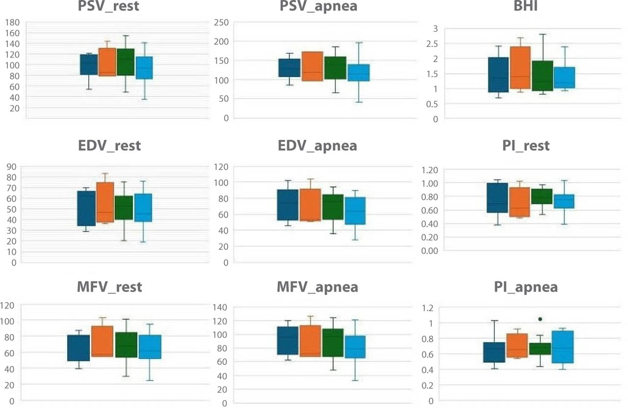
Fig. 1.
Subanalysis of cerebral hemodynamic parameters in patients treated with different types and dosages of anti-CGRP monoclonal antibodies at rest and after apnea. Dark blue – Fremanezumab 225 mg monthly; Orange – Fremanezumab 675 mg every three months; Green – Galcanezumab 120 mg monthly; Light blue – Erenumab 140 mg monthly. PSV – peak systolic velocity (cm/s); EDV – end-diastolic velocity (cm/s); MFV – mean flow velocity (cm/s); BHI – breath-holding index (%/s); PI – pulsatility index
Exploratory outcomes: MIDAS response and safety profile
The median MIDAS score before initiating anti-CGRP mAb therapy was 81.5. After three months of treatment, the median score decreased to 25.5 and remained stable at the time of TCCD evaluation. Based on a ≥50% reduction in MIDAS score at three months, 85% of patients were classified as clinical responders. No significant differences in TCCD-derived parameters were found between responders and non-responders (Fig. 2). Furthermore, no adverse events related to anti-CGRP mAbs were reported by any patient during the entire treatment period.
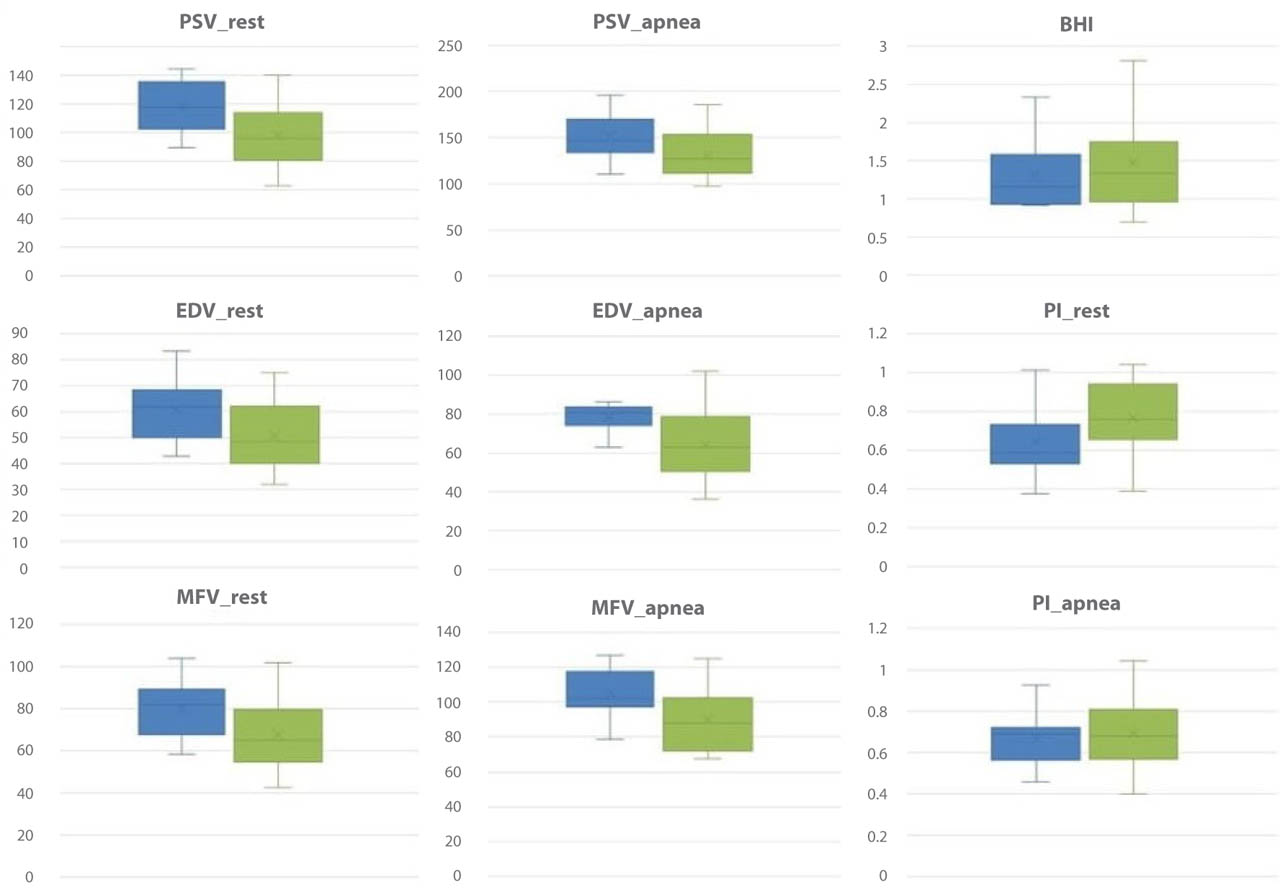
Fig. 2.
Comparison of TCCD-derived cerebrovascular parameters between responders and non-responders to anti-CGRP mAbs. Blue – responders; Green – nonresponders; PSV – peak systolic velocity (cm/s); EDV – end-diastolic velocity (cm/s); MFV – mean flow velocity (cm/s); BHI – breath-holding index (%/s); PI – pulsatility index
Discussion
Cerebral autoregulation maintains stable cerebral blood flow despite fluctuations in perfusion pressure, primarily through dynamic changes in arteriole diameter. This complex mechanism is governed by metabolic, neurogenic, endothelial, and myogenic responses. CVR, defined as the capacity of cerebral arterioles to dilate or constrict in response to vasoactive stimuli, is a reliable marker of cerebrovascular function and a potential predictor of stroke risk(10). Hypercapnia, a potent vasodilatory stimulus, induces an increase in local hydrogen ion concentration, thereby promoting vasodilation(18). Consequently, the BHT combined with TCD ultrasonography has become a well-established surrogate for evaluating CVR(11). TCCD provides a non-invasive and reliable method to indirectly assess cerebrovascular reserve capacity by enabling visualization of intracranial vessels through B-mode and color Doppler imaging with angle correction, allowing accurate and reproducible measurements of flow velocities(19). Recent studies have confirmed the validity of TCCD for CVR assessment via BHT, even without simultaneous capnography or bilateral insonation(20,21,22).
CGRP is one of the most potent vasodilatory neuropeptides, acting primarily on medium- and small-caliber arteries(23). It plays a key role in regulating regional cerebral blood flow (CBF) under physiological and pathological conditions(5). CGRP is also central in migraine pathophysiology. Upon activation of trigeminal C-fibers by cortical spreading depression or hypothalamic triggers, CGRP is released, promoting vasodilation and sensitization of Aδ fibers, thereby amplifying pain transmission(24).
Concerns have been raised regarding the cerebrovascular safety of anti-CGRP mAbs, especially in light of CGRP's protective vascular role. Preclinical studies suggest that inhibition of the CGRP pathway could impair cerebrovascular responses(9). Given that CGRP receptors are predominantly located on the abluminal side of cerebral vessels(25), CGRP-targeting therapies may need to cross the blood-brain barrier (BBB) to exert direct cerebrovascular effects. However, the large molecular size of mAbs limits their BBB permeability(26). On the other hand, the canonical CGRP receptor (the CLR/RAMP1 complex, which is the selective target of erenumab) has been observed in non-BBB-protected areas, including intra- and extracranial vessels(24). Furthermore, CGRP receptor mRNA has been detected in the endothelium of major cerebral arteries and distal microvessels(27), raising the possibility that any cerebrovascular action of these therapies may occur via luminal receptors or through more indirect mechanisms.
Although rare, adverse events have been reported. A case of ischemic stroke following erenumab administration was documented in 2019, and more recently, two cases of reversible cerebral vasoconstriction syndrome were reported in patients treated with fremanezumab and erenumab. Nonetheless, data from large RCTs evaluating anti-CGRP mAbs do not indicate a significant increase in cerebrovascular risk(8).
Altamura et al. demonstrated preserved CVR after erenumab therapy using the BHT with TCD(15). To our knowledge, no studies have investigated this topic using the BHT in patients treated with mAbs that directly target CGRP. The present study extends those findings to include galcanezumab and fremanezumab, evaluating CVR via BHT with TCCD.
Our results support the notion that the cerebral hemodynamic response to hypercapnia is not significantly impaired in migraine patients treated with mAbs targeting the CGRP ligand or receptor. Specifically, BHI values were comparable to those observed in healthy controls, and no critically low BHI suggestive of increased cerebrovascular risk was detected. The significant differences observed in the EDV and PI of the MCA at rest between groups should be interpreted with caution. Although the study was not powered to explore the determinants of these findings, they may reflect interindividual variability in baseline vascular tone or microvascular resistance rather than a consistent treatment-related effect. A sub-analysis categorizing patients by different types and dosages of anti-CGRP mAbs revealed no significant differences in cerebrovascular parameters across the various therapies. Similarly, no differences in CBFv were found between clinical responders and non-responders, contrasting with the modest CBFv reductions reported by Carter et al. (9) Methodological discrepancies and the timing of measurements may explain this divergence.
The limited BBB penetration of mAbs and their predominant action at extraparenchymal sites could explain these results. Moreover, Edvinsson et al. highlighted that CGRP's vasodilatory effects occur mainly via extraluminal activation of perivascular fibers rather than through endothelium-dependent mechanisms, suggesting minimal interference with CVR(28).
In agreement with this, Dzator et al. reported that CVR in the anterior circulation may remain unaffected in migraine patients(29), supporting our findings. Importantly, our study excluded patients with major vascular risk factors and used a validated TCCD protocol, enhancing reliability.
Several limitations must be acknowledged. First, the single-center design of the study limits the sample size, and the cross-sectional nature precludes speculation on potential interindividual changes following treatment initiation. Cerebrovascular reactivity was assessed at a single time point only; therefore, longitudinal changes over time cannot be evaluated, and no conclusions can be drawn regarding potential long-term cerebrovascular risk. However, the aim of the study was to assess cerebrovascular risk-related physiological markers by evaluating the BHI of the anterior circulation in this patient group, and further longitudinal analyses fall beyond the scope of this objective. Use of a technique more aligned with clinical practice, such as TCCD, was preferred over the standard simultaneous assessment of MCA and PCA on both sides with capnography(30), prioritizing patient comfort and feasibility but potentially lacking physiological standardization, as the absence of direct CO2 monitoring may have introduced variability in the hypercapnic stimulus and consequently affected the accuracy and reproducibility of CVR estimation. The effect of eptinezumab was not assessed because it was not available at our center during the study period. Additionally, while the predominantly female cohort reflects the demographic distribution of migraine in both the general and clinical populations(1), this gender imbalance may limit generalizability. Despite these considerations, this is the first investigation to assess cerebral hemodynamic response to hypercapnia in patients treated with anti-CGRP mAbs antibodies beyond erenumab, including galcanezumab and fremanezumab, implementing an alternative method widely validated in the literature to ensure reliable data collection(19,20,21,22).
Conclusions
The findings of this study suggest that anti-CGRP therapy is not associated with a BHI indicative of increased cerebrovascular risk, thereby mitigating concerns about potential adverse effects. Importantly, this study highlights the feasibility and reliability of TCCD ultrasonography as a non-invasive, patient-friendly tool to assess CVR in real-world clinical practice. Longitudinal multicenter studies with larger cohorts are warranted to confirm and extend these observations.
Notes
[4] Conflicts of interest Conflict of interest
The authors do not report any financial or personal connections with other persons or organizations, which might negatively affect the contents of this publication and/or claim authorship rights to this publication.
[5] Contributed by Author contributions
Original concept of study: MarP. Writing of manuscript: GB, PA. Analysis and interpretation of data: PA, IC, MarP. Final approval of the manuscript: VG, IC, MauP, MarP. Collection, recording and/or compilation of data: GB. Critical review of manuscript: PA, IC, MauP, MarP.