
Introduction
Shear wave elastography (SWE) is an ultrasound technique that quantitatively measures tissue stiffness by generating short, focused acoustic pulses and then measuring the velocity of the induced shear waves as they propagate transversely through the tissue; the measured velocity is converted to kilopascal values that are proportional to the shear modulus of the tissue(1,2,3).
In hepatology, SWE has gained histological validation and is now widely used as a biopsy-sparing method for staging liver fibrosis and tracking its progression over time(4,5,6).
Renal studies in patients with chronic kidney disease do not show any consistent correlation between elastographic measurements and histological changes or renal function(7,8). Investigations of pediatric vesicoureteral reflux and other obstructive uropathies are likewise inconclusive, with elasticity measurements showing no agreed relation to reflux grade, cortical scarring, or postoperative outcome(9,10,11,12,13,14).
Methodological heterogeneity is a likely source of these discrepancies: study protocols differ in terms of patient hydration, positioning (supine or prone), breathing (free breathing or breath-hold), probe orientation (longitudinal or transverse to the long axis of the kidney), and corticomedullary differentiation(7,15).
Normal stiffness ranges for healthy pediatric kidneys have not yet been established(16,17). Current EFSUMB recommendations lack a dedicated renal SWE protocol and, together with other guidelines, emphasize that absolute values are not interchangeable across platforms and that renal cut-off points still require validation through collaborative studies before clinical thresholds can be proposed(18,19,20).
Research questions addressed in this study
Does probe orientation (longitudinal vs. transverse) influence renal SWE measurements?
Does breath control (breath-hold vs. free breathing) alter measured stiffness?
How does patient hydration status affect renal elasticity values?
Do cortical and medullary regions exhibit reproducible stiffness differences?
Materials and methods
Study design and ethical approval
This prospective single-center study was approved by the Bio-ethics Committee of the Medical University of Lublin (KE-0254/158/06/2022) and conducted in accordance with the Declaration of Helsinki.
Participants
Ninety-six healthy volunteers (both kidneys examined) were prospectively enrolled and assigned to four methodological sub-studies (Tab. 1).
Tab. 1.
Methodology of sub-studies
| Sub-study | Participants (n) | Sex (F/M) | Age, median (range), years | Key protocol and comparison |
|---|---|---|---|---|
| Probe orientation | 35 pediatric volunteers | 14/21 | 12 (7–18) | Same kidney scanned longitudinally and then transversely; paired comparison of IQR/M ratios |
| Breathing impact | 20 pediatric volunteers | 10/10 | 13 (6–17) | Baseline measurements during quiet free breathing, then during a ~5-s mid-expiratory breath-hold; paired comparison of median stiffness and IQR/M |
| Hydration | 12 young adults | 8/4 | 29 (24–30) | Baseline after 4-h fluid restriction and micturition, then SWE repeated at 20 and 40 min after 700 mL water intake; concurrent bladder-volume tracking – baseline 20 min before water intake, repeated at the moment of intake and 20 and 40 min after intake to measure the increase in diuresis |
| Cortex vs. medulla | 29 pediatric volunteers | 10/19 | 13 (6–17) | Separate ROIs placed in the cortex and pyramids; ROI depth recorded; paired cortex-versus-medulla stiffness comparison, assessment of the impact of measurement depth |
Inclusion criteria:
normal renal appearance on baseline B-mode ultrasound;
no history of renal disease or systemic illness affecting renal per-fusion;
body mass index <30 kg/m2;
ability to cooperate with simple breath-hold instructions.
Exclusion criteria:
previous urological surgery;
congenital urinary-tract malformations;
acoustic window too poor for reliable SWE acquisition.
Three sub-studies (probe orientation, respiratory phase, cortex versus medulla) were conducted in pediatric volunteers aged 6–18 years; the hydration protocol involved young adults (24–30 years) because of its 60-minute duration. No laboratory tests were obtained; screening ultrasound confirmed morphologically normal kidneys in all cases.
Sample-size rationale
Paired-sample power calculations (α = 0.05, 80% power, within-subject SD ≈0.5–0.8 kPa) showed that at least 22, 20, and 18 kidneys were required for the orientation, respiratory phase, and corticomedullary comparisons, respectively; these numbers were achieved. The hydration series, designed as a feasibility pilot study, included 12 adult participants.
Ultrasound technique
All scans were performed on a Toshiba Aplio i700 with a 3.5 MHz convex i8Cx probe and the abdominal SWE preset. One radiologist with >5 years of ultrasonography experience conducted all examinations with participants in a prone position. A circular 5-mm region of interest (ROI) was centered in the mid-renal parenchyma (in the cortex for all sub-studies and in both cortex and medulla separately in the corticomedullary differentiation sub-study) – between the renal capsule and the pyramid base – avoiding vessels and the renal pelvis.
Each measurement series comprised five independent shear wave acquisitions; for each series, the median stiffness and the interquartile-range-to-median ratio (IQR/M) were recorded. The EFSUMB quality threshold (IQR/M ≤0.30) was applied in all sub-studies except the probe-orientation experiment; consequently, 11 participants were excluded from the corticomedullary differentiation series, five from the breathing series, and one from the hydration series. Conversely, the probe-orientation sub-study deliberately retained all frames, irrespective of IQR/M, to capture the full spectrum of variability attributable to probe rotation. Group summaries in tables are reported as the mean ± SD of these subject-level medians. All tests were performed on subject-level medians.
Mean skin-to-ROI depth (2–7 cm) was recorded for correlation analysis in the cortex-versus-medulla sub-study. In the orientation sub-study, each kidney was scanned first longitudinally and then transversely; all other sub-studies used the longitudinal plane only.
In the hydration experiment, participants fasted for 4 hours and emptied their bladder immediately before the test, whereas in the other studies no special preparation was required.
Elastography measurements were performed during a mid-expiratory breath-hold, except for the free-breathing acquisitions included in the breathing sub-study.
Statistical analysis
Data were analyzed in Statistica 13.3 (TIBCO Software Inc.). Normality was assessed with the Shapiro–Wilk test. Paired comparisons (cortex vs. medulla; longitudinal vs. transverse orientation; quiet breathing vs. breath-hold) were performed using the Wilcoxon signed-rank test. Hydration measurements (three time points) were analyzed with Friedman's ANOVA. Spearman's ρ was used to assess correlations between ROI depth and stiffness. A two-sided p <0.05 was considered statistically significant.
Results
Probe orientation
Data from 35 pediatric volunteers show that holding the probe in the longitudinal plane yielded lower variability than in the trans-verse plane: mean IQR/M decreased from 0.477 to 0.362 in the right kidney (Wilcoxon p = 0.0037) and from 0.431 to 0.310 in the left kidney (p = 0.014). Mean stiffness (kPa, computed as the mean of subject-level medians) changed little between planes – right: 6.2 ± 1.5 (transverse) vs. 6.1 ± 1.4 (longitudinal) kPa; left: 6.0 ± 1.6 (trans-verse) vs. 5.9 ± 1.5 kPa (longitudinal), both p >0.5. The acquisition technique is shown in Fig. 1.

Fig. 1.
ROI placements in transverse (A) and longitudinal (B) planes of the kidney from the dorsal approach, measurements were taken in the cortex of the mid-portion of the kidney
Quiet breathing versus breath-hold
Data from 20 pediatric volunteers show that a brief mid-expiratory breath-hold did not significantly alter measurement variability or renal stiffness: mean IQR/M was 0.154 (quiet breathing) vs. 0.180 (breath-hold) in the right kidney (Wilcoxon p = 0.84) and 0.192 (quiet breathing) vs. 0.168 (breath-hold) in the left kidney (p = 0.88). Mean stiffness (kPa, computed as the mean of subject-level medians) changed little between respiratory states – right: 6.1 ± 1.1 (quiet breathing) vs. 6.2 ± 1.3 (breath-hold) kPa; left: 5.8 ± 0.8 (quiet breathing) vs. 5.7 ± 0.9 (breath-hold) kPa – with Wilcoxon tests for subject-level medians non-significant (right p = 0.91; left p = 0.73).
Hydration effect
Oral hydration (700 mL water) caused a statistically significant increase in median renal stiffness in both kidneys (right χ2 = 8.27, p = 0.016; left χ2 = 13.15, p = 0.0014). Numerical results are summarized in Tab. 2 and Fig. 2.
Tab. 2.
Hydration sub-study (n = 12 adults): effect of drinking 700 mL water on renal SWE
| Kidney | Parameter | Baseline mean (±SD) of subject-level medians, kPa | +20 min, kPa | +40 min, kPa | Friedman χ2 (2 df) | p-value |
|---|---|---|---|---|---|---|
| Right | Stiffness (kPa) | 5.3 ± 0.7 | 6.7 ± 0.8 | 6.6 ± 0.9 | 8.27 | 0.016 |
| IQR/M ratio | 0.25 ± 0.05 | 0.23 ± 0.06 | 0.22 ± 0.05 | – | – | |
| Left | Stiffness (kPa) | 5.3 ± 0.6 | 6.4 ± 0.7 | 6.7 ± 0.8 | 13.15 | 0.0014 |
| IQR/M ratio | 0.24 ± 0.06 | 0.23 ± 0.05 | 0.23 ± 0.05 | – | – |
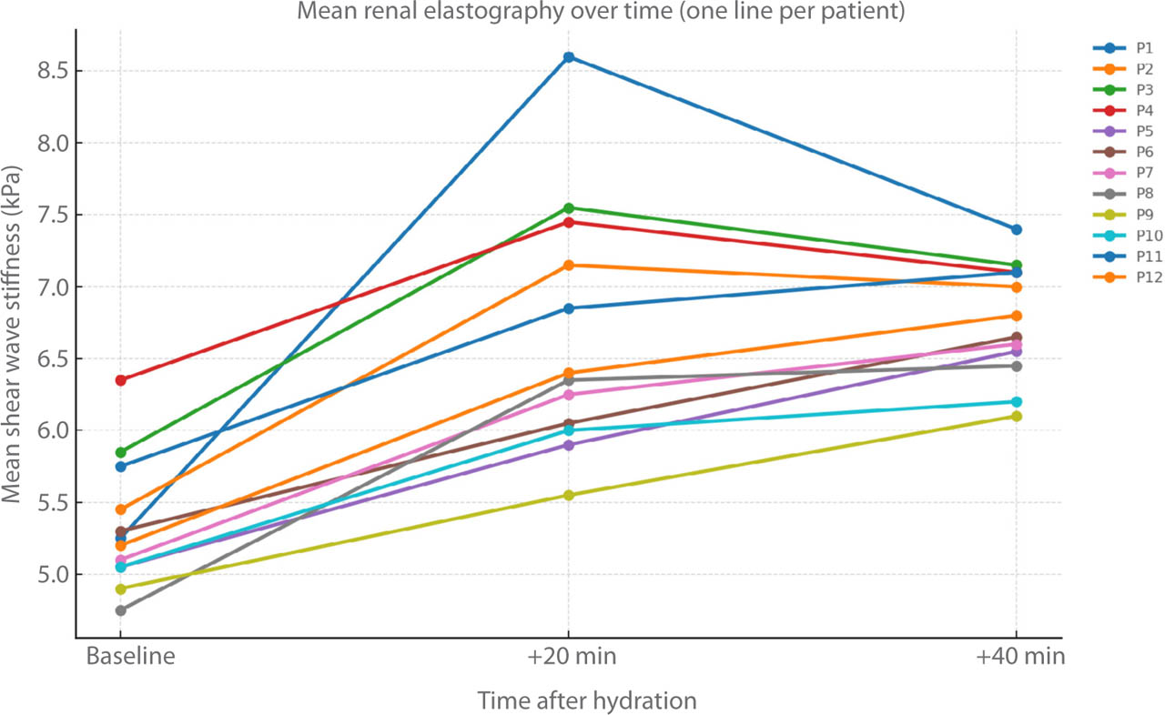
Fig. 2.
Average renal shear wave stiffness (kPa) before and after hydration in 12 adult volunteers. Each line represents the mean value of both kidneys for one participant. Measurements were taken at baseline, 20 minutes, and 40 minutes following oral fluid intake. The plot demonstrates a consistent post-hydration increase in renal cortex stiffness
Concomitant bladder volume monitoring showed urine flow increasing from 1.07 mL/min at rest to 1.83 mL/min in the first 20 min after fluid intake and 4.71 mL/min during the following 20 min, in line with recognized physiological ranges for baseline and water-loaded diuresis(21,22).
Corticomedullary differentiation in SWE
Data from 29 pediatric volunteers show that cortical stiffness exceeded medullary stiffness at matched depth in both kidneys: mean stiffness (kPa, computed as the mean of subject-level medians) was 8.3 ± 1.9 (cortex) vs. 7.0 ± 1.7 (medulla) in the right kidney (Wilcoxon p = 0.0059) and 7.7 ± 1.8 (cortex) vs. 6.7 ± 1.6 (medulla) in the left kidney (p = 0.037).
ROIs were acquired at comparable mid-parenchymal depths. Mean depth (computed as the mean of subject-level medians) was: left cortex 3.88 ± 0.93 cm (range 2.5–5.5), left medulla 4.49 ± 0.91 cm (2.9–6.3), right cortex 3.61 ± 0.95 cm (2.4–5.8), and right medulla 4.13 ± 1.03 cm (2.6–6.4). There was no depth-related effect on stiffness (Spearman's ρ from −0.24 to +0.25; all p ≥0.19).
Discussion
Probe orientation
In a pig model, Gennisson et al. demonstrated that shear modulus was ≈ 12% higher in the outer cortex, 28% in the inner cortex, and 32% in the medulla when the shear wave travelled parallel rather than perpendicular to tubular bundles(23).
Leong et al. confirmed this effect: in a sheep-kidney phantom cortical stiffness rose from 7.2 ± 1.1 kPa (perpendicular to tubular bundles) to 11.8 ± 1.3 kPa (parallel), and in 10 healthy volunteers from 2.99 kPa to 5.30 kPa – a 77% increase(24).
Diffusion-tensor MRI reveals that renal tubules and vessels are radially aligned. In 10 healthy adults, Ries et al. reported fractional anisotropy (FA) values of 0.22 ± 0.12 in the cortex and 0.39 ± 0.11 in the pyramids, confirming a highly anisotropic microarchitecture(25).
Saini et al. found even higher FA (cortex 0.39 ± 0.026, medulla 0.496 ± 0.030) and showed that parenchymal disease lowers FA, underscoring the clinical importance of fiber orientation(26).
Because the kidney is ellipsoidal, the local angle between a laterally propagating shear wave and the tubules depends on probe orientation. With the transducer aligned to the long axis, the wave moves cranio-caudally across the mid-zone ROI and meets radial tubules at a nearly constant 90° angle. Rotating the probe into the short axis forces the wave to sweep mediolaterally; the crossing angle then fluctuates from near parallel to perpendicular within a single frame, increasing variability.
Accordingly, in the present study, the ROI was placed in the mid-portion of the kidney and the renal cortex was measured in both planes. The long-axis view produced a markedly lower IQR/M, while median kPa remained unchanged, indicating that this orientation minimizes anisotropic bias and improves repeatability in pediatric renal SWE.
Breath impact
There are no guidelines for breathing during renal elastography. The review by Iyama et al. noted that breathing should be held during the study because the kidneys move during respiration(7). Four-dimensional computed tomography illustrates the basic kinematics: during quiet respiration, the central renal mass moves up and down by 12–25 mm in children <9 years of age and by 21–52 mm in older children(27).
Studies of abdominal organs clarify how respiration affects shear wave elastography. Hong et al. used a mid-expiratory breath-hold and found that the incidence of quality errors (stability index <90%) decreased from 17% during free breathing to 7% during the pause, while the percentage of unreliable measurements decreased from 16.7% to 8.3% during free breathing and from 14.8% to 0% after the breath-hold – showing that breath-hold improves data reliability(28).
Goertz et al., examining 30 healthy adult volunteers, found that liver stiffness remained statistically unchanged across deep inspiration, deep expiration, and Valsalva maneuvers(29).
In contrast, Kaya and Gürün demonstrated that deep-inspiration breath-hold caused an approximately 40% increase in pancreatic stiffness, reinforcing warnings against measurements during deep inspiration(30).
Postek et al. examined renal SWE in 56 spontaneously breathing neonates, and 85–89% of measurements met the IQR/M quality threshold of ≤0.30(31).
In the present study of 20 children, a five-second mid-expiratory breath-hold did not demonstrate a statistically significant effect on the median values of the measurement series or on the IQR/M values for both kidneys relative to quiet breathing. However, the short pause stabilized the kidney and facilitated localization of the ROI in the renal cortex. Therefore, when the child is cooperative, incorporating this breath-hold remains a practical approach to reducing motion artifacts and improving ROI placement reproducibility.
Hydration status
In a magnetic resonance elastography (MRE) study, Dittmann et al. found that drinking 1 L of water caused only a small increase in renal stiffness (1.93 ± 0.22 → 1.97 ± 0.23 m·s−1; ≈ +2%), with cortical stiffness remaining higher than medulla stiffness(32).
Gandhi et al., using a segmented MRE approach, observed a significant increase in the stiffness of different renal regions after hydration, ranging from 3.6% to 7.5%, accompanied by an increase in bladder volume(33).
Animal models confirm that renal elasticity is highly sensitive to hemodynamic changes. In rabbits, Liu et al. progressively occluded the renal vein or artery and monitored compartment-specific shear modulus using SWE, vein ligation tripled renal cortical stiffness (16 → 55 kPa) and doubled medullary values, whereas progressive arterial constriction had the opposite effect, reducing cortical modulus to ~11 kPa and medullary modulus to ~8 kPa(34).
Ex vivo work by Muttray et al. increased hydrostatic pressure in 20 porcine kidneys from 0 to 90 mmHg and noted an increase in shear wave velocity from 1.47 m s−1 to 2.24 m s−1, with the central parenchyma responding more strongly than the peripheral parenchyma(35).
These studies demonstrate that both vascular inflow and intrapelvic pressure can significantly alter measured stiffness, providing a basis for the hydration effects observed in humans and underscoring the need to document hydration status when interpreting SWE.
There are only limited data on how hydration affects renal SWE. Gao et al. reported that after 30 adult volunteers drank 1 L of water, cortical shear modulus rose by ≈ 40 %, increasing from 9.34 kPa to 13.05 kPa at 60 minutes (p <0.001)(36).
For the present hydration sub-study, adults (n = 12) were purposely recruited because a 60-min protocol would have overloaded children. After participants emptied their bladders, the mean baseline cortical stiffness was 5.3 kPa bilaterally; then, drinking 700 mL of water increased the median at 40 min to 6.6 kPa on the right side and 6.7 kPa on the left side (+≈ 25%, p <0.05). Concurrent ultrasonographic examination of the urinary bladder documented a marked increase in urine production, confirming effective hydration.
Although these results were obtained in adults, they indicate that even moderate fluid loading can alter elastographic values in the pediatric population, underscoring the need for standardized prehydration in both children and adults.
Corticomedullary differentiation
In the study of pediatric patients by Bhatia et al., renal cortical stiffness exceeded medullary values by approximately 22–28% in infants <1 year of age, 1–29% in children 1–5 years of age, and 9–37% in children >5 years of age(37).
Leong's systematic review – including 26 papers on adult renal SWE – shows wide variation in ROI localization relative to the renal medulla and cortex(38).
Pediatric data from the present study reflect this literature: cortical stiffness exceeded medullary values by ≈15–19% on both sides (p <0.05). Taken together, the evidence suggests that renal SWE should always define ROI localization relative to the renal medulla and cortex; combining cortex and medulla in the same ROI compromises the reliability of measurements.
Limitations
Several constraints should be borne in mind when interpreting these findings. First, the four sub-studies were intentionally designed with different quality-control approaches. The breathing, hydration, and corticomedullary comparisons applied the EFSUMB IQR/M ≤0.30 threshold; in contrast, the orientation experiment retained every frame to reveal the full dispersion introduced by probe rotation and respiratory motion. This methodological asymmetry sacrifices strict cross-study comparability in favor of answering distinct mechanistic questions (probe orientation) and should be considered when juxtaposing absolute kPa values.
Second, the hydration series was undertaken only in young adults because the 60-minute protocol and repeated bladder scans were not practical in children. Whether pediatric kidneys respond to fluid loading in the same way remains untested; the hydration effect we observed (≈25% rise in median stiffness) may therefore over- or underestimate the pediatric response.
Third, all examinations were performed by a single sonographer on one ultrasound platform; this maximizes internal consistency but limits generalizability across operators and vendors. Future multi-center work with inter-operator assessment is required before clinical cutoffs can be proposed.
Fourth, the present study relied on sonographic appearance to confirm “healthy” kidneys and did not include laboratory panels or histology. Subclinical nephron alterations cannot be excluded, particularly in adolescents, and could have contributed to inter-individual variability.
Finally, the breathing experiment involved only a brief mid-expiratory breath-hold. Given the inclusion of children (≥6 years), breath-hold quality can be variable; we therefore prioritized mid-expiratory holds and avoided extremes of respiration. Deep inspiration, expiration, or real-time quality gating were not tested, each of which may affect shear wave consistency differently. Likewise, the probe-orientation test did not explore oblique angles between the true long- and short-axis views, so the optimum insonation corridor may differ from the two orthogonal planes investigated here.
These limitations underscore the exploratory nature of the present work and highlight the need for larger, multicenter studies – ideally with age-stratified hydration protocols, uniform quality filters, and cross-vendor validation – to develop a robust renal SWE protocol applicable across the pediatric and adult spectrum.
Conclusions and practical implications
Our results confirm that several seemingly minor technical and physiological factors can alter renal SWE in children sufficiently to mask differences resulting from pathology:
Probe orientation. Placing the transducer along the long axis of the kidney reduces measurement variability (lower IQR/M), making it the default choice for quality and repeatability of measurements.
Respiration. Renal SWE values did not differ between quiet breathing and a brief mid-expiratory breath-hold. Deep inspiration was not assessed; any influence mediated by inspiratory increases in intra-abdominal pressure lies beyond the present data.
Hydration. Drinking 700 mL of water increased cortical stiffness by ~25% in the adult group. Scans should be scheduled after a specified fasting interval or with documented fluid intake.
Corticomedullary differentiation. The renal cortex is stiffer than the medulla. Mixing cortex and medulla in a single region of interest may mask clinically significant changes.
A suggested protocol for renal shear wave elastography is presented in Fig. 3.
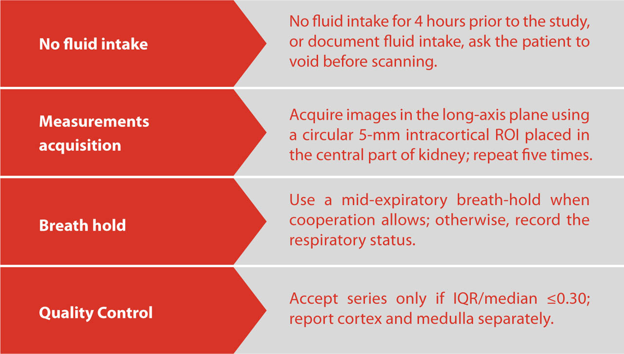
Fig. 3.
Suggested protocol for renal shear wave elastography
Acknowledgements
The authors thank all individuals who contributed to the creation of the paper. An AI-assisted writing tool (ChatGPT, OpenAI; model GPT-5 Thinking; accessed Aug 2025) was used to refine English phrasing; the text was subsequently verified for accuracy and integrity by the authors, who assume full responsibility for the final content.