
Introduction
Early identification of patients who may require ventilatory support is crucial for optimal resource allocation and timely clinical intervention in high-acuity settings such as the emergency department (ED). Diaphragm ultrasound has emerged as an effective tool for evaluating diaphragmatic structure and function(1). One of the key parameters derived from this imaging modality is the diaphragm thickening fraction (DTF), which quantifies the percentage change in diaphragm thickness from end-expiration to end-inspiration. This metric directly reflects the diaphragm's contractile performance and intrinsic strength(2). In spontaneously breathing patients, DTF shows significant promise, as it reliably captures the extent of diaphragm shortening without being confounded by external mechanical influences(3,4). Accordingly, the present study was designed to assess the clinical utility of DTF by examining its relationship with the initiation of ventilatory support – both non-invasive (e.g., continuous positive airway pressure (CPAP) or bilevel positive airway pressure (BiPAP)) and invasive – during the ED stay and throughout hospitalization. A secondary aim was to establish an optimal, data-driven DTF cutoff value to accurately identify patients at heightened risk of respiratory decompensation.
Materials and methods
Adult patients (aged ≥18 years) admitted with acute respiratory failure to the ED of Baggiovara Hospital in Modena, Italy, between January 1, 2023, and December 31, 2024 (C.E. 467/2023/OSS/AOUMO SIRER ID 6296 – DSF2023; NCT05869045), were prospectively enrolled.
Acute respiratory failure was defined by the presence of hypoxemia, characterized by oxygen saturation measured by pulse oximetry (SpO2) ≤92% or partial pressure of arterial oxygen (PaO2) ≤60 mmHg at room air, together with clinical sign of respiratory distress, including at least one of the following: increased respiratory effort (use of accessory musculature, paradoxical breathing, or thoracoabdominal asynchrony) or tachypnea (respiratory rate >25 breaths/min). All patients underwent arterial blood gas analysis, and PaO2/FiO2 ratios ≤300 mmHg were recorded to stratify respiratory failure severity. Oxygenation refers to respiratory support delivered in the ED and during hospitalization, respectively, and includes any supplemental oxygen (low-flow) and high-flow nasal cannula when used; non-invasive ventilation (NIV) and invasive mechanical ventilation were captured separately as study outcomes. The choice between NIV and invasive ventilation was made by treating clinicians according to usual care and institutional practice, and not solely on baseline clinical severity.
Because escalation decisions incorporate dynamic response and safety considerations (including contraindications to NIV and clinician risk–benefit judgment), some unmeasured determinants may have influenced ventilation allocation.
Diaphragm ultrasound examinations were conducted using the ESAOTE MYLAB XPRO30 ultrasound system (Esaote S.p.A., V. Siffredi, 58; 16,010 Genova, Italy) equipped with a 7–12 MHz linear probe, with data acquisition performed within 6 hours of ED admission. To maintain data quality and relevance, patients with incomplete ultrasound measurements, a history of chest wall surgeries that could interfere with diaphragm visualization, neuromuscular disorders, or chronic home NIV use were excluded.
Board-certified sonographers and emergency physicians with at least three years of critical care experience conducted all ultrasound examinations. These operators were not involved in the clinical management of patients. Patients were positioned in a semi-recumbent posture (30°–45°), and the linear probe was placed in the right mid-axillary line to assess the right hemidiaphragm, identified in the zone of apposition at the 8th or 9th intercostal space along the mid-axillary line.
M-mode was employed to evaluate diaphragmatic thickening over three consecutive breaths, from which the thickening fraction was derived. Diaphragm thickness was measured at end-inspiration and end-expiration. Measurements were obtained over at least three consecutive tidal breaths, and average values were used to ensure precision. The diaphragm thickening fraction was computed using the formula: DTF = ((T_ins – T_exp) / T_exp) × 100 (Fig. 1).
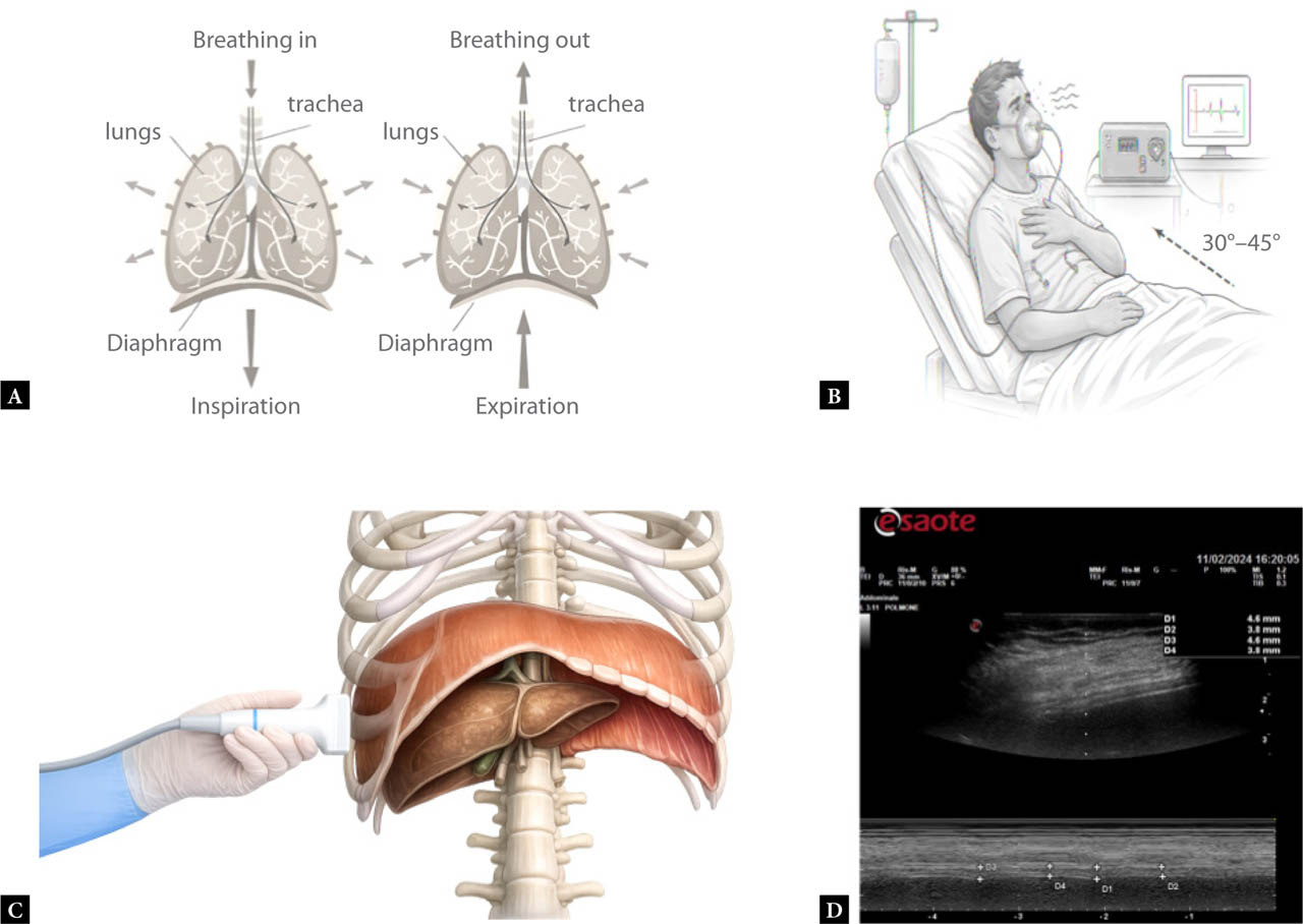
Fig. 1.
Ultrasound protocol for the assessment of diaphragm thickening fraction (DTF). A. Schematic representation of diaphragmatic motion during inspiration and expiration: during inspiration, the diaphragm thickens and moves caudally, whereas during expiration it becomes thinner and ascends. B. Patient positioning in a semi-recumbent posture (30–45°) for standardized diaphragm ultrasound acquisition. C. Probe placement using a high-frequency linear transducer (7–12 MHz) along the right mid-axillary line at the 8th–9th intercostal space to visualize the diaphragm within the zone of apposition. D. M-mode ultrasound image illustrating measurements of diaphragm thickness at end-inspiration (T_ins) and end-expiration (T_exp), averaged over three consecutive tidal breaths. These measurements were used to calculate the diaphragm thickening fraction according to the formula: DTF = ((T_ins– T_exp) / T_exp) × 100
In addition to ultrasound parameters, data collection included patient demographics, comorbidity profiles (quantified by the Charlson Comorbidity Index), and oxygenation status via the PaO2/FiO2 ratio. The clinical decision to initiate ventilatory support during the ED stay and at any point during hospitalization was documented. To contextualize the prognostic value of DTF, other clinical outcomes such as hospital length of stay (LOS) and in-hospital mortality were also recorded.
Statistical analysis
Continuous variables are reported as median (interquartile range, IQR) or mean (SD), as appropriate, and categorical variables as counts and percentages. Between-group comparisons were performed using the Wilcoxon rank-sum test for continuous variables and the chi-square test or Fisher's exact test for categorical variables, as appropriate.
The discriminative performance of DTF for the primary outcome (initiation of NIV or invasive mechanical ventilation during hospitalization) was assessed using receiver operating characteristic (ROC) analysis and the area under the ROC curve (AUC). AUC 95% confidence intervals (CIs) were computed using DeLong's method. The optimal DTF threshold was identified using the Youden index, and corresponding sensitivity, specificity, and 95% CIs were calculated using exact binomial methods.
The association between DTF and the primary outcome was evaluated using regression modeling for a binary outcome; model estimates are reported as regression coefficients and exponentiated odds ratios (ORs) with corresponding p values. Missing data were handled using complete-case analysis. All analyses were performed using R (version 4.5.0; R Foundation for Statistical Computing, Vienna, Austria). A two-sided p value <0.05 was considered statistically significant.
Results
Out of 72 patients initially screened, 56 met all inclusion criteria and were included in the final analysis. The entire cohort exhibited a median DTF of 25.6% with an IQR of 17.5–35.2%. Among these 56 patients, 22 (39%) required ventilatory support (NIV or invasive mechanical ventilation), whereas 34 (61%) did not. Patients requiring ventilatory support had a higher proportion of males (73% vs 41%; p = 0.029) and showed lower DTF (median 20.93% vs 29.8%; p = 0.006) and lower PaO2/FiO2 ratio (216 vs 282 mmHg; p = 0.003), along with a higher respiratory rate (29 vs 20 breaths/min; p = 0.024) (Tab. 1). DTF was consistently lower in patients requiring ventilatory support compared with those not requiring ventilatory support (Fig. 2). Other baseline variables, including age and Charlson Comorbidity Index, were similar between groups, whereas length of stay tended to be longer among patients requiring ventilatory support (14 vs 10 days; p = 0.050).
Tab. 1.
Baseline characteristics and diaphragmatic ultrasound parameters by ventilation status
| Variable | Population (n = 56) | No ventilatory support (n = 34) | Ventilatory support (n = 22) | p-value |
|---|---|---|---|---|
| Sex (male), n (%) | 30 (54%) | 14 (41%) | 16 (73%) | 0.029 |
| Age (years), median (IQR) | 80 (75–87) | 78 (74–87) | 84 (75–88) | 0.351 |
| Charlson Comorbidity Index, median (IQR) | 6 (5–8) | 6 (5–8) | 6 (5–8) | 0.531 |
| Dyspnea, n (%) | 55 (98%) | 33 (97%) | 22 (100%) | 1.000 |
| Cough, n (%) | 15 (27%) | 9 (27%) | 6 (27%) | 1.000 |
| Fever, n (%) | 16 (29%) | 11 (32%) | 5 (23%) | 0.550 |
| End-inspiratory diaphragm thickness (mm), median (IQR) | 3.4 (2.7–5.0) | 3.40 (3.1–5.2) | 3.0 (2.4–4.5) | 0.052 |
| End-expiratory diaphragm thickness (mm), median (IQR) | 2.5 (2.0–3.4) | 2.6 (2.0–3.5) | 2.3 (1.9–3.3) | 0.430 |
| Diaphragmatic thickening fraction (DTF) (%), median (IQR) | 25.6 (17.5–35.2) | 29.8 (21.1–37.2) | 20.93 (10.6–26.8) | 0.006 |
| PaO2/FiO2 ratio (mmHg), median (IQR) | 258 (216–318) | 282 (244–350) | 216 (169–248) | 0.003 |
| Systolic blood pressure (mmHg), median (IQR) | 130 (115–150) | 132 (110–155) | 130 (120–145) | 0.904 |
| Diastolic blood pressure (mmHg), median (IQR) | 80 (65–90) | 80 (65–85) | 85 (64–90) | 0.283 |
| Heart rate (bpm), median (IQR) | 92 (79–110) | 95 (79–110) | 89 (82–104) | 0.610 |
| SpO2 (%), median (IQR) | 90 (85–94) | 91 (88–94) | 88 (84–94) | 0.289 |
| Respiratory rate (bpm), median (IQR) | 21 (18–32) | 20 (18–22) | 29 (22–35) | 0.024 |
| Body temperature (°C), median (IQR) | 36.5 (36.0–37.3) | 36.7 (36.0–37.6) | 36.1 (36.0–37.0) | 0.164 |
| Oxygenation in the ED | 50 (89%) | 30 (88%) | 20 (91%) | 1.000 |
| Oxygenation (hospital) | 44 (79%) | 25 (74%) | 19 (86%) | 0.329 |
| LOS (days), median (IQR) | 12 (7–17) | 10 (5–14) | 14 (8–21) | 0.050 |
| Death, n (%) | 7 (13%) | 4 (12%) | 3 (14%) | 1.000 |
[i] Values are reported as median (IQR) or n (%). P-values refer to between-group comparisons (no ventilatory support vs ventilatory support (NIV/IMV)). ED – emergency department; IMV – invasive mechanical ventilation; IQR – interquartile range; LOS – length of stay; NIV – non-invasive ventilation; SpO2, peripheral oxygen saturation; PaO2/FiO2 – arterial oxygen partial pressure to inspired oxygen fraction ratio
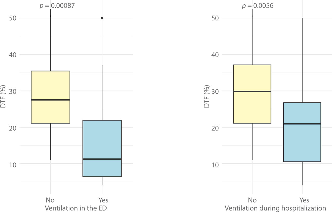
Fig. 2.
Comparison of diaphragm thickening fraction (DTF) by ventilatory support status. Yellow box: No ventilatory support; Blue box: Ventilatory support (noninvasive ventilation or invasive mechanical ventilation); Left panel: Comparison of DTF in the emergency department (ED) based on ventilation status (p = 0.00087); Right panel: Comparison of DTF during hospitalization based on ventilation status (p = 0.0056)
In a logistic regression model, higher DTF was independently associated with lower odds of requiring ventilatory support (β = −0.075, p = 0.008), corresponding to an OR of 0.93 (95% CI 0.88–0.98) per 1% increase in DTF. Moreover, receiver operating characteristic (ROC) analysis yielded an area under the curve (AUC) of 0.72 (95% CI 0.58–0.87), and the optimal DTF cutoff was determined to be 22.1%. At this threshold, sensitivity for predicting the need for ventilatory support was 59%, and specificity was 71% (Fig. 3).
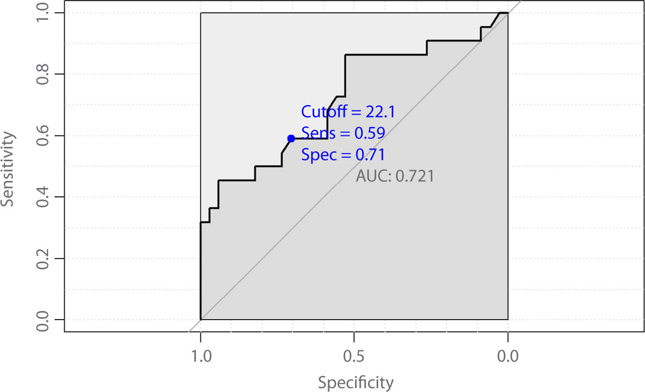
Fig. 3.
Receiver operating characteristic curve for diaphragm thickening fraction (DTF) in predicting the need for ventilatory support (non-invasive ventilation or invasive mechanical ventilation) from emergency department presentation through hospitalization. The area under the curve (AUC) was 0.72 (95% CI 0.58–0.87). The optimal DTF cutoff identified using the Youden index was 22.1%, corresponding to a sensitivity of 0.59 and a specificity of 0.71 (blue point). The diagonal reference line represents no discriminative ability
Discussion
The findings of this study underscore the potential value of DTF as an early biomarker for identifying patients at risk of respiratory de-compensation. A low DTF indicates diminished diaphragm contractile performance and, consequently, reduced respiratory muscle reserve(5,6). In the clinical context, an acute respiratory failure patient presenting with a DTF below the identified cutoff of 22.1% warrants closer monitoring and may benefit from earlier respiratory interventions. Importantly, the outcome of the present study captured the need for any ventilatory support (non-invasive or invasive), thereby reflecting clinically relevant escalation rather than modality-specific failure.
The non-invasive and rapid nature of diaphragm ultrasound makes it ideally suited for the ED setting, where timely assessments are crucial. The ability to quickly evaluate diaphragmatic performance may enable clinicians to make earlier decisions about initiating ventilatory support, thereby potentially improving patient outcomes through early intervention(7,8). Although the present study did not demonstrate statistically significant associations between DTF and longer-term outcomes such as hospital LOS or in-hospital mortality, its strength lies in predicting immediate ventilatory needs.
Furthermore, incorporating DTF into routine clinical evaluation could provide a more nuanced understanding of respiratory function when used in tandem with other parameters, such as the PaO2/FiO2 ratio. A composite scoring approach might enhance patient risk stratification and support more personalized care pathways, especially for critically ill patients(9).
Recent evidence supports the role of DTF as a clinically relevant marker beyond this specific setting, particularly in relation to NIV outcomes. In patients with de novo acute respiratory failure treated with NIV in the ED, Mercurio et al. reported that both DTF and the respiratory rate/DTF ratio were associated with NIV success, suggesting that diaphragm performance may capture an early “load– capacity” imbalance that precedes overt clinical failure(10). This concept is especially pertinent in the ED, where initial hypoxemia or hypercapnia may not fully reflect impending deterioration, and where physiological reserve – rather than gas exchange alone – often drives escalation decisions. Feasibility also appears favorable: Cammarota et al. showed that diaphragm ultrasound can be reliably performed in highly dyspneic patients with acute hypercapnic respiratory failure, supporting its practicality as a bedside monitoring tool even in unstable ED patients(11).
From a decision-making standpoint, DTF should not be viewed as a standalone trigger for escalation but rather as a complementary component of a multimodal risk-stratification approach. Existing clinical tools designed to predict NIV failure, such as the HACOR scale (evaluated in acute cardiogenic pulmonary oedema), may help contextualize ultrasound findings and facilitate earlier identification of patients who require closer surveillance or expedited escalation to invasive ventilation(12). Finally, a recent systematic review emphasized that both lung ultrasound indices and diaphragm ultrasound metrics – particularly DTF – may contribute to predicting NIV outcomes, reinforcing the rationale for incorporating ultrasound-based physiology into early risk-assessment pathways(13).
Several limitations should be acknowledged. The modest sample size in the present study restricts the generalizability of findings, and despite examinations being performed by experienced sonographers, the potential for inter-observer variability in ultrasound measurements cannot be overlooked. Decisions to initiate NIV versus proceed to invasive mechanical ventilation are multifactorial (e.g., trajectory under initial support, work of breathing, mental status and airway protection, hemodynamics, and local resources/protocols), so residual confounding by indication cannot be excluded in this observational design. Additionally, exploring the value of DTF alongside other emerging ultrasound-derived indices may support a more integrated approach to evaluating respiratory muscle function(14). Future multicenter studies should validate DTF thresholds across diverse etiologies of respiratory failure, quantify inter-operator reliability, and determine whether DTF-guided strategies can improve clinically meaningful outcomes.
Conclusions
Early DTF assessment in the ED may help identify patients with acute respiratory failure who are more likely to require ventilatory support (non-invasive or invasive) during their ED stay and throughout hospitalization. Larger prospective studies are warranted to validate clinically meaningful thresholds and determine the incremental value of DTF beyond conventional clinical and gas-exchange parameters.
Acknowledgements
The authors are grateful to the emergency department nursing and medical staff of Baggiovara Hospital (Modena, Italy) for their support in patient enrolment and bedside ultrasound examinations.
The authors used a large language model (ChatGPT (OpenAI); accessed in 2025) to assist with language editing and to refine the structure of the manuscript. All study design, data collection, statistical analyses, and interpretation of the results were performed and critically verified by the authors, who take full responsibility for the scientific content of this article.
Notes
[2] Ethical approval and consent
The study protocol was approved by the local Ethics Committee (C.E. 467/2023/OSS/AOUMO SIRER ID 6296 – DSF2023) and registered at ClinicalTrials.gov (NCT05869045). Written informed consent was obtained from all participants or their legal representatives in accordance with the Declaration of Helsinki.
[3] Conflicts of interest Conflict of interest
The authors do not report any financial or personal relationships with other individuals or organizations that could inappropriately influence this work.
[4] Data availability
The datasets generated and analyzed during the current study are available from the corresponding author on reasonable request.
[5] Contributed by Author contributions
Original concept of study: CCG, AA. Writing of manuscript: DO. Analysis and interpretation of data: CCG, AA, MLG, DO. Final approval of the manuscript: DO. Collection, recording and/or compilation of data: AA, MLG, GS, EO, FO. Critical review of manuscript: GS, DO.