Fig. 1
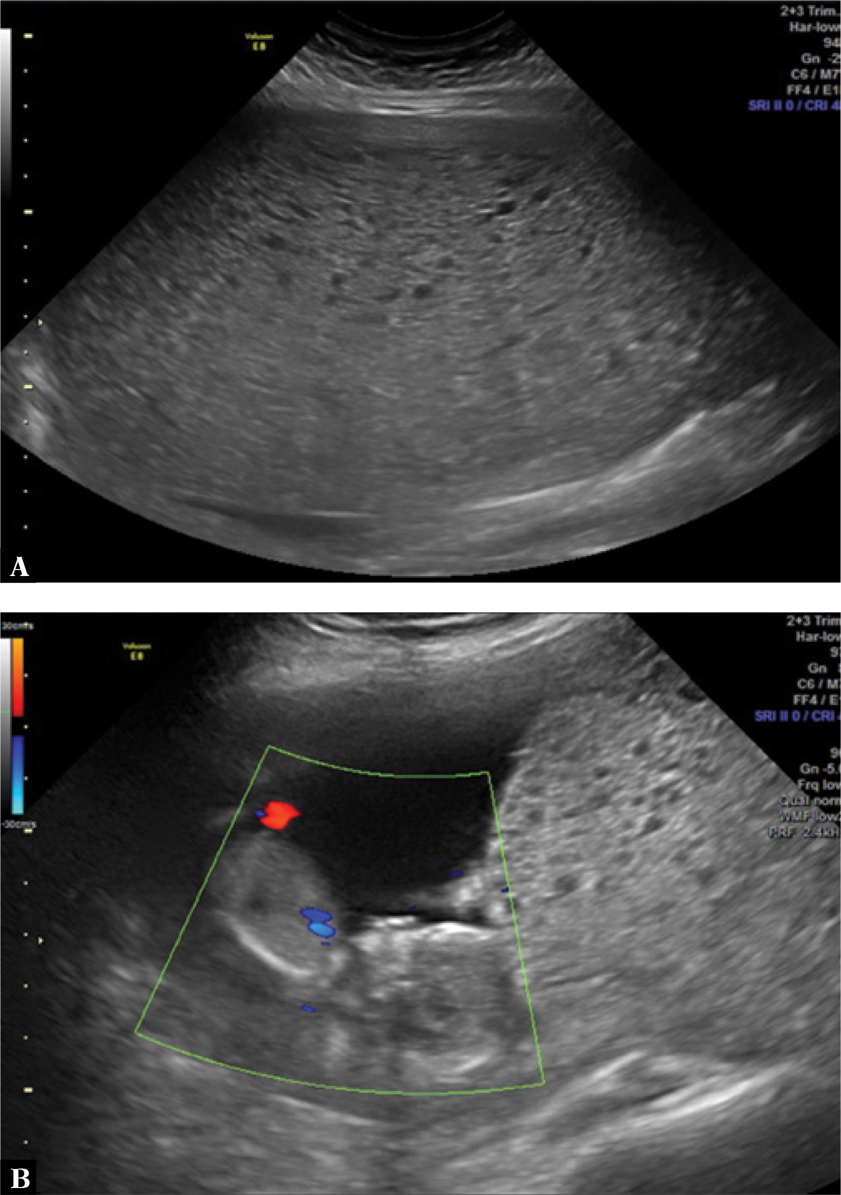
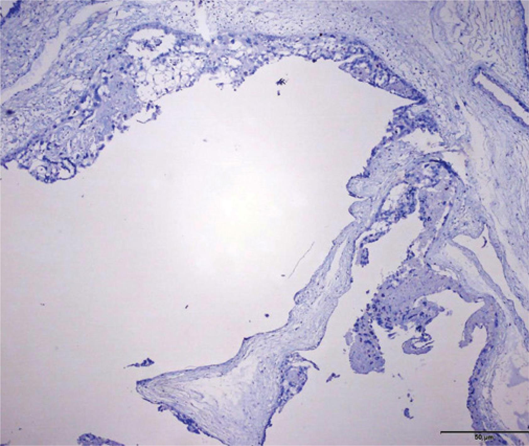
Fig. 2
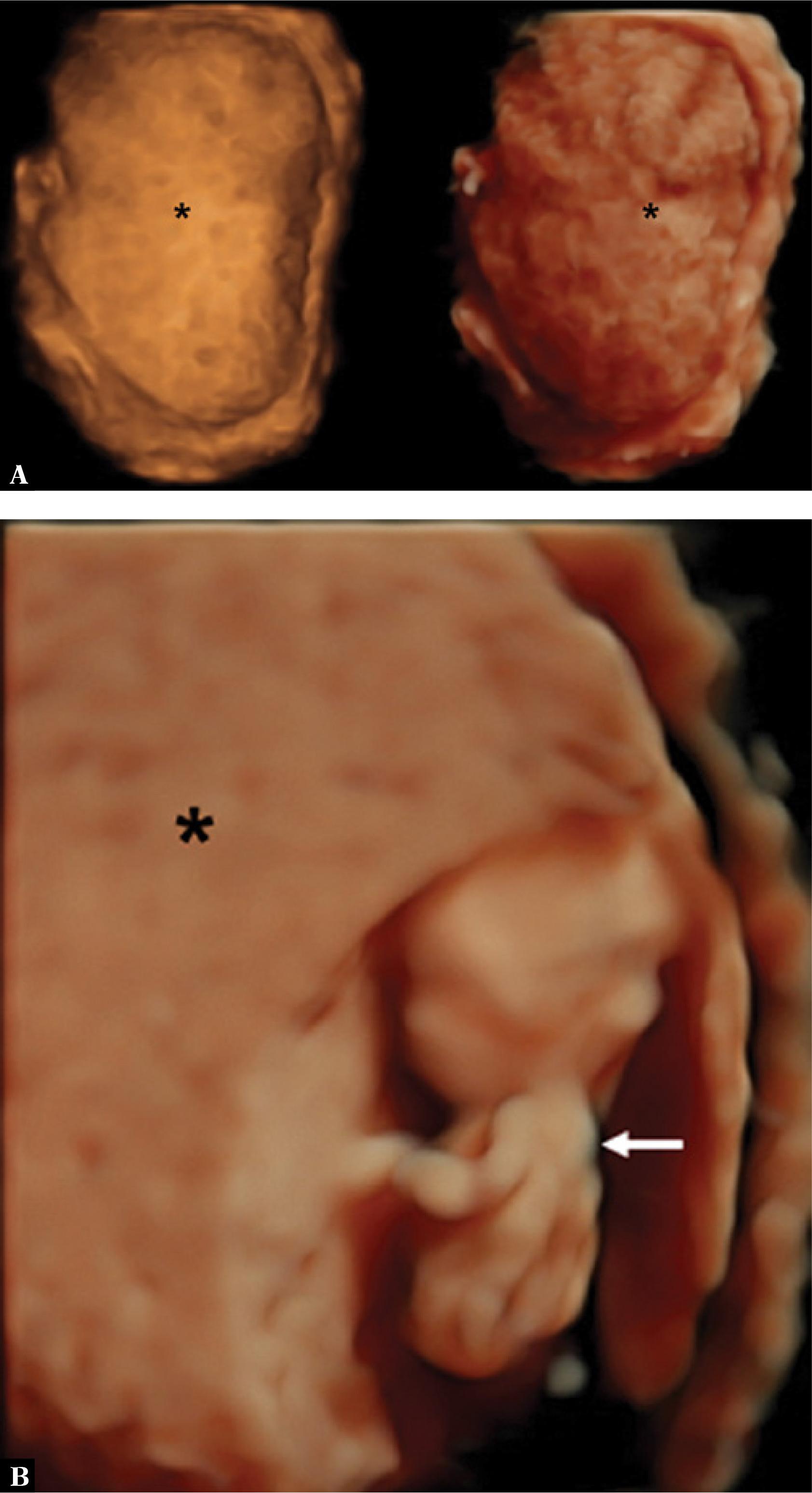
Fig. 3
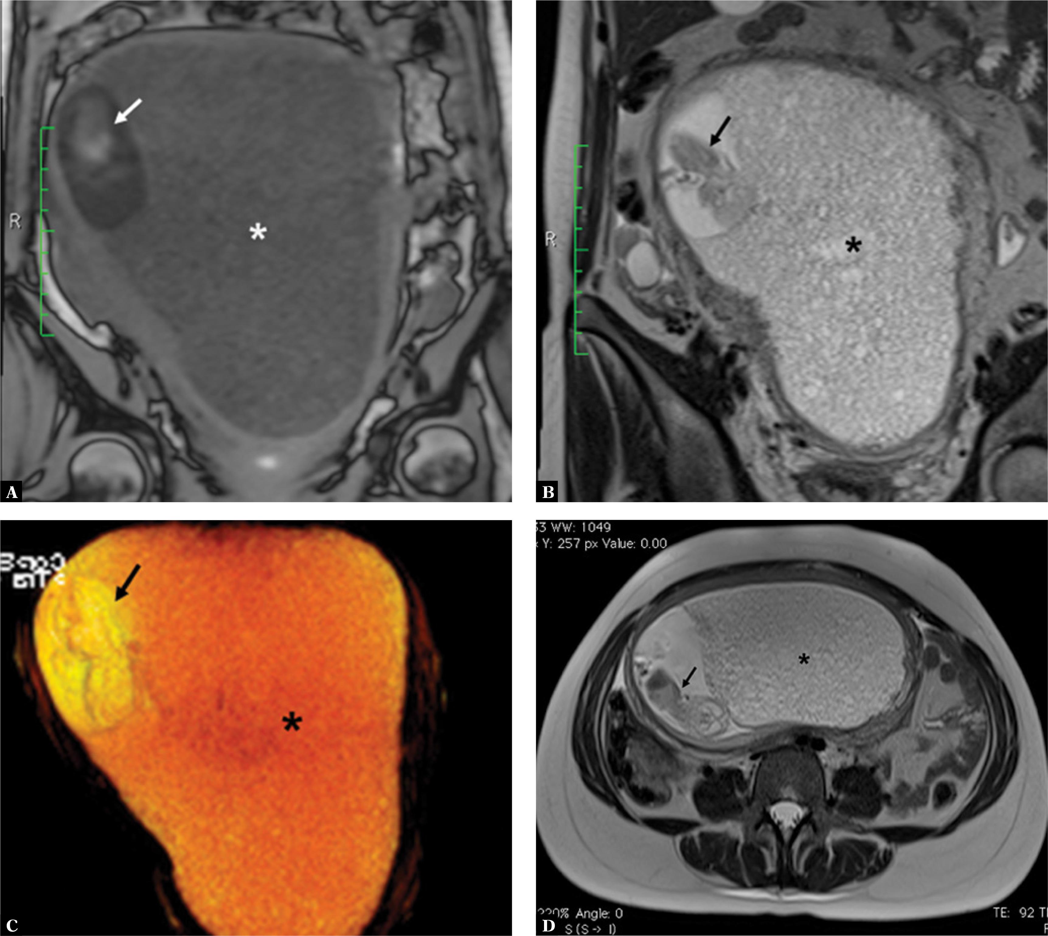
Fig. 4
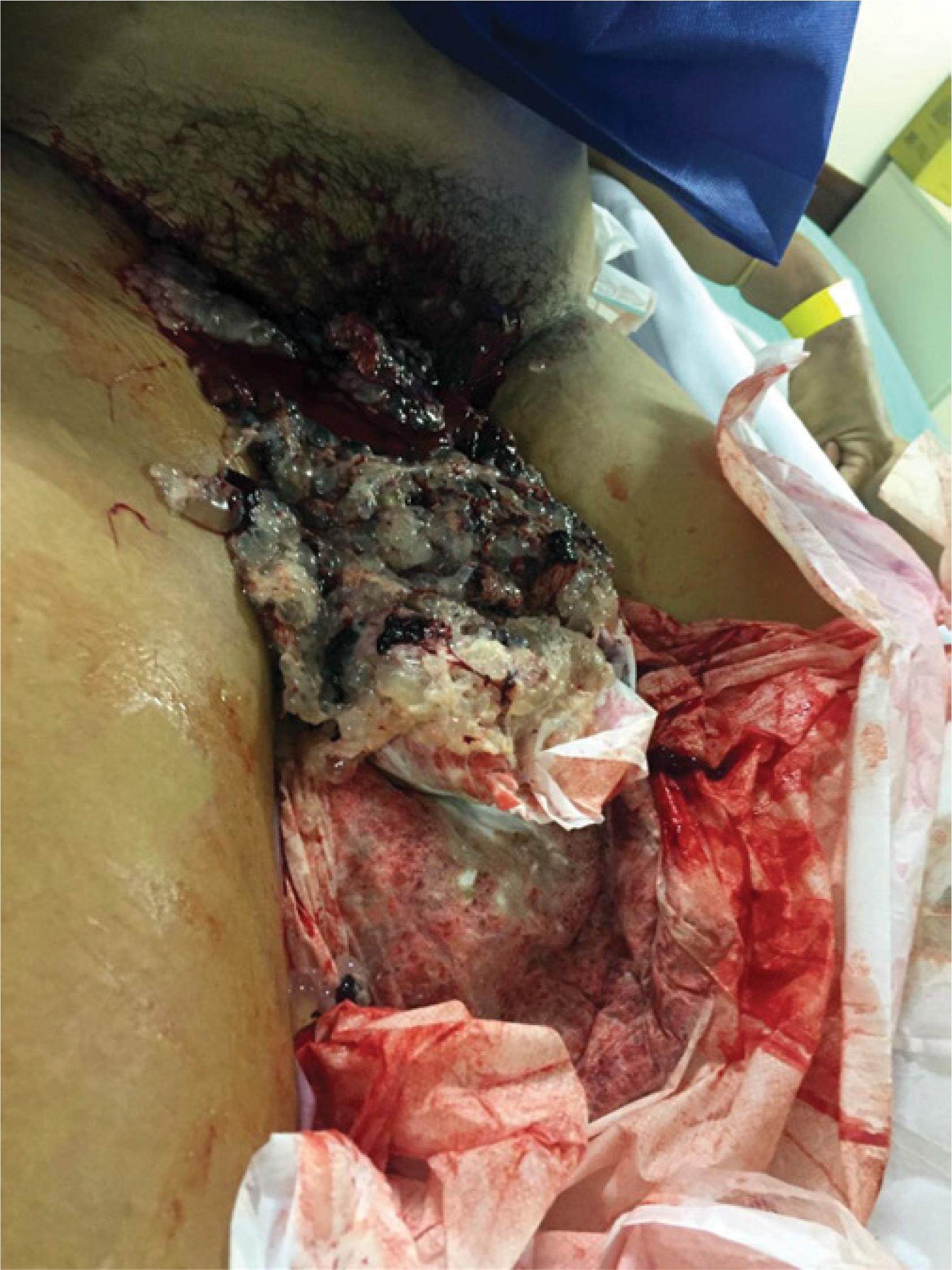
Fig. 5
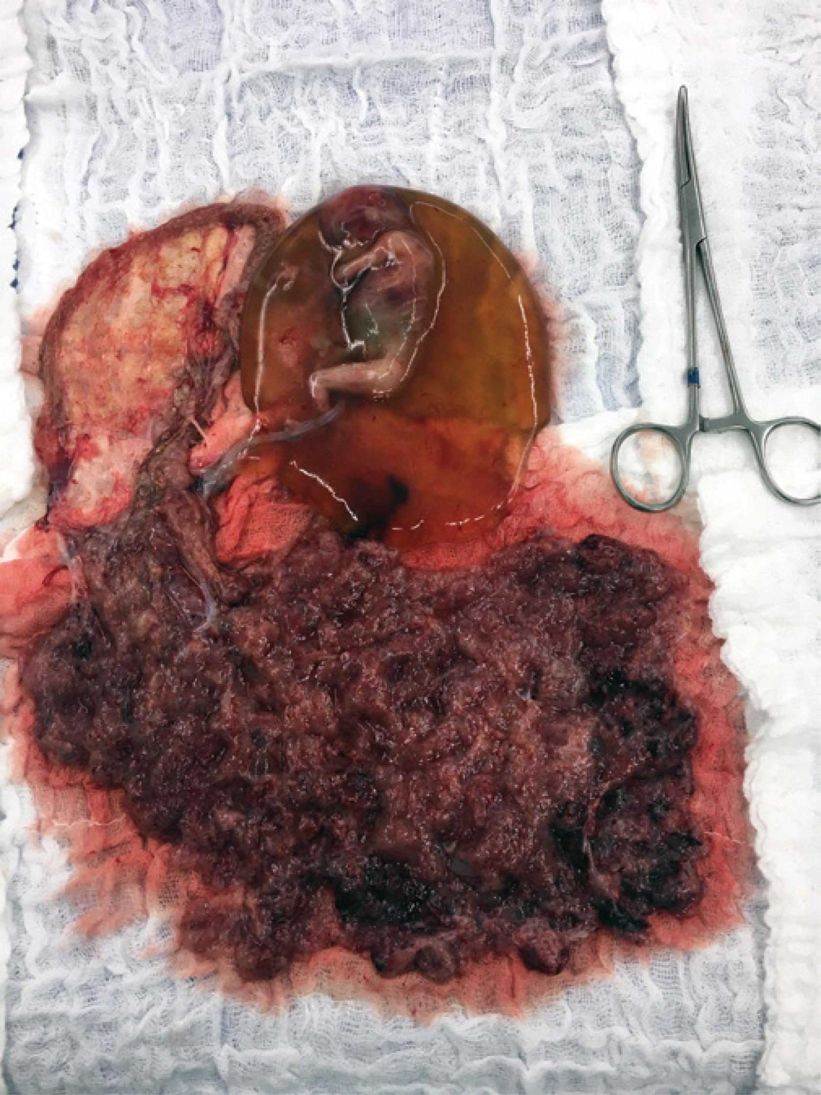
Fig. 6

© 2017 Antonio Braga, Bruna Obeica, Heron Werner, Sue Yazaki Sun, Joffre Amim Júnior, Jorge Rezende Filho, Edward Araujo Júnior, published by MEDICAL COMMUNICATIONS Sp. z o.o.
This work is licensed under the Creative Commons Attribution-NonCommercial 3.0 License.