
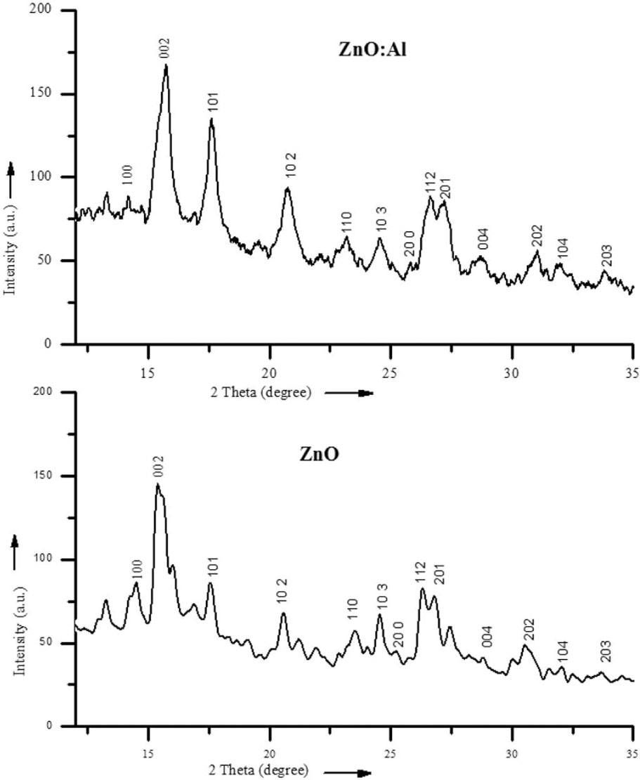
Fig. 1
XRD spectra of undoped and doped ZnO.
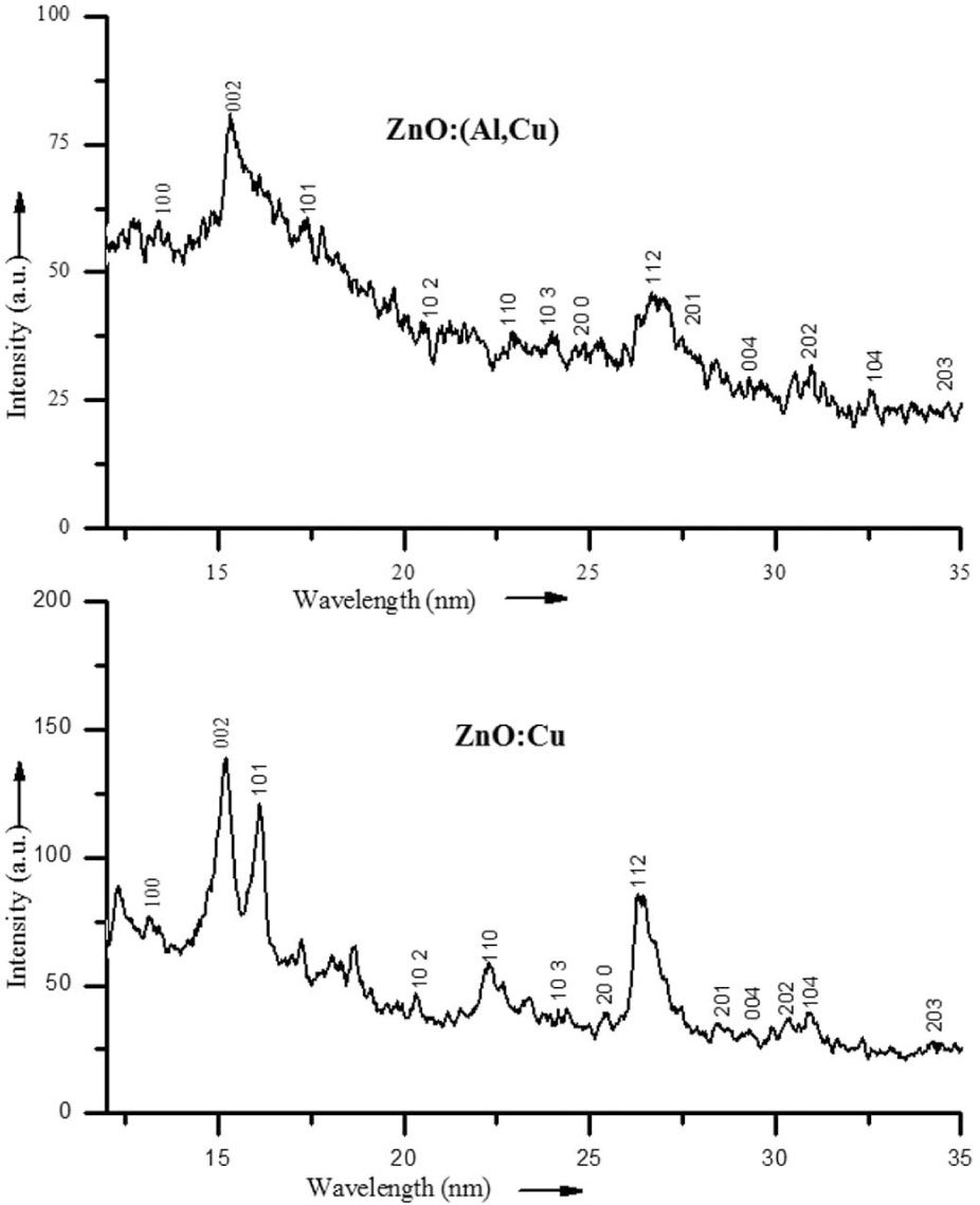
Fig. 2
XRD spectra of doped and co-doped ZnO.
Table 1
Change of d values corresponding to lattice planes.
| Standard d value [nm] | Observed d value of ZnO | Observed d value of ZnO:Al | Observed d value of ZnO:Cu | Observed d value of ZnO(Al,Cu) | (h k l) |
|---|---|---|---|---|---|
| 0.28204 | 0.28047 | 0.28760 | 0.273697 | 0.270137 | 100 |
| 0.26062 | 0.26073 | 0.25919 | 0.265534 | 0.265534 | 002 |
| 0.24806 | 0.24116 | 0.23220 | 0.253398 | 0.251864 | 101 |
| 0.19141 | 0.19838 | 0.19745 | 0.183749 | 0.188419 | 102 |
| 0.16284 | 0.16554 | 0.16620 | 0.155873 | 0.162570 | 110 |
| 0.14793 | 0.14919 | 0.14316 | 0.144454 | 0.152229 | 103 |
| 0.13810 | 0.13471 | 0.13234 | 0.137529 | 0.134283 | 112 |
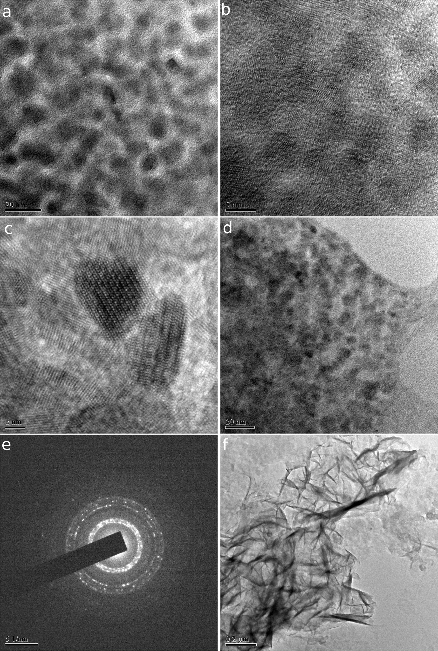
Fig. 3
HRTEM images of as-prepared samples: (a), (b), (c), (d), (e) and (f).
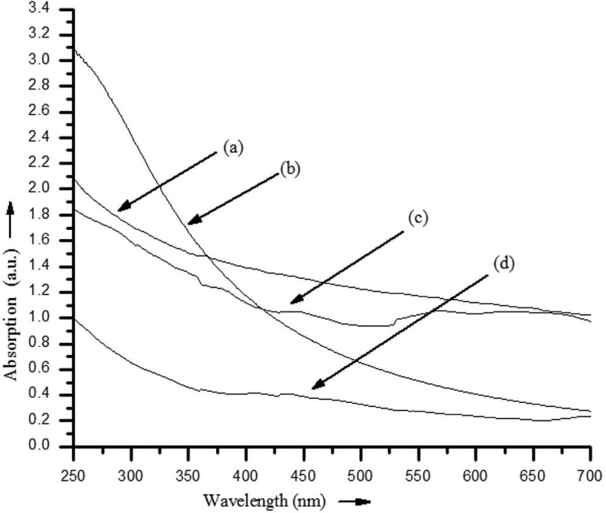
Fig. 4
UV-Vis absorption spectra of synthesized nano samples: (a) ZnO, (b) ZnO:Al, (c) ZnO:Cu and (d) ZnO:( Al,Cu).
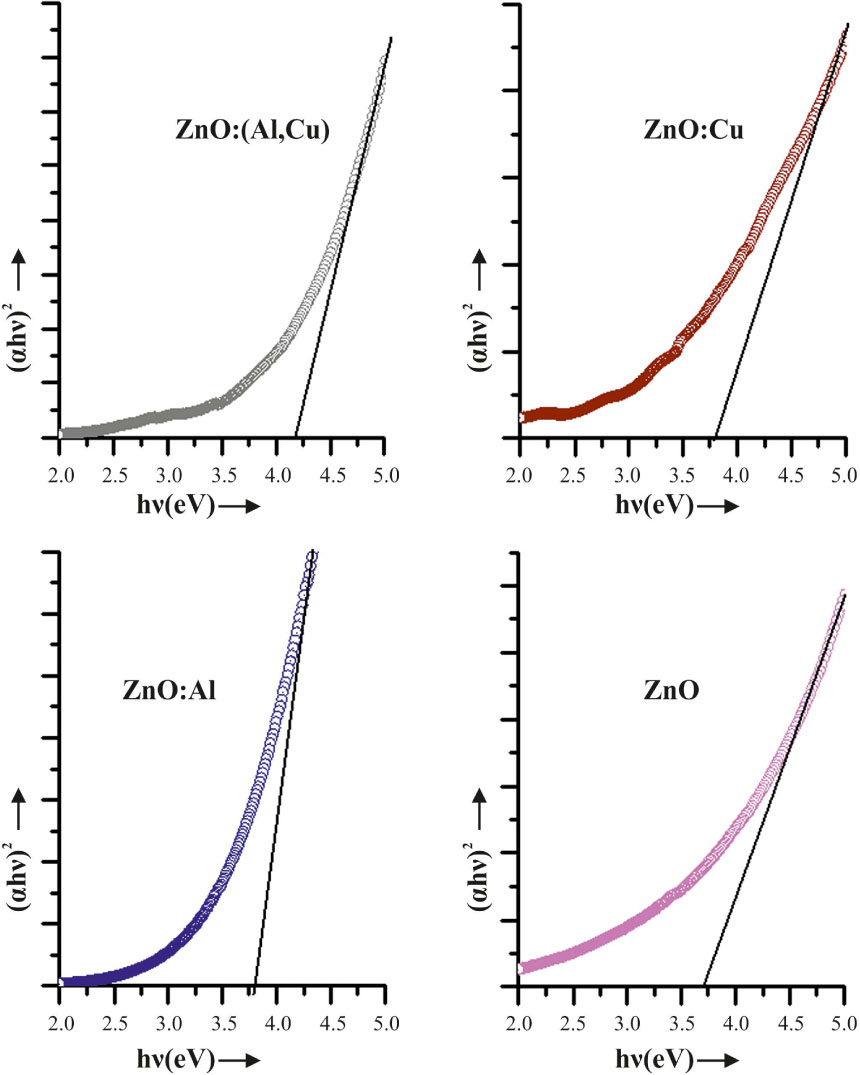
Fig. 5
Tauc plot of (αhν)2 vs. photon energy (eV) of ZnO:Al,Cu; ZnO:Cu; ZnO:Al and pure ZnO.
Table 2
Particle sizes and energy band gaps of ZnO quantum dots.
| Sample | Particle size from XRD [nm] | Particle size from HRTEM [nm] | Energy band gap [eV] | Blue shift [eV] | Particle size from EMA [nm] |
|---|---|---|---|---|---|
| ZnO | 6.25 | 5.3 | 3.7 | 0.33 | 5.18 |
| ZnO:Al | 5.67 | 4.1 | 3.8 | 0.43 | 4.52 |
| ZnO:Cu | 9.39 | 5.0 | 3.78 | 0.41 | 4.62 |
| ZnO:Al,Cu | 7.46 | 4.2 | 4.13 | 0.76 | 3.39 |
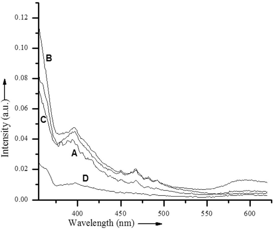
Fig. 6
PL spectra of synthesized nano-samples of (A) ZnO, (B) ZnO:Al, (C) ZnO:Cu and (D) ZnO:(Al,Cu).
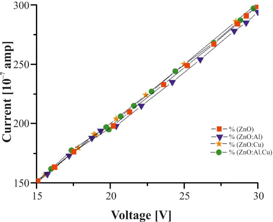
Fig. 9
I-V characteristics of ZnO nanostructures.
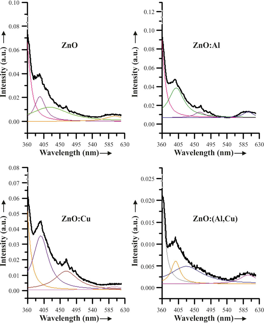
Fig. 7
Deconvoluted photoluminescence peaks of undoped and doped ZnO.
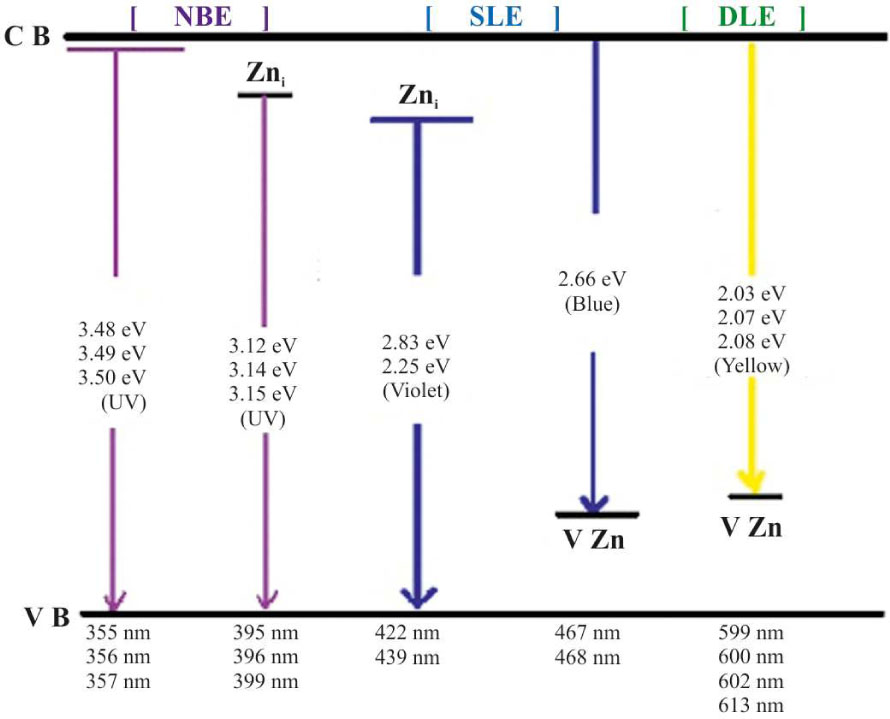
Fig. 8
Schematic energy level diagram of ZnO nanoparticles showing emission mechanism.