
Introduction
Dilated cardiomyopathy (DCM) is a primary disorder of heart muscle defined by dilatation and systolic dysfunction that cannot be explained by secondary causes, such as ischemic, valvular, viral, autoimmune, alcohol/toxin-driven, or hypertensive heart disease. The prevalence of DCM is estimated at 0.4% of the general population, or 1 in 250 adults, and it is the leading cause of nonischemic heart failure as well as sudden cardiac death.1 Among the three primary types of cardiomyopathies—DCM, hypertrophic cardiomyopathy, and arrhythmogenic cardiomyopathy—DCM is the most genetically diverse, and approximately 25% to 35% of DCM cases have an identifiable genetic cause.2,3 Presentation and response to treatment vary widely based on the specific underlying genetic variant, and increasing evidence suggests that polygenic mechanisms and environmental factors influence the clinical course.4 In this review, we discuss the genetic variants known to cause DCM, indications for genetic screening, clinical management, novel therapies, and future directions for research.
Defining Dilated Cardiomyopathy
According to the 2023 European Society of Cardiology (ESC) Guidelines for the management of cardiomyopathies, the diagnosis of DCM is based on evidence of left ventricular (LV) dilatation and systolic dysfunction. The left ventricle is considered dilated when the LV end-diastolic diameter is > 58 mm in men and > 52 mm in women or when the indexed LV end-diastolic volume is ≥ 75 mL/m² in men and ≥ 62 mL/m² in women.5 DCM may have genetic and non-genetic causes, with significant gene-environment interactions. Common non-genetic etiologies of DCM, which may represent a second hit, include viral infection, alcohol use, chemotherapy, tachyarrhythmias, autoimmune disorders, endocrinopathies, and pregnancy.1
Non-Dilated Left Ventricular Cardiomyopathy
The 2023 ESC Guidelines have introduced a new cardiomyopathy phenotype termed non-dilated left ventricular cardiomyopathy (NDLVC), which is defined by the presence of fibrous and/or fatty replacement of the LV myocardium in the absence of ischemic heart disease and without LV dilation.5 Patients with global LV hypokinesis without evidence of scarring or dilatation are also classified as having NDLVC.5
Overlap with Arrhythmogenic Cardiomyopathy
There is considerable overlap between DCM and arrhythmogenic cardiomyopathy (ACM). ACM was originally termed arrhythmogenic right ventricular cardiomyopathy (ARVC) and was characterized by a predominant right ventricle (RV) dysfunction associated with high risk of life-threatening ventricular arrhythmias. Over the past 10 years, the definition of ACM has been broadened to include right-dominant ARVC, left-dominant (arrhythmogenic left ventricular cardiomyopathy), and biventricular forms, all characterized by an arrhythmic risk disproportionate to the degree of systolic dysfunction. The redefinition of ACM has led to a considerable overlap with DCM, as several forms of genetic DCM present with ventricular arrhythmias disproportionate to the degree of LV dilation and dysfunction, mainly in carriers of pathogenic genetic variants in Lamin (LMNA), Desmoplakin (DSP), Phospholamban (PLN), RNA-binding motif protein 20 (RBM20), and Filamin C (FLNC).3
Genetic Causes of DCM
Although over 50 genes have been associated with the development of DCM, establishing clear genetic causality remains challenging in many cases. Indeed, a pathogenic gene variant is identified in only 40% of familial DCM cases, indicating that gene-gene and gene-environment interactions may also play a role in heritable DCM.1 Additionally, truncating variants in the Titin gene (TTNtv) are prevalent in 1% to 3% of the general population, but only 0.4% of the population develops DCM, consistent with incomplete penetrance of pathogenic variants.1 Most of the identified DCM causal genes have an autosomal dominant inheritance, although X-linked, autosomal recessive, and mitochondrial inheritance patterns have also been observed.6 As noted, penetrance is often incomplete and appears also to be dependent on age.
Genes Associated with DCM
Despite the complexity of genetic DCM, variants in several genes have strong evidence for pathogenicity in DCM as defined by the Clinical Genome Resource (ClinGen), a National Institutes of Health funded resource that collects human genomic data and evaluates the evidence linking genes and genetic variants to human diseases. ClinGen is widely used in clinical genetics as it provides standardized assessments of the strength of gene-disease relationships.7 The strength of the association between genetic variants and DCM is defined as definitive, strong, moderate, or limited based on the ClinGen gene-disease validity classification, which assesses the evidence for the association.8 Genes with a definitive, strong, or moderate association with DCM are listed in Table 1.9
Table 1
Genes associated with dilated cardiomyopathy per ClinGen classification.9 AD: autosomal dominant; AR: autosomal recessive
| GENE | INHERITANCE MODE | LEVEL OF EVIDENCE | |
|---|---|---|---|
| BAG3 | BAG Cochaperone 3 | AD | Definitive |
| DES | Desmin | AD | Definitive |
| DSP | Desmoplakin | AD | Definitive |
| FLNC | Filamin C | AD | Definitive |
| LMNA | Lamin A/C | AD | Definitive |
| MYH7 | Myosin Heavy Chain 7 | AD | Definitive |
| RBM20 | RNA Binding Motif Protein 20 | AD | Definitive |
| SCN5A | Sodium Voltage-Gated Channel Alpha Subunit 5 | AD | Definitive |
| TMEM43 | Transmembrane Protein 43 | AD | Definitive |
| TNNC1 | Troponin C1, Slow Skeletal and Cardiac Type | AD | Definitive |
| TNNT2 | Troponin T2, Cardiac Type | AD | Definitive |
| TTN | Titin | AD | Definitive |
| NRAP | Nebulin-Related Anchoring Protein | AR | Strong |
| PPA2 | Inorganic Pyrophosphatase 2 | AR | Strong |
| PRDM16 | PR/SET Domain 16 | AD | Strong |
| TBX20 | T-Box Transcription Factor 20 | AD | Strong |
| ACTC1 | Actin Alpha Cardiac Muscle 1 | AD | Moderate |
| JPH2 | Junctophilin 2 | AR (DCM) AD (HCM) | Moderate |
| MYLK3 | Myosin Light Chain Kinase 3 | AD | Moderate |
| NEXN | Nexilin F-Actin Binding Protein | AD | Moderate |
| PLEKHM2 | Pleckstrin Homology and RUN Domain Containing M2 | AR | Moderate |
| PLN | Phospholamban | AD | Moderate |
| TNNI3 | Troponin I3, Cardiac Type | AD | Moderate |
| TNNI3K | TNNI3 Interacting Kinase | AD | Moderate |
| TPM1 | Tropomyosin 1 | AD | Moderate |
| VCL | Vinculin | AD | Moderate |
Phenotypes of Common Genetic DCM
Pathogenic variants associated with DCM occur in genes encoding components of the cytoskeleton, desmosomes, sarcomeres, calcium transporters or channels, nuclear envelope, and RNA regulation. The most common genes causing DCM include TTN, LMNA, DSP, and FLNC. The following sections summarize the phenotypic features associated with DCM resulting from pathogenic variants in these genes, while Table 2 describes the phenotypes of other rarer genetic dilated cardiomyopathies.1,3,10,11,12
Table 2
| GENE | % OF GENETIC DCM | PHENOTYPE | AGE OF ONSET |
|---|---|---|---|
| TTN | 60% | Relatively good prognosis and low arrhythmia burden | 40–70 |
| DSP | 15% | Biventricular, myocarditis-like “hot phases” | 20–49 |
| LMNA | 15% | High burden of supraventricular arrhythmias and AV conduction abnormalities | 25–55 |
| FLNC | 5% | Arrhythmogenic, initial presentation often similar to myocarditis | 25–60 |
| MYH7 | 5% | Severe, often progresses to end-stage HF with poor response to standard therapies | 20–49 |
| RBM20 | 5% | Highly arrhythmogenic and penetrant | 20–50 |
| TNNT2 | 5% | – | 25–55 |
| BAG3 | < 5% | Early onset, high burden of ventricular arrhythmia | 20–50 |
| DES | < 5% | Frequent AV conduction abnormalities, highly penetrant | 35–60 |
| NRAP | < 5% | Autosomal recessive, often with severe pediatric onset disease10 | 9 months–47 years |
| PPA2 | < 5% | Often triggered by viral illness or alcohol use, can present with sudden cardiac death in pediatric patients11 | |
| PRDM16 | < 5% | Highly variable | 0–60 |
| TBX20 | < 5% | Relatively nonaggressive, with low rate of adverse cardiovascular events12 | – |
| TMEM43 | < 5% | High burden of ventricular arrhythmias, high risk for end-stage heart failure | – |
| TNNC1 | < 5% | Relatively low risk of ventricular arrhythmias | 25–55 |
| SCN5A | < 5% | High burden of supraventricular arrhythmias | 15–25 |
Titin Truncating Variants (TTNtv)
Truncating variants in the TTN gene, which encodes the giant sarcomere protein titin, are the most common genetic cause identified in DCM. TTNtv-associated DCM presents later in life compared to other genetic DCMs and has a relatively good prognosis, with a low arrhythmic risk. The age of onset is between 40 and 70 years.1 TTNtv DCM has the highest rate of LV reverse remodeling (LVRR) (over 50% of cases) in response to treatment with guideline directed medical therapies (GDMT) compared with the other genetic DCMs.13 Asymptomatic carriers of TTNtv are at increased risk of developing DCM after exposure to mechanical and chemical stresses, such as pregnancy, alcohol use, and chemotherapy.14,15,16
Lamin (LMNA) Pathogenic Variants
LMNA encodes lamin A and lamin C proteins, which are components of the nuclear lamina.3 LMNA-associated DCM is highly penetrant compared to other types of genetic DCM and has the greatest risk of progressive, refractory heart failure and a high burden of conduction defects and arrhythmias, including atrioventricular blocks, atrial fibrillation, and lethal ventricular arrhythmias.6,13 Moreover LMNA-DCM has the lowest rates of LVRR in response to GDMT among the genetic DCMs.17
Desmoplakin (DSP) Pathogenic Variants
The first description of a myocardial disease linked to DSP mutations referred to patients with Carvajal syndrome, an autosomal recessive cardio-cutaneous syndrome, including DCM, palmoplantar keratoderma and wooly hair.18 DCM associated with DSP variants has a strong overlap with ACM with frequent ventricular arrhythmias. The age of onset ranges from 20 to 49.1 DSP cardiomyopathy is also characterized by acute episodes of inflammation similar to myocarditis.19
Filamin C (FLNC) Pathogenic Variants
Filamin C is a key structural protein in the sarcomere. FLNC genetic variants have been associated with skeletal myopathies as well as isolated cardiomyopathies, both dilated and hypertrophic. FLNC-associated cardiomyopathies have a high risk of ventricular arrhythmias and sudden cardiac death as well as high rates of myocardial fibrosis.20
Factors Contributing to the Expression of Genetic DCM
Burden of Genetic Variants
It has been theorized that common genetic variants associated with LV structure and function, when clustered together, contribute to an increased risk of developing DCM in carriers of the rare pathogenic and likely pathogenic variants.4
Two-hit Hypothesis
It is thought that many cases of genetic DCM follow the two-hit hypothesis: an underlying genetic variant makes a patient prone to developing DCM when exposed to a second trigger-factor. This second factor could be a toxin such as chemotherapy or alcohol, or another medical condition such as myocarditis, pregnancy, or thyroid disorders. The two-hit hypothesis is supported by familial clustering of postpartum cardiomyopathy, alcohol-related cardiomyopathy, and chemotherapy-related cardiomyopathy.1
Inflammation
The progression of dilated cardiomyopathy appears to depend on an inflammatory cycle. Volume overload, endothelial dysfunction, and oxidative stress triggered by either the direct consequences of the underlying genetic variant or by a secondary environmental insult lead to myocardial injury. Myocardial injury triggers an inflammatory response with cytokine release and immune cells that promotes extracellular matrix remodeling, collagen deposition, and fibrosis. Over time, this process culminates in progressive left ventricular dilatation.1
On endomyocardial biopsy, the inflammation of genetic DCM is similar to that of myocarditis, and clinically an association is seen in patients with acute and chronic myocarditis and genetic variants associated with DCM and ACM.21 Certain genes are associated with recurrent myocarditis-like inflammatory episodes or “hot phases.” DSP is the most common, but FLNC and DES cardiomyopathy can also present this way.22
Sex Differences
In general, men with genetic DCM have a worse prognosis in terms of transplant-free and overall survival. In a multicenter observational longitudinal study of 1,716 patients with DCM (510 with pathogenic or likely pathogenic variants), men with genetic DCM had the highest rates of all-cause mortality, transplantation, heart failure hospitalizations, and malignant ventricular arrhythmias compared with women with genetic DCM and all patients with non-genetic DCM. The burden of these major adverse events was associated with specific underlying genotype in men, but this association was not noted in women. Conversely, the rate of LVRR correlated with genotype in women but not in men.23
Evidence suggests than men with BAG3-DCM have earlier onset of disease than women,6 and male sex is one of the four criteria for implantable cardiac defibrillator (ICD) placement in patients with LMNA-associated cardiomyopathy.24 The one exception is DSP-associated cardiomyopathy, which affects more women than men and is often more severe in women.19
Management of Genetic DCM
The American Heart Association (AHA), ESC, and expert consensus documents5,25 emphasize the importance of referring patients with cardiomyopathies, particularly those with inherited disease, to specialized multidisciplinary centers. These centers provide integrated expertise in genetic evaluation, family screening, arrhythmia management, and advanced heart failure therapies, facilitating comprehensive diagnosis, risk stratification, and individualized management.
Genetic Testing
The ESC 2023 cardiomyopathy guidelines suggest as a class 1B recommendation that all patients with DCM should undergo genetic testing to guide risk stratification and prognostication.5 On the other hand, the AHA assigns a Class IIa recommendation for genetic testing, limiting its use to selected patients with non-ischemic cardiomyopathy.25
The AHA recommends genetic testing for patients with suspected or confirmed inherited cardiomyopathies, using panels that include genes associated with clinically relevant cardiomyopathy phenotypes. Given the overlap between DCM and ACM, comprehensive panels incorporating genes associated with both conditions may improve diagnostic yield, although they can increase cost and the likelihood of identifying variants of uncertain significance.25 A 2022 study demonstrated that more restricted panels targeting genes associated with either DCM or ACM could miss clinically relevant genetic variants in approximately 11% of patients when compared to panels including both ACM and DCM genes.26
While AHA and ESC guidelines on management of genetic cardiomyopathies do not recommend a specific number of genes to test for in a comprehensive panel, this study suggests that panel encompassing genes associated with both DCM and ACM may provide sufficient diagnostic coverage when gene selection is guided by the clinical phenotype, family history, and other relevant clinical features.26 Most of the genetic panels used in clinical practice include 80 to 120 genes.1 Factors that increase the pre-test possibility of having genotype positive DCM include family history of DCM, presence of skeletal myopathy, absence of other comorbidities, and low QRS amplitude.3,6
Diagnostics
In addition to the standard transthoracic echocardiogram, patients with dilated cardiomyopathy should undergo cardiac magnetic resonance imaging as part of their initial evaluation, mainly for myocardial tissue characterization following gadolinium contrast administration, which can identify fibrosis, scar formation, and active myocardial injury. Certain genetic cardiomyopathies present with unique late gadolinium enhancement (LGE) patterns. For example, in LMNA-cardiomyopathy, the LGE is typically localized to the basal interventricular septum, while DSP-DCM usually shows inferior subepicardial LGE. However, LGE distribution is often nonspecific, and TTNtv variants have not been shown to exhibit a distinctive LGE pattern.6
Risk Stratification
Given the diverse phenotypes of genetic DCM, individualized risk stratification tools are needed. The LMNA VTA SCD (Lamin A/C ventricular tachyarrhythmia sudden cardiac death) risk calculator (https://lmna-risk-vta.fr) is a validated tool to predict the 5-year risk of lethal ventricular arrhythmia for patients with LMNA cardiomyopathy and can be used to help select patients for primary prevention ICD.27 The PLN 5-year VA (Phospholamban 5-year Ventricular Arrhythmia Risk Calculator, https://plnriskcalculator.shinyapps.io/final_shiny/) similarly predicts percentage risk of lethal arrhythmia in the next 5 years for patients with the p.arg14del PLN variant.28 The DSP specific risk stratification tool (https://www.dsp-risk.com) is also available as well as the FLNC (Filamin C gene) risk stratification tool (https://flnctv.shinyapps.io/RiskCalculator/), for which the external validation is currently in progress. However, there is no risk stratification algorithm to predict the development of heart failure in asymptomatic patients with genetic variants.
Preventive Care
There is no good evidence to guide management of asymptomatic carriers of DCM genetic variants. The clinical consensus is to perform surveillance cardiac imaging every 2 to 3 years.6 Reduction in global longitudinal strain is the imaging finding that most strongly correlates with development of DCM and poor prognosis in the gene positive-phenotype negative subjects.6
The EARLY-GENE (Early Treatment With Candesartan vs Placebo in Genetic Carriers of Dilated Cardiomyopathy) trial (NCT05321875), a prospective phase 3 randomized double-blind placebo-controlled study that is currently ongoing, is investigating whether candesartan could prevent progression to DCM in asymptomatic carriers of DCM-associated variants.
Familial Screening
The AHA guidelines recommend that first-degree relatives of patients diagnosed with cardiomyopathy that is either inherited or has an identified DCM-associated gene variant undergo genetic screening and counseling.26 The ESC guidelines recommend genetic counseling and cascade testing of first-degree relatives of probands and that genetic screening be considered for second-degree relatives if the proband is deceased.5
Standard Treatment
Basic treatment for genetic DCM should include referral to an advanced heart failure specialist and the standard of care for heart failure with reduced ejection fraction (< 50%), including GDMT and consideration of ICD for primary prevention of sudden cardiac death following the ESC or AHA guidelines. Figure 1 summarizes the general management of genetic DCM.
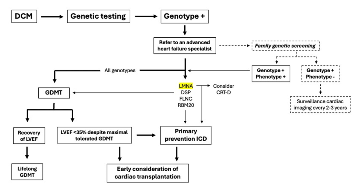
Figure 1
Standard treatment for genetic dilated cardiomyopathy. DCM: dilated cardiomyopathy; GDMT: guideline-directed medical therapy; CRT-D: cardiac resynchronization therapy defibrillator; LVEF: left ventricular ejection fraction; ICD: implantable cardioverter defibrillator
Guideline-directed Medical Therapy
No trials have been conducted specifically to look at the response to GDMT in patients with genetic DCM. Based on trials including patients with all forms of DCM, there is some evidence to suggest that overall genetic DCM may have a less robust response to medications targeting the neurohormonal axis than non-genetic DCM.29 Among the genetic DCMs, TTNtv-DCM has shown the best response to GDMT, with LVRR greater than 50%.13 In contrast, the sarcomeric variants MYH7 and TNNT2 have shown a poor response to GDMT, with LVRR occurring in only 23% to 28% of patients.13 LMNA has the worst response to GDMT, with LVRR occurring in less than 20% of patients.29
Despite the lack of evidence to guide medical management of genetic DCM with heart failure, the consensus is to treat with standard GDMT and continue it even if LVEF is recovered.6
Implantable Cardiac Defibrillator
The DANISH trial (Danish Study to Assess the Efficacy of ICDs in Patients with Non-ischemic Systolic Heart Failure on Mortality) looked at the benefit of ICD for primary prevention in patients with nonischemic DCM and found that there was no great benefit in older patients.30 However, these results likely cannot safely be applied to patients with genetic DCM given they are typically younger and can have ventricular arrhythmia disproportionate to and even before the development of systolic dysfunction and dilatation. Some evidence suggests that cardiac fibrosis and global longitudinal strain on imaging may be better parameters to guide ICD placement.6
In certain arrhythmogenic DCM phenotypes, the underlying genetic variant is an indication for ICD placement independent of other factors. This applies to LMNA, FLNC, DSP, and RBM20.3
Cardiac Resynchronization Therapy
Cardiac resynchronization therapy is indicated for patients with symptomatic DCM, LVEF less than 35%, and QRS duration greater than 130 ms. Cardiac resynchronization therapy has specifically been shown to improve outcomes in LMNA cardiomyopathy.31
Exercise
There is little evidence to guide exercise recommendations for patients with genetic DCM. In patients with LVEF less than 45%, extensive fibrosis on imaging or biopsy, or frequent ventricular arrhythmias, high-intensity exercise should be discouraged.32
Transplantation
Patients with genetic DCM actually have favorable outcomes after heart transplantation compared to the overall population of transplant candidates, likely due to their younger age at presentation and relative lack of comorbidities.33
Novel Therapies
Targeted therapies for DCM and gene therapies in particular are promising, but none have yet been shown to have enough benefit to be incorporated into clinical practice.
Targeted Molecular Therapy
Myosin modulators have been evaluated for use in DCM. The selective cardiac myosin activator omecamtiv mecarbil has been studied in heart failure patients, including a small portion of patients with DCM, but this drug has not been approved by the FDA due to unfavorable risk-benefit ratio.34
Another myosin modulator, danicamtiv, has shown promise in a recent phase 2 trial comparing DCM patients with genetic variants to DCM patients without genetic variants. Patients with TTN and MYH7 variants showed the best response to danicamtiv, with increase in cardiac contractility.35
An inhibitor of p38 mitogen activated protein kinase targeted at LMNA DCM had promising phase 1 and phase 2 results, but the phase 3 trial (REALM-DC) did not show efficacy.36 Crenolanib, a PDGFRA inhibitor, has been shown to improve contractility and reduce arrhythmogenicity in preclinical studies on induced pluripotent stem cells derived cardiomyocytes (IPSC-CMs) with FLNC truncating variants.37 Further studies are necessary in animal models before testing crenolanib as a repurposed drug in FLNC-DCM patients.
Gene Therapy
Concepts of gene therapy for DCM include gene replacement, gene silencing, and direct genome editing. Gene replacement is simpler for small genes with loss of function mutations. In contrast, gene silencing requires development of short hairpin RNA specific to each individual genetic variant, which is not feasible given the genetic diversity of DCM. Direct genome editing using CRISPR-Cas9 technology has been effective in treating sickle cell disease, but applying it to cardiomyopathies is more complicated because of the challenges associated with safe, efficient, and cardiomyocyte-specific delivery of the editing machinery.38
Gene replacement therapy to the heart can be delivered via adeno-associated virus, serotype-9. The disadvantages of adenovirus delivery are decrease of efficacy over time, the risk of triggering an immunologic inflammatory response that can be severe, and the prevalence of neutralizing antibodies in many patients due to prior adenovirus exposure.38 Intracoronary delivery of gene therapies is a possible way to reduce side effects of gene therapy by allowing a lower dose but is more invasive than systemic delivery.1 Additionally, adenovirus delivery is size-limited, typically < 5 kb, and does not work for larger genes.
Next Steps
Further research is needed to better understand the genetic mechanisms and molecular pathways underlying the pathogenesis of DCM, including genome-wide association studies to investigate the cumulative burden of the variants hypothesis. More studies on the interaction between environment and genetics are needed, focusing particularly on the observed genetic predisposition in some subjects to develop DCM in alcohol abuse, chemotherapy exposure, pregnancy, and age. Additionally, further research into the impact and safety of exercise is important to guide patient-centered care. The development of novel risk stratification algorithms specific to the genetic cause, especially for predicting heart failure, is critical to guide preventive care for phenotype negative genotype positive patients and prognostication for symptomatic genetic DCM patients.
Conclusion
Genetic DCM is a diverse group of cardiomyopathies with complex underlying genetic mechanisms. Current treatment for heart failure in DCM follows international society guidelines, while precision medicine risk stratification tools are now available for prevention of life-threatening ventricular arrhythmias. Finally, although novel gene therapies are exciting, the heterogeneity of genetic DCM makes these treatments especially difficult to design, and they are far from being incorporated into clinical practice. Ongoing efforts to elucidate the impact of environment and gene-gene interactions is critical to guide proactive management of patients with genetic DCM and preventative care of carriers.
Key Points
Genetic dilated cardiomyopathy (DCM) has complex underlying genetic mechanisms including gene-environment interactions and polygenic interactions.
There is considerable overlap between genetic DCM and arrhythmogenic cardiomyopathy.
Titin gene truncating (TTN) variants constitute the majority of genetic DCM cases and, interestingly, they are also detectable in 2% to 3% of ostensibly healthy persons.
Lamin A/C and Desmoplakin variants are also common causes of genetic DCM, with poor prognosis, limited response to standard treatment, and high burden of arrhythmias.
Treatment is limited to standard heart failure care at this time, as no gene therapies or targeted molecular therapies have shown enough benefit to be incorporated into clinical practice.