
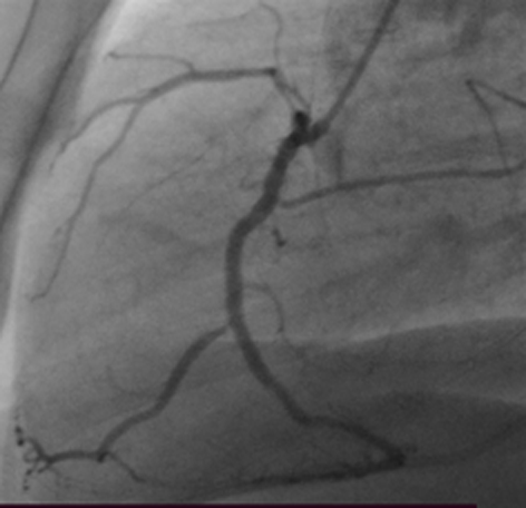
Figure 1
Coronary angiography after suspicion for coronary perforation post-percutaneous coronary intervention. No extravasation of contrast from the coronary arteries is seen.
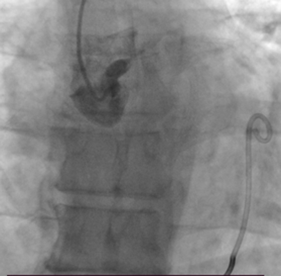
Figure 2
Contrast introduction into the ascending aorta reveals signs of perforation of the noncoronary aortic sinus.
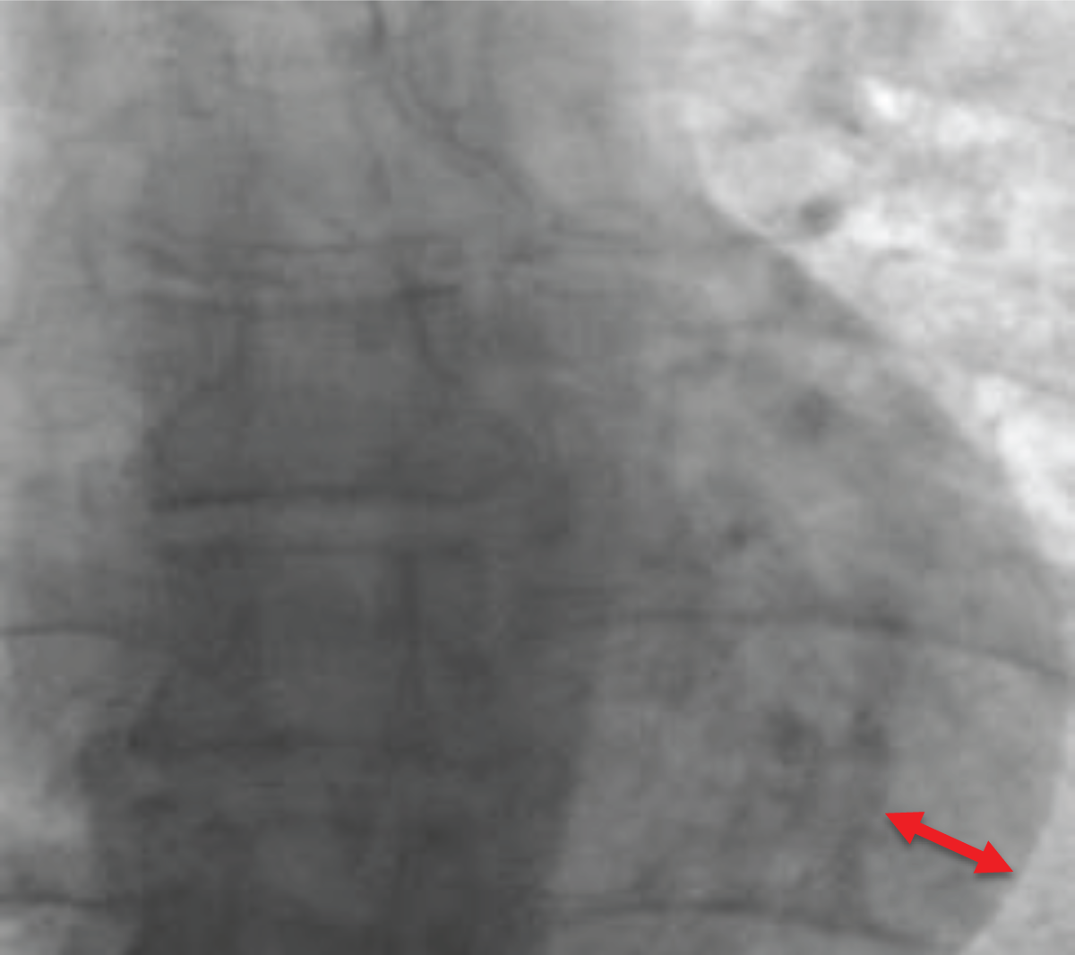
Figure 3
Fluoroscopic sign of an acute cardiac tamponade. Arrow indicates accumulation of blood within pericardium.
Question 1
Consider the options and find the answer in this video quiz; see also at https://youtu.be/AbDi9EjCa9I.
Question 2
Consider the options and find the answer in this video quiz; see also at https://youtu.be/JwEiuFhFB44.
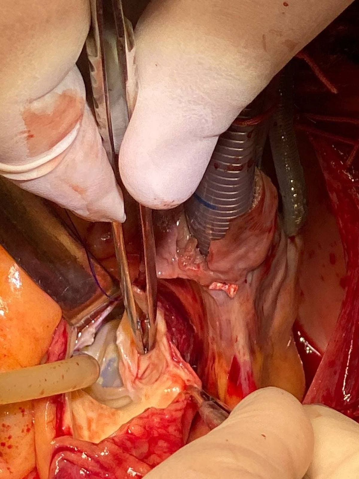
Figure 4
Intraoperative visualization of the perforation of the noncoronary aortic sinus.
Question 3
Consider the options and find the answer in this video quiz; see also at https://youtu.be/cUQr009258g.