
Introduction
In the current era, complications during percutaneous coronary intervention (PCI) occur in fewer than 1% of interventions. However, some of these complications are serious and require timely and skilled management to avoid substantial morbidity and even mortality. One of the most concerning complications is coronary perforation during PCI, which may occur during 0.5% of cases1 and can result in cardiac tamponade. The subsequent case illustrates an even more concerning complication of PCI and its challenging management.
Case Presentation
A 47-year-old man was admitted for elective stenting of the left anterior descending first diagonal branch (D1), circumflex, and right coronary arteries. The PCI was performed using right radial access. The brachial artery was tortuous, making catheterization of the left main coronary artery challenging.
At the completion of PCI, mild hypotension and tachycardia were noted, requiring low doses of ionotropic support. Emergent coronary angiography was repeated secondary to a suspicion of coronary perforation in the area of stent employment; however, no extravascular coronary jet was revealed (Figure 1). Manual contrast ejection into the aortic root identified noncoronary sinus perforation (Figure 2). Fluoroscopic examination demonstrated accumulation of blood within the pericardium that, combined with the hemodynamic instability, was indicative of acute cardiac tamponade (Figure 3).
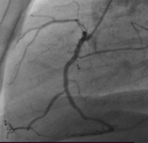
Figure 1
Coronary angiography after suspicion for coronary perforation post-percutaneous coronary intervention. No extravasation of contrast from the coronary arteries is seen.
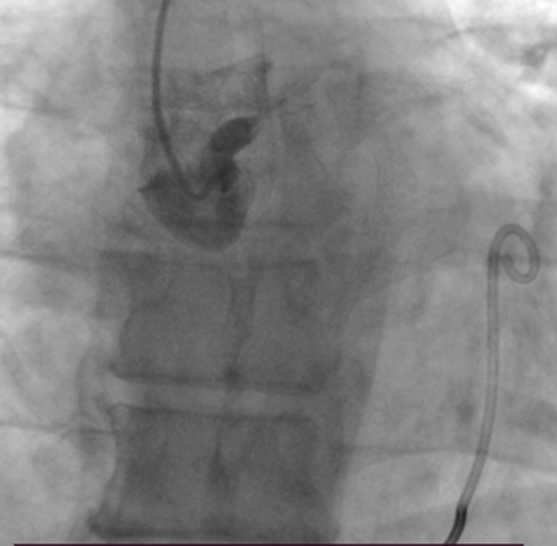
Figure 2
Contrast introduction into the ascending aorta reveals signs of perforation of the noncoronary aortic sinus.
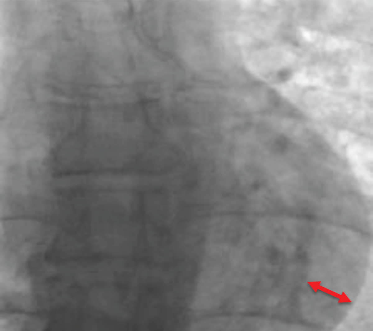
Figure 3
Fluoroscopic sign of an acute cardiac tamponade. Arrow indicates accumulation of blood within pericardium.
Question 1
What are the common causes and risk factors for aortic or coronary perforation during percutaneous coronary intervention (PCI)?
Use of anticoagulants and beta blockers during PCI
Advanced age, vascular tortuosity, heavy calcification, and prior cardiac surgery
Poor hydration and low blood pressure before the procedure
Lack of imaging guidance during PCI
High cholesterol and sedentary lifestyle
Question 1
Consider the options and find the answer in this video quiz; see also at https://youtu.be/AbDi9EjCa9I.
Question 2
What is the appropriate patient management in acute cardiac tamponade in a rapidly decompensating patient with no surgeon on call and no blood in the catheterization laboratory?
Immediate transfer of patient to the intensive care unit (ICU)
Reinfusion of blood from the pericardium directly into the femoral vein using a closed system
Administering intravenous fluids and vasopressors
Clamshell thoracotomy performed by an interventional cardiologist
Question 2
Consider the options and find the answer in this video quiz; see also at https://youtu.be/JwEiuFhFB44.
Emergent pericardiocentesis was performed. However, in the catheterization laboratory, there were no devices available for closure of the iatrogenic perforation nor for filtering the blood from pericardium for direct reinfusion. Therefore, a 6F pigtail catheter was placed into the pericardium for blood evacuation and another 6F catheter was introduced into the right femoral vein. A “Leuer” stopcock was connected to create a closed system of tubing. A 50-mL syringe was then used to facilitate blood evacuation from the pericardium and reinfusion directly into the femoral vein for 40 minutes, allowing the patient to be transferred to the operating room for surgical repair of the aortic perforation.
In the operating room, the patient underwent median sternotomy and heparin administration for cardiopulmonary bypass, and autologous hemotransfusion was ceased. Intraoperatively, 0.3 × 0.3 cm perforation of the noncoronary sinus was confirmed (Figure 4) and repaired with 5.0 polypropylene utilizing a “sandwich technique.” The patient was extubated on the following day and discharged home 1 week later with no additional postprocedural complications.
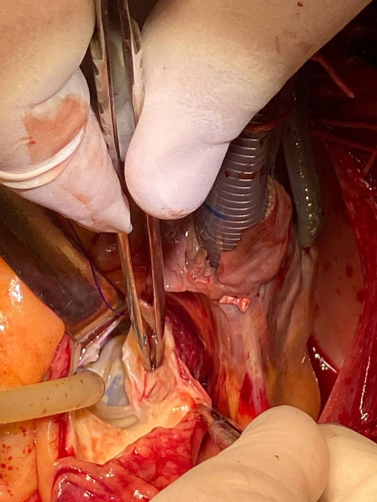
Figure 4
Intraoperative visualization of the perforation of the noncoronary aortic sinus.
Question 3
What role does timely surgical intervention play in patient survival after iatrogenic aortic perforation?
It is optional if autologous reinfusion is performed
It prevents the need for pericardiocentesis
It ensures complete recovery without any complications
It provides definitive repair and is critical for survival after initial stabilization with autologous reinfusion
Question 3
Consider the options and find the answer in this video quiz; see also at https://youtu.be/cUQr009258g.
Discussion
Coronary and aortic perforation during PCI are exceedingly rare and serious complications of the procedure. Advanced age, coronary tortuosity, calcification of the vessel, and previous cardiac surgical procedures can all predispose to weakness of the vessel wall and rupture of the vessel with even minimal manipulation. Other potential related complications include aortic dissection, coronary dissection, coronary dissection-associated myocardial infarction, and cardiac tamponade. One-third of such patients require emergency surgery, with an associated mortality rate of 50%.2
The classical approach for treatment of this complication is pericardiocentesis along with either percutaneous stenting or surgical repair of the perforated coronary artery. In this case report, a 47-year-old man who underwent elective PCI complicated by aortic perforation was emergently resuscitated with pericardiocentesis and autologous blood reinfusion followed by surgical repair.
A case series published in 2019 by Fiocca and colleagues describes the use of autologous blood reinfusion during iatrogenic acute hemorrhagic cardiac tamponade in 30 patients over a span of 10 years at one hospital. Importantly, autologous blood reinfusion was administered in a similar manner to the technique reported in our current case report, with blood drained from the pericardium and reinfused through the femoral vein, with a mortality rate of 10% when autologous blood reinfusion was used.3 Fiocca and colleagues concluded, “In the complex clinical scenario of acute tamponade occurring during catheter-based cardiac procedures, autotransfusion of pericardial blood through a femoral vein is safe and effective. It can be a useful trick up the sleeve of the interventional cardiologist.” Our case report is novel because it documents that this life-saving technique can even help salvage a patient with an acute iatrogenic aortic perforation.
Conclusion
Autologous blood reinfusion is an effective treatment in the emergent setting for acute cardiac tamponade, including tamponade associated with iatrogenic aortic perforation. In our presented case, no other options existed to treat the life-threatening bleeding and rapid cardiac deterioration seen in our patient. Despite concerns associated with the reintroduction of unfiltered blood into the systemic circulation, autologous blood reinfusion helped stabilize and salvage our critically ill patient. In this case report, we document that autologous blood reinfusion is a safe and effective bridge to definitive treatment in the setting of PCI associated with iatrogenic aortic perforation, especially in the absence of available percutaneous closure devices and immediately available on-call cardiothoracic surgeons. As illustrated here, knowledge and proper implementation of this technique can be lifesaving.
CME Credit Opportunity
Houston Methodist is accredited by the Accreditation Council for Continuing Medical Education (ACCME) to provide continuing medical education for physicians.
Houston Methodist designates this enduring material for a maximum of 1 AMA PRA Category 1 Credit™. Physicians should claim only the credit commensurate with the extent of their participation in the activity. Click to earn CME credit: https://learn.houstonmethodist.org/MDCVJ-21.1-1705.
Competing Interests
The authors have no competing interests to declare.