
Figure 1
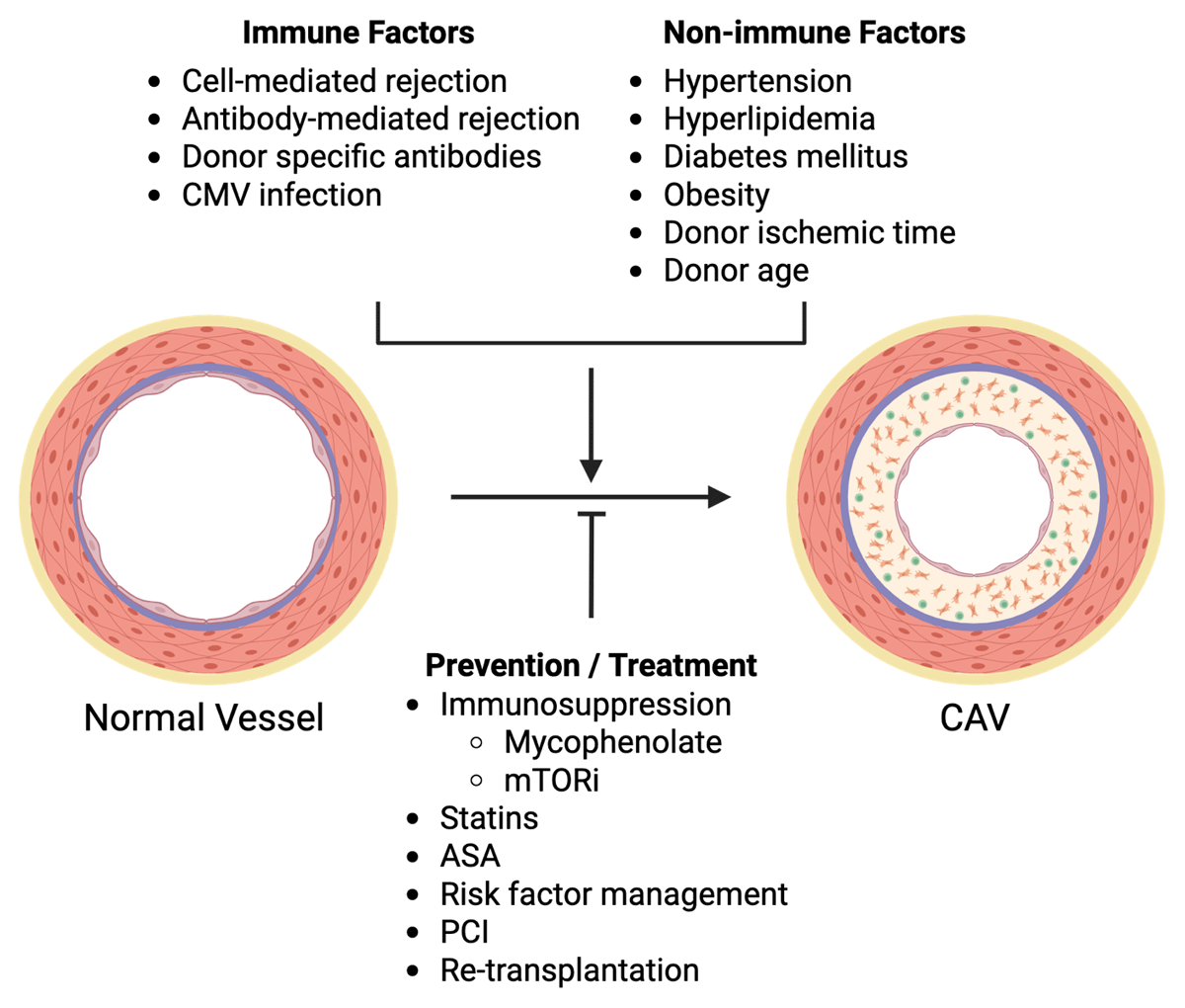
Risk factors and treatment of cardiac allograft vasculopathy. Multiple immune risk factors and nonimmune risk factors contribute to the development and progression of cardiac allograft vasculopathy (CAV), which is defined by intimal hyperplasia due to immune infiltration and immune signaling/activity, myofibroblast proliferation, and collagen deposition, ultimately restricting coronary luminal diameter. CMV: cytomegalovirus; mTORi: mammalian target of rapamycin inhibitors (including sirolimus and everolimus); ASA: aspirin; PCI: percutaneous coronary intervention. Image created with Biorender
Table 1
Guideline-based grading of CAV by coronary angiography and intravascular ultrasound.
| CAV SEVERITY | ISHLT GRADING BY CA* | STANFORD CLASSIFICATION BY IVUS-DERIVED INTIMAL THICKNESS** |
|---|---|---|
| None/Minimal | CAV0: No lesion with normal allograft function | I: < 0.3 mm and < 180° |
| Mild | CAV1: LM < 50% or primary vessel < 70% or branch stenosis < 70% with normal allograft function | II: < 0.3 mm and > 180° |
| Moderate | CAV2: LM < 50% and single primary vessel > 70% or at least 2 branch vessels > 70% with normal allograft function | III: 0.3-0.5 mm or > 0.5 mm and < 180° |
| Severe | CAV3: LM > 50% or 2 primary vessels > 70% or branch vessels > 70% in all coronary distributions; or CAV1 or CAV2 with allograft dysfunction | IV: > 1.0 mm or > 0.5 mm and > 180° |
Table 2
Imaging modalities for the detection of cardiac allograft vasculopathy.
| MODALITY | ADVANTAGE | DISADVANTAGE | GUIDELINE INDICATION* | GUIDELINE RECOMMENDATION* |
|---|---|---|---|---|
| Invasive | ||||
| Coronary angiography | Widely available Traditional gold standard for epicardial CAV | Low sensitivity for early CAV Iodinated contrast Procedural risk | Diagnosis and grading based on ISHLT guidelines | Class 1 |
| Intravascular ultrasound | Improved detection of early CAV Vessel anatomy | Moderate resolution (compared to OCT) Increased cost Procedural risk | Early detection of donor-derived CAD and early progressive CAV | Class 2a |
| Optical coherence tomography | Improved detection of early CAV Detailed vessel anatomy with highest resolution | Additional iodinated contrast Increased cost Procedural risk | Early detection of donor-derived CAD | Class 2a |
| Coronary physiology | Improved sensitivity for small vessel CAV | Increased cost and time Procedural risk | Intracoronary flow to detect small vessel CAV | Class 2a |
| Noninvasive | ||||
| Cardiac positron emission tomography | Lower radiation than coronary angiography No iodinated contrast Accuracy to diagnose moderate-severe CAV, and possibly early mild CAV | Lower availability than other modalities Requires expertise for interpretation | Noninvasive detection of CAV and prognostication | Class 2a |
| Cardiac magnetic resonance imaging | No radiation or iodinated contrast Accuracy to diagnose moderate-severe CAV Early detection of myocardial fibrosis | Low sensitivity for mild CAV Lower availability than other modalities Requires significant expertise for interpretation Gadolinium-based contrast may be discouraged in ESRD | Noninvasive detection of CAV and prognostication | Class 2b |
| Stress echocardiography | Widely available Low cost No radiation or iodinated contrast | Low sensitivity for the detection of CAV | Prognostication | Class 2a |
| Coronary computed tomography angiography | Alternative to ICA for epicardial disease Able to detect moderate-severe CAV | No information on small vessel disease Requires lower heart rate for optimal images Iodinated contrast | CAV detection in ≥ 2 mm epicardial vessels | Class 2a |
[i] *Based on the 2023 ISHLT guidelines for the care of heart transplant recipients;54 CAD: coronary artery disease; CAV: cardiac allograft vasculopathy; OCT: optical coherence tomography; ESRD: end-stage renal disease; ICA: invasive coronary angiography
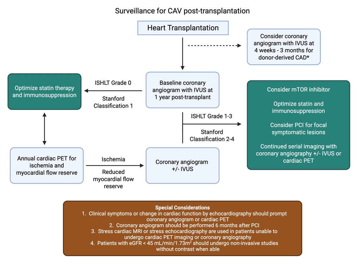
Figure 2
Diagnostic algorithm for the surveillance of cardiac allograft vasculopathy (CAV) post-transplantation. This is an adaptation of the CAV surveillance protocol at our institution. Following heart transplantation, CAV screening typically starts at 1-year post-transplantation with coronary angiography with IVUS to assess for early CAV. Early coronary angiography between 4 weeks and 3 months post-transplantation can be considered if there are risk factors for donor-derived coronary artery disease. For patients with ISHLT grade 0 or Stanford classification 1 CAV (see Table 1), optimization of statin therapy and immunosuppression is preferred. These patients are screened for progression of CAV with serial noninvasive imaging, such as cardiac PET (alternatives include cardiac MRI or stress echocardiography, depending on institutional expertise). If serial cardiac PET imaging reveals ischemia or reduced myocardial flow reserves, patients undergo coronary angiography with or without IVUS. When ISHLT grade 1-3 or Stanford classification 2-4 CAV is detected, mTOR inhibition is considered, PCI may be performed for symptomatic, focal, lesions, and serial imaging is continued with either coronary angiography or cardiac PET imaging. Certain special considerations are listed above. CAV: cardiac allograft vasculopathy; IVUS: intravascular ultrasound; ISHLT: International Society for Heart and Lung Transplantation; PET: positron emission tomography; mTOR: mammalian target of rapamycin; PCI: percutaneous coronary intervention. Image created with Biorender