
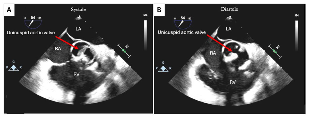
Figure 1
Intraoperative transesophageal echocardiogram in 2-dimenstional short-axis view showing calcification of the aortic valve with severe aortic stenosis. (A) At systole: aortic valve open; (B) at end diastole. RV: right ventricle, LA: left atrium, RA: right atrium
Video 1
Intraoperative transesophageal echocardiogram in 2D short-axis view showing calcification of the aortic valve with severe aortic stenosis; see also at https://youtu.be/2WuRKqMCZ_o.
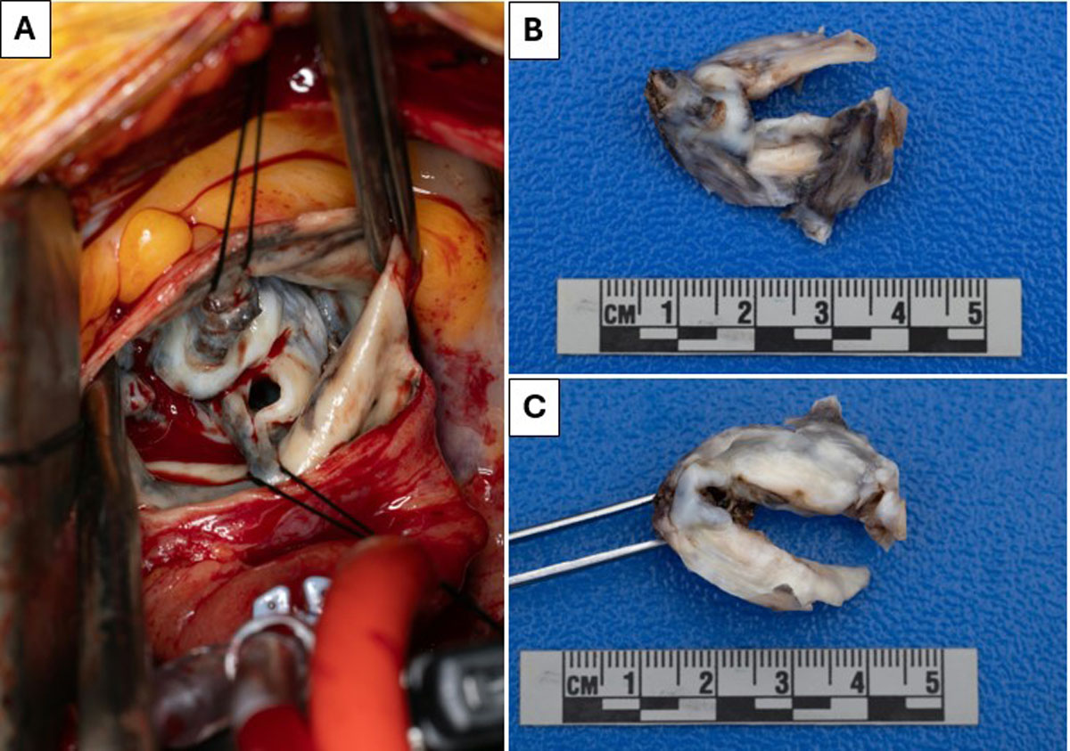
Figure 2
(A) Intraoperative calcified and blackened unicuspid aortic valve. (B, C) Macroscopic view of the unicuspid aortic valve post-excision.
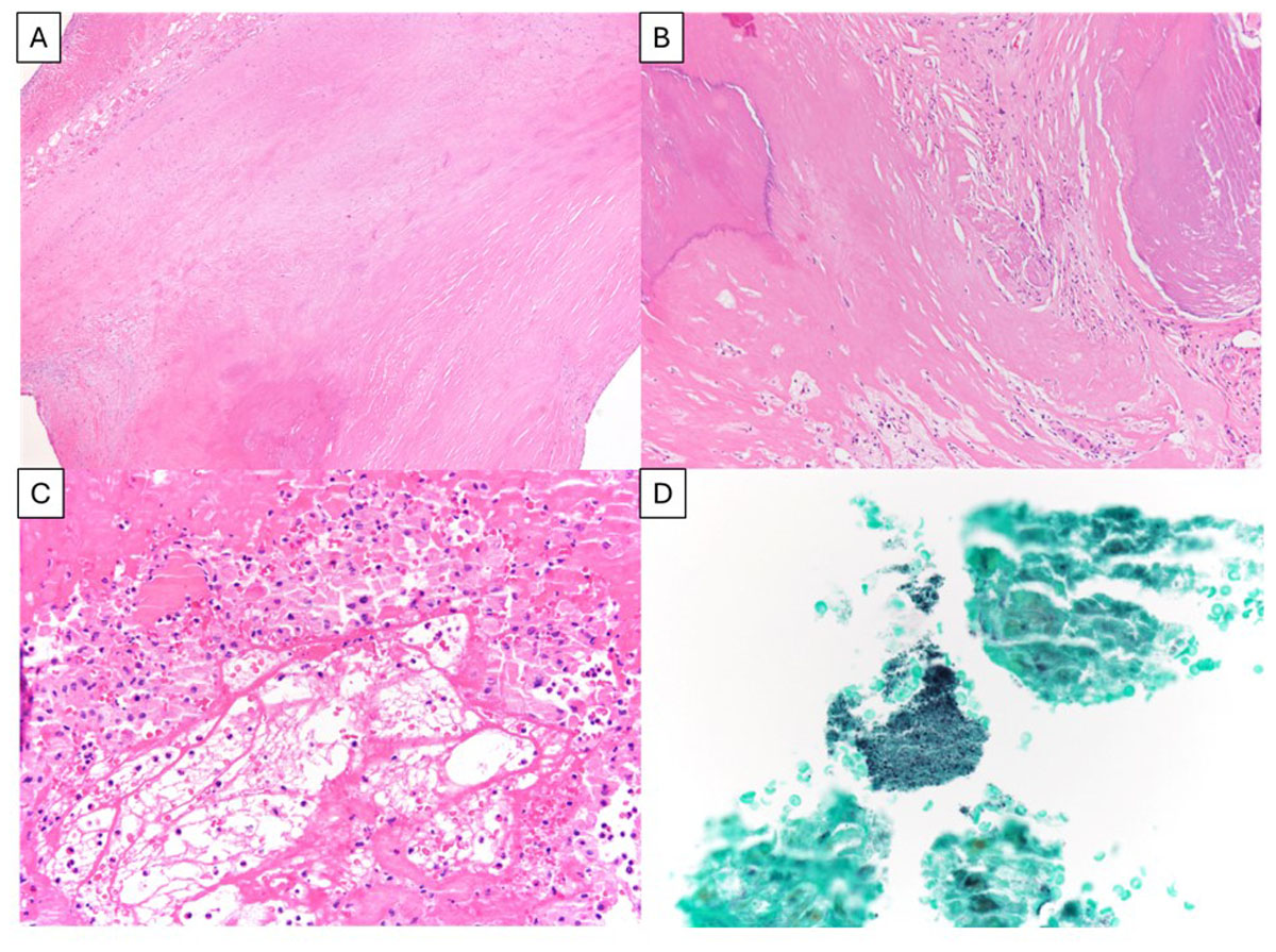
Figure 3
Dense fibrosis of the aortic valve. (A) Original magnification with 40x and (B) 100x calcified deposits. Acute, fibrinous endocarditis was also present, with bacterial colonies highlighted by Grocott–Gömöri’s methenamine silver stain at (C, D) 200x and 400x, respectively. A-C: hematoxylin and eosin staining