
Background
Blue-black discoloration of connective tissue can be associated with prolonged minocycline use, rarely involving cardiac tissue. Minocycline is often used for gram-positive and gram-negative bacterial infections as well as severe acne. Although long-term use of minocycline is common, the number of reported cases of pigmentation caused by the drug is exceedingly rare, and most describe this pigmentation after > 10 years of minocycline administration. The effect on cardiovascular tissue is often an incidental intraoperative finding, as in this case. The question remains whether this pigmentation can impact the functional capacity of the valves. Most reported cases did not demonstrate functional impairment of blackened valves. However, some suggested that the discoloration may impact valve visibility both preoperatively and intraoperatively by distorting the normal appearance of the valves, thus hindering proper excision and placement of sutures.
This case uniquely demonstrates significant cardiovascular hyperpigmentation following only 2 years of minocycline therapy. Future studies on minocycline-induced hyperpigmentation are essential to better understand its impact on rapid valvular calcification and degeneration. Detailed reports of cardiovascular issues related to minocycline use are necessary, as awareness of the condition is limited.
Introduction
Blue-black discoloration of tissues can impact various organ systems, including the joints, kidneys, and skin, with different underlying pathogenesis. Minocycline-induced hyperpigmentation (MIH), refers to the development of such discoloration following prolonged use of minocycline.1 In this case report we describe the first case seen with significant cardiac hyperpigmentation after only 2 years of daily minocycline use. We also discuss potential differentials that need to be considered when encountering this phenomenon in the operating room.
Clinical Vignette
A 47-year-old female with a history of cardiology follow-up since age 20 and known aortic valve disease presented to our institution complaining of worsening exertional dyspnea. Her past medical history was significant for acne, for which she was medicated with minocycline 100 mg per oral daily for approximately 2 years. On physical examination, it was noted that her upper and lower teeth had an underlying blue hue. Her echocardiogram showed calcified aortic valve leaflets and trivial aortic regurgitation. There was severe aortic stenosis with a mean transaortic gradient of 46 mm Hg, a calculated valve area of 0.85 cm2, and a left ventricular ejection fraction (LVEF) of 68%. The cusps were poorly visualized, raising suspicion for a bicuspid aortic valve.
Considering her symptoms and aortic stenosis, it was agreed to proceed with an elective aortic valve replacement (AVR) (Figure 1).
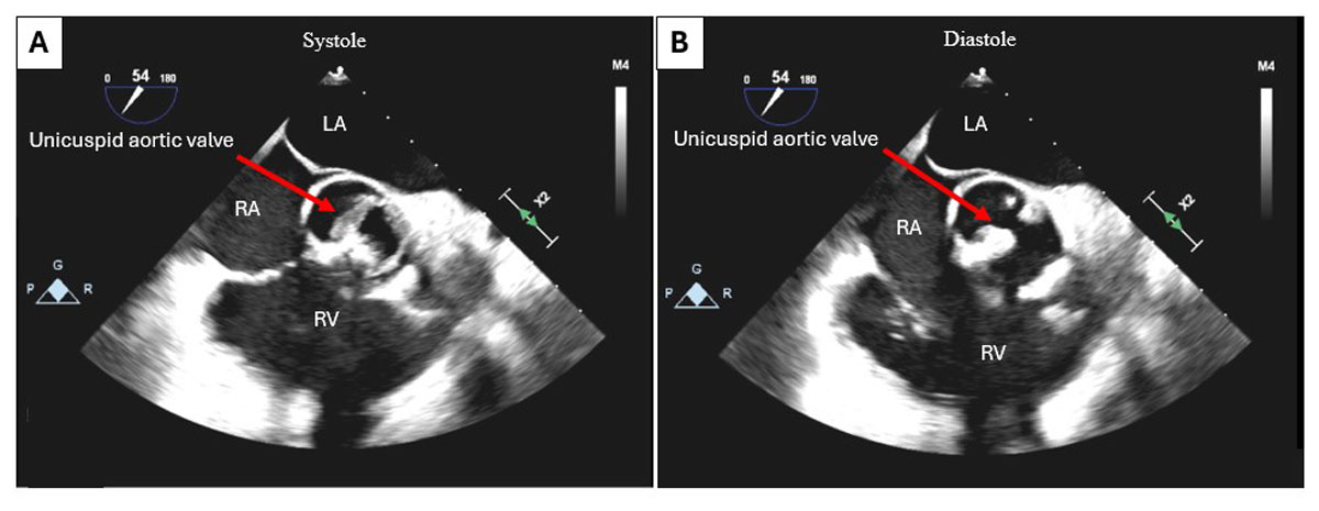
Figure 1
Intraoperative transesophageal echocardiogram in 2-dimenstional short-axis view showing calcification of the aortic valve with severe aortic stenosis. (A) At systole: aortic valve open; (B) at end diastole. RV: right ventricle, LA: left atrium, RA: right atrium
In September 2024, the patient underwent minimally invasive AVR with a 21-mm On-X mechanical valve (Artivion, Inc.) and sternal plating. An intraoperative transesophageal echocardiogram was performed and confirmed preserved LVEF, severe aortic valve stenosis, and indeterminate number of cusps with mild aortic regurgitation (Figure 1; Video 1). The intraoperative visualization of the aortic valve revealed a unicuspid nature, with extensive black discoloration on the valve and endocardium of left ventricle and aorta (Figure 2). The operation had no complications, and the patient was extubated in the operating room and transferred to the intensive care unit for postoperative management.
Video 1
Intraoperative transesophageal echocardiogram in 2D short-axis view showing calcification of the aortic valve with severe aortic stenosis; see also at https://youtu.be/2WuRKqMCZ_o.
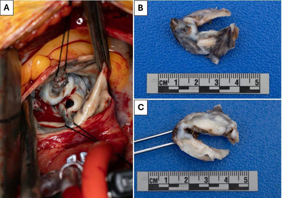
Figure 2
(A) Intraoperative calcified and blackened unicuspid aortic valve. (B, C) Macroscopic view of the unicuspid aortic valve post-excision.
Outcomes and Follow-Up
On light microscopy using hematoxylin and eosin staining, examination of the excised valves showed dense fibrosis of the aortic valve with calcified deposits as seen in Figure 3. On day 6, the patient was eligible for home discharge; the patient is now asymptomatic and well, regularly following up with cardiology.
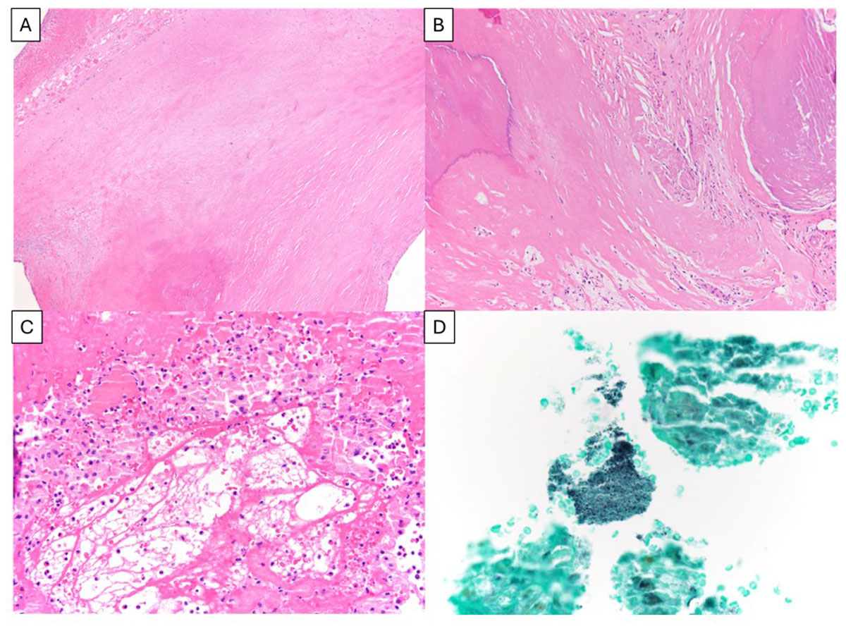
Figure 3
Dense fibrosis of the aortic valve. (A) Original magnification with 40x and (B) 100x calcified deposits. Acute, fibrinous endocarditis was also present, with bacterial colonies highlighted by Grocott–Gömöri’s methenamine silver stain at (C, D) 200x and 400x, respectively. A-C: hematoxylin and eosin staining
Discussion
Minocycline belongs to the tetracycline family of antibiotics and is often used for gram-positive and gram-negative bacterial infections as well as severe acne.2 Although long-term use of minocycline is common, the number of reported cases of pigmentation caused by the drug is exceedingly rare.3 Binetz et al. published the first article that described minocycline’s effect on thyroid tissue discoloration in animal models.4 Recently, a few case reports were published describing a rare phenomenon whereby MIH involved the cardiovascular structures.1,2,3,5,6,7,8,9
The effect on the cardiovascular tissue is often an intraoperative incidental finding, as in this case. In 2022, O’Conell et al. described a case of a 71-year-old male who underwent an AVR with an incidental finding of blackened aortic valve and aorta.7 Another case report highlighted a 29-year-old liver transplant patient who was found to have a discolored aorta and cardiac valves.5
Tsunekawa et al. in 2014 described a case of a 65-year-old female with hyperpigmented aortic valve and the sinuses of Valsalva from long-term minocycline use.2 Most of the available literature describes MIH after prolonged minocycline use reported after > 10 years of minocycline administration.1,2,3,5,6,7,8,9
When encountering such a case, several differentials can be considered. One possibility that is well documented in literature is ochronosis, or the black discoloration of tissues caused by alkaptonuria. Alkaptonuria is an uncommon autosomal recessive metabolic disease characterized by a deficiency in homogentisate oxidase. Although less common, cardiovascular ochronosis can occur and has shown a predominance in patients with calcific aortic stenosis.10 It has been suggested that the presence of the ochronotic pigment leads to greater calcification and causes issues with valve function.11 Another differential to be considered is heavy metal tissue deposition.1
The question remains whether this pigmentation can impact the functional capacity of the valves. Tsunekawa et al. proposed that this MIH does not have detrimental effects, as their patient did not have any problems with the prosthetic valve a year after the AVR.2 Some suggested that the discoloration may impact valve visibility both preoperatively, as in our case, and intraoperatively by distorting the normal appearance of the valves, which hinders proper excision and placement of sutures.7 In misidentification instances, treatment may begin based on an inaccurate diagnosis. Since some conditions on the differential list necessitate prompt surgical intervention, misdiagnosing this otherwise harmless condition could harm patients.
Consequently, detailed reports of cardiovascular issues related to minocycline use are essential to ensure this possibility is considered in future cases, as awareness of the condition is limited.
Conclusion
Healthcare providers should include MIH in their differential diagnosis for patients with a history of chronic minocycline use who exhibit superficial signs of black or blue pigmentation and display black discoloration in cardiovascular structures.
Competing Interests
The authors have no competing interests to declare.