
INTRODUCTION
Pulmonary hypertension (PH) is a notable complication of chronic parenchymal lung disease. Chronic lung disease (CLD)-related PH is most commonly caused by chronic obstructive pulmonary disease and interstitial lung diseases. Other rarer parenchymal lung diseases can also cause PH, including combined pulmonary fibrosis and emphysema (CPFE), lymphangioleiomyomatosis, sarcoidosis, and pulmonary Langerhans cell histiocytosis. Lung diseases fall under group 3 (due to CLD and/or chronic hypoxia) and group 5 (unclear and/or multifactorial mechanisms) within the most recent PH clinical classification system (see PH clinical classifications in the manuscript by Beshay et al. on page 9 of this issue).1,2
Based on the 2019 update of the 6th World Symposium on Pulmonary Hypertension, PH is defined as having a mean pulmonary artery pressure (mPAP) ≥ 20 mm Hg in the supine position at rest as measured on right heart catheterization (RHC).2 Similar to those with group 1 PH due to pulmonary arterial hypertension (PAH), patients with lung-disease–related PH have a precapillary hemodynamic profile that includes an elevated pulmonary vascular resistance (PVR) > 3 Wood Units (WU) and a normal pulmonary arterial wedge pressure (PAWP) or left ventricular end-diastolic pressure ≤ 15 mm Hg.3 When present, PH causes more severe symptoms and exercise intolerance and portends a worse prognosis in patients with CLD.4
It can be clinically challenging to differentiate patients with PH due to lung disease from those with PAH and comorbid mild lung disease (Figure 1). The principle of managing PH due to CLD is primarily to optimize treatment of the underlying disorder and to identify and manage other potential contributing factors such as concurrent left heart dysfunction, sleep-disordered breathing, and chronic thromboembolic disease.3 However, histopathologic evidence of pulmonary vascular remodeling and similar precapillary hemodynamics in patients with CLD-associated PH and those with PAH5,6 has spurred interest in the use of PAH therapies in group 3 PH. It is important to note that oral therapies for PAH have not been approved for use in PH due to CLD, and there is a lack of evidence to support their efficacy. Furthermore, there is concern that by inducing vasodilation in poorly ventilated areas of the lung, PAH therapies can worsen ventilation-perfusion matching and further exacerbate arterial hypoxemia.7 Therefore, caution must be exercised in the use of PAH therapies in patients with CLD-associated PH. Treatment decisions should be carefully considered solely in specialized PH referral centers with extensive experience in the use of these therapies or in the context of clinical trials. This article reviews the current evidence pertaining to specific therapies for PH associated with CLD.
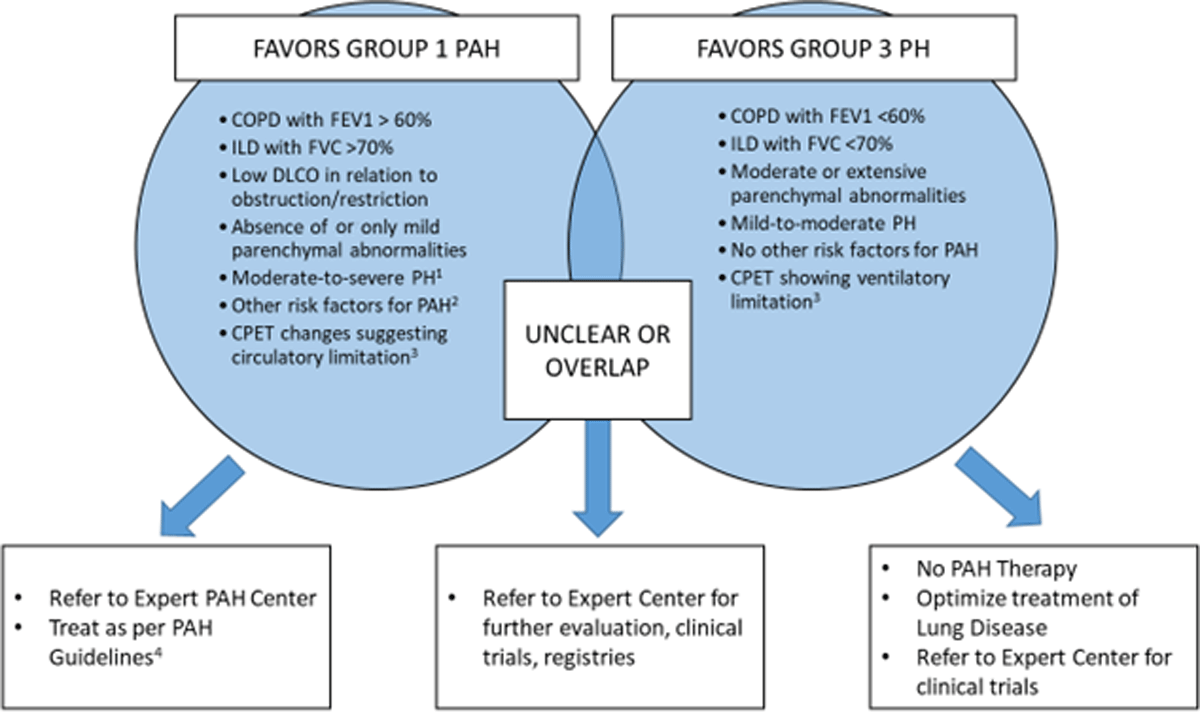
Figure 1
Differentiation between group 1 pulmonary hypertension (PH) and group 3 PH.1 Figure courtesy of authors. COPD: chronic obstructive pulmonary disease; FEV1: forced expiratory volume in 1 second; FVC: forced vital capacity; ILD: interstitial lung disease; PAH: pulmonary arterial hypertension; DLCO: carbon monoxide diffusing capacity test; CPET: cardiopulmonary exercise test
1 Moderate-to-severe PH defined as mean pulmonary arterial pressure ≥ 35 mm Hg, or ≥ 25 mm Hg with a cardiac index < 2.0 L/min/m2
2 Other risk factors include connective tissue disease, portal hypertension, human immunodeficiency virus infection, exposure to definite or probable medications that cause group 1 PH, genetic mutations, congenital heart disease
3 Features of circulatory limitation include preserved breathing reserve, low oxygen pulse, low cardiac output/VO2 slope, no change or decrease in PaCO2 during exercise. Features of ventilatory limitation include low breathing reserve, normal oxygen pulse, normal cardiac output/VO2 slope, increase in PaCO2 during exercise, dynamic hyperinflation.
PULMONARY HYPERTENSION ASSOCIATED WITH CHRONIC OBSTRUCTIVE PULMONARY DISEASE
According to international guidelines, optimization of underlying chronic obstructive pulmonary disease (COPD) is the primary approach for managing COPD-associated PH.8 The pathophysiologic mechanisms of PH in COPD include airflow limitation, hyperinflation and compression of alveolar capillary vessels, alveolar hypoxia, vasoconstriction, and destruction or remodeling in the pulmonary vascular bed, which can all be present in varying degrees, ultimately leading to elevated PVR and mPAP.9,10,11 Supplemental oxygen therapy remains a key component of COPD management in patients with resting hypoxemia (PaO2 < 60 mm Hg or < 55 mm Hg in the presence of PH) because continuously administered oxygen for at least 18 hours per day has been shown to increase survival, improve PVR and cardiac function, and possibly slow the progressive increase in mPAP.12 Patients with right heart failure and edema may be managed with diuretics and sodium restriction. Due to the poor associated prognosis, moderate-to-severe PH in the setting of COPD is considered a criterion for lung transplantation in otherwise eligible patients.13
There is significant interest in the use of PAH therapies for PH in COPD, particularly in patients with severe PH, defined as mPAP ≥ 35 mm Hg or mPAP ≥ 25 mm Hg with a cardiac index < 2.0 L/min/m2.3,8 Severe PH is rare in COPD and often caused by concomitant left ventricular dysfunction or other pulmonary diseases; however, approximately 1% of cases have severe PH in the absence of another confounding factor. These patients may potentially have a “pulmonary vascular phenotype” of COPD and exhibit severe PH and a cardiovascular limitation to exercise without severe airflow obstruction.14,15 Therefore, pulmonary vasodilator therapies could be of interest in this subgroup population with severe hemodynamic impairment and lower concern for ventilation-perfusion mismatch and hypoxemia, although evidence to support the use of PAH therapies in this context is limited.14,15,16
Most randomized controlled trials (RCTs) evaluating PAH therapies in COPD-associated PH have tested phosphodiesterase-5 inhibitors (PDE5i), which have mostly demonstrated equivocal results, with no mortality or consistent functional benefit (Table 1).17,18,19,20,21,22,23 An RCT conducted by Vitulo et al. compared sildenafil with placebo to determine the long-term hemodynamic effects of sildenafil in COPD patients with severe PH. They demonstrated a significant reduction in PVR as well as significant improvements in dyspnea and quality of life scores, both of which were secondary outcomes. However, there was no change in PaO2 or 6-minute walk distance (6MWD).21 A recent meta-analysis by Hao et al. noted modest improvements in 6MWD in patients treated with sildenafil but no change in reported dyspnea or functional symptoms.22 Further studies of sildenafil or other PDE5i would be of interest in the specific subgroups of patients with mild COPD and severe PH.
Table 1
Randomized trials of therapies proposed in the management of chronic obstructive pulmonary disease (COPD)-associated pulmonary hypertension (PH). Table courtesy of authors. PDE5: phosphodiesterase type-5; TID: three times a day; sPAP: systolic pulmonary artery pressure; 6MWD: 6-minute walk distance; VO2: oxygen uptake; QoL: quality of life; mPAP: mean pulmonary artery pressure; BNP: brain natriuretic peptide; PVR: pulmonary vascular resistance; WU: Wood units; BODE: body mass index, obstruction, dyspnea, exercise capacity index; CI: cardiac index; SF-36: Short Form 36; BID: twice daily
| AUTHOR | REF | N | DRUG | SEVERITY OF PH | FOLLOW-UP PERIOD | END POINT(S) |
|---|---|---|---|---|---|---|
| PDE5 INHIBITORS | ||||||
| Rao et al., 2011 | 17 | 33 | Sildenafil 20 mg TID vs placebo | sPAP > 40 mm Hg on echo | 12 weeks | 6MWD: +190 m vs placebo sPAP: decreased |
| Lederer et al., 2012 | 18 | 10 | Sildenafil 25 mg TID vs placebo | No PH | 4 weeks | 6MWD: no effect VO2: no effect QoL: worsened |
| Blanco et al., 2013 | 19 | 60 | Sildenafil 20 mg TID vs placebo in patients undergoing pulmonary rehabilitation | mPAP 31 ± 5 mm Hg | 12 weeks | Cycle endurance time: no
effect 6MWD: no effect VO2: no effect |
| Goudie et al., 2014 | 20 | 120 | Tadalafil 120 mg daily vs placebo | sPAP 42 ± 10 mm Hg (as estimated on echocardiogram) | 12 weeks | sPAP: decreased –12.3 mm
Hg 6MWD: no effect QoL: no effect BNP: no effect |
| Vitulo et al., 2017 | 21 | 28 | Sildenafil 20 mg TID vs placebo | mPAP 25 mm Hg in 68% mPAP 39 ± 8 mm Hg PVR 7 ± 2.6 WU | 16 weeks | PVR: –1.38 WU CI: +0.42 L/min/m2 BODE: improved 6MWD: no effect SF-36: +9.85 units |
| ENDOTHELIN RECEPTOR ANTAGONISTS | ||||||
| Stoltz et al., 2008 | 23 | 30 | Bosentan 62.5 mg BID for 2 weeks followed by 125 mg BID vs placebo | mPAP at rest ≥ 30 mm Hg | 12 weeks | 6MWD: -10 m vs placebo |
| Valerio et al., 2009 | 24 | 32 | Bosentan 125 mg BID vs placebo | mPAP 37 ± 5 mm Hg | 18 months | 6MWD: +65 m mPAP: – 6 mm Hg PVR: –50 dynes·sec·cm–5 BODE index: improved |
Treatment with endothelin receptor antagonists such as bosentan, ambrisentan, or macitentan is not supported by existing evidence.23,24 Similarly, while studies have been conducted on calcium-channel blockers, inhaled nitrous oxide, inhaled and intravenous prostacyclins, and guanylate cyclase stimulators in the context of COPD-associated PH, no studies support their routine use in these patients and they are not approved by regulatory bodies for this indication.3
Inhaled vasodilators have theoretical advantages in terms of avoiding worsening ventilation-perfusion mismatch, but the data are mixed. In patients with COPD and severe PH, inhaled nitric oxide (NO) may actually worsen gas exchange by inhibiting hypoxic vasoconstriction.25,26 Conversely, more recent studies suggest some benefit of NO inhalation on pulmonary vascular function in mild COPD without PH and in patients with PH.27,28 In nonrandomized studies of COPD patients with PH, inhaled iloprost (a prostacyclin analogue) improved hemodynamics and ventilatory efficiency without a detrimental effect on gas exchange.29,30
PULMONARY HYPERTENSION ASSOCIATED WITH INTERSTITIAL LUNG DISEASES
As with COPD, the treatment of PH associated with interstitial lung diseases (ILD) consists of optimizing the underlying condition.3 ILD-related PH is most frequently seen in patients with idiopathic pulmonary fibrosis (IPF). The main mechanism of PH in ILD is due to parenchymal fibrosis and vascular bed destruction, although other concomitant factors can exist, including venous thromboembolism, sleep-disordered breathing, and left heart dysfunction with postcapillary PH.31,32,33,34 The only approved pharmacologic therapies for treating IPF are the antifibrotic agents pirfenidone and nintedanib, which slow the rate of disease progression but have shown no effect on the development or progression of pulmonary vascular disease.
There has been much effort in testing PAH-targeted therapies for patients with IPF and other ILDs (Table 2). Unfortunately, multiple RCTs have demonstrated no benefit of endothelin receptor antagonists.35,36,37,38 Rather, there have been major safety concerns, as one trial using ambrisentan was terminated early due to increased risk of harm in the treatment group.37 The recent RISE-IIP study (riociguat for idiopathic interstitial pneumonia-associated pulmonary hypertension) was a phase 2b placebo-controlled RCT examining the effectiveness of riociguat (a soluble guanylate cyclase stimulator) in patients with ILD-related PH. This study was also terminated early because it demonstrated serious adverse events and increased mortality in the treatment group (11 deaths with riociguat and 3 with placebo)39; therefore, riociguat is not recommended for ILD-related PH.
Table 2
Randomized trials of therapies proposed in the management of interstitial lung disease–associated pulmonary hypertension (PH). Table courtesy of authors. IPF: idiopathic pulmonary fibrosis; BID: twice daily; RVSP: right ventricular systolic pressure; 6MWD: 6-minute walk distance; QoL: quality of life; FVC: forced vital capacity; TID: three times daily; ILD: interstitial lung disease; IIP: idiopathic interstitial pneumonia; IBW: ideal body weight
| AUTHOR | REF | N | LUNG DISEASE | DRUG | PH PATIENTS INCLUDED? | FOLLOW-UP PERIOD | END POINTS |
|---|---|---|---|---|---|---|---|
| ENDOTHELIN RECEPTOR ANTAGONISTS | |||||||
| King et al., 2008 | 35 | 158 | IPF | Bosentan 62.5 mg BID for 4 weeks followed by 125 mg BID vs placebo | Yes: mild PH on echo (RVSP < 50 mm Hg) included | 12 months | 6MWD: no difference Disease progression: favors bosentan Lung function: no difference Dyspnea and QoL: favors bosentan |
| King et al., 2011 | 36 | 616 | IPF | Bosentan 62.5 mg BID for 4 weeks followed by 125 mg BID vs placebo | Yes | 20 months | Time to IPF worsening or death: no
difference QoL: no difference Dyspnea: no difference Change in FVC: no difference |
| Raghu et al., 2013 | 37 | 494 | IPF | Ambrisentan 10 mg daily vs placebo | Yes, but excluded patients on other long-term PH therapies | 34 weeks | Time to IPF progression: terminated early
due to higher disease progression in ambrisentan
group Hospitalizations: increased in ambrisentan group 6MWD: no difference Lung function decline: no difference |
| Raghu et al., 2013 | 38 | 494 | IPF | Macitentan 10 mg vs placebo | Yes | 12 months | Change in FVC: no difference Time to IPF worsening or death: no difference Dyspnea: no difference Adverse events: no difference |
| SOLUBLE GUANYLATE CYCLASE STIMULATORS | |||||||
| Nathan et al., 2019 | 39 | 147 | IIP | Riociguat, up to 2.5 mg TID vs placebo | Yes | 26 weeks | 6MWD: no difference Time to clinical worsening: no difference |
| PDE5 INHIBITORS | |||||||
| Zisman et al., 2010 | 40 | 180 | IPF | Sildenafil 20 mg TID vs placebo | Yes | 12 weeks | 6MWD improvement of > 20%: no significant difference |
| Han et al., 2013 | 41 | 119 | IPF | Sildenafil 20 mg TID vs placebo | Yes | 12 weeks | 6MWD improvement of > 20%: no
significant difference QoL improvement: no significant difference |
| Behr et al., 2020 | 42 | 177 | IPF | Sildenafil 20 mg TID with pirfenidone vs. placebo with pirfenidone | Yes | 52 weeks | Progression-free survival: no significant
difference 6MWD decline < 15%: no significant difference |
| INHALED MEDICATIONS | |||||||
| Nathan et al., 2020 | 43 | 41 | Fibrosing ILD | Inhaled NO 30–45 μg/kg IBW/h vs. placebo | Subjects stratified as low-, intermediate- and high-probability of PH. Subjects included if PH proven on RHC. | 8 weeks | Moderate/ vigorous physical activity: statistically significant improvement |
| Waxman et al., 2020 | 47 | 326 | Any ILD | Treprostinil 60–72 μg inhaled vs. placebo | Yes | 16 weeks | Mean change from baseline in peak 6MWD
through week 16: +31.12 m (95% CI, 16.85–45.39) NT-proBNP: improved Time to clinical worsening: 39% reduction |
The STEP-IPF (Sildenafil Trial of Exercise Performance in Idiopathic Pulmonary Fibrosis) RCT evaluated sildenafil in IPF patients but was not specifically designed to enroll patients with IPF and PH.40 Although the STEP-IPF primary end point was not achieved, there was a signal for improved exercise tolerance and quality of life in the subgroup with baseline RV dysfunction.41 In contrast, a recent RCT demonstrated no benefit in the addition of sildenafil to pirfenidone in IPF patients with no firm diagnosis of PH.42 As with COPD patients, there is concern that pulmonary vasodilation with PDE5i may aggravate hypoxemia in patients with ILD. Finally, a recent phase 2b/3 RCT demonstrated a modest improvement in moderate/vigorous physical activity in patients with fibrotic ILD who are at risk for PH and who were treated with pulsed inhaled NO at 30 μg/kg ideal body weight/h over an 8-week period.43 Although the clinical implications of this remain unclear, inhaled vasodilators may have particular advantages and may mitigate concerns over worsening ventilation-perfusion matching.
Research on the management of ILD-associated PH has targeted patients with systemic sclerosis (SSc) due to its association with group 1 PAH. SSc causes PH through multiple mechanisms, including PAH (group 1), postcapillary PH secondary to left ventricular fibrosis and diastolic dysfunction (group 2), precapillary PH secondary to ILD (group 3), and pulmonary veno-occlusive disease.44 Although PAH therapies are effective in SSc-induced PAH, several observational studies have shown no benefit from PAH-targeted therapies when significant ILD is present. No improvements were seen in functional class or 6MWD, and even the presence of ILD is known to portend a worse prognosis.44,45 A recent observational study demonstrated modest hemodynamic improvements in SSc ILD-associated PH when treated with PAH therapies, but no significant improvement was seen in 6MWD, and only 13% of patients experienced improved symptoms.46 This dissociation between hemodynamic benefits, symptoms, and exercise capacity indicates that PH may not be the key mechanism of limitation in ILD patients because symptoms and exercise limitation are frequently multifactorial and related to restrictive lung physiology, impaired gas exchange, and peripheral factors (eg, deconditioning, myopathy, arthritis). Therefore, there is no clear evidence that patients with SSc and moderate-to-severe ILD with PH benefit from PAH therapies.
Research on inhaled PAH-targeted therapies may yield more promising results for improved exercise tolerance in patients with ILD-associated PH, and the therapeutic landscape may change in the coming years. The INCREASE (Safety and Efficacy of Inhaled Treprostinil in Adult PH With ILD Including CPFE) RCT tested the effect of inhaled treprostinil (a prostacyclin analogue) in patients with ILD. There were 326 participants with ILD of various etiologies randomized to inhaled treprostinil or a placebo four times daily. After 16 weeks of treatment, inhaled treprostinil improved placebo-corrected 6MWD (+31.12 m, 95% CI, 16.85–45.39) while also offering statistically significant improvements in secondary outcomes including N-terminal pro-brain natriuretic peptide and a 39% reduction in time to clinical worsening. Importantly, the effect of inhaled treprostinil on 6MWD was consistent across age groups, between men and women, and between patients with idiopathic interstitial pneumonias and connective tissue disease. There were some important insights from subgroup analyses that may help with patient selection. First, the effect of inhaled treprostinil was a 40.8 m (95% CI 24.1–57.6) improvement in 6MWD in patients with a PVR ≥ 4 WU but tended to be null in patients with a PVR < 4 WU. Additionally, there was a less impressive effect in those with 6MWD > 350 m at baseline and in patients with CPFE. The most frequent adverse events in the inhaled treprostinil group were cough (43.6%), headache (27.6%), and dyspnea (25.2%), which were similar to placebo. Throat irritation (12.3%) and oropharyngeal pain (11%) were more common with inhaled treprostinil, however. Serious adverse events, such as respiratory failure, pneumonia, or death, occurred in 23.3% of patients with inhaled treprostinil and 25.8% of patients with placebo.47
While there is now evidence supporting inhaled PAH therapies, data are still insufficient to recommend oral PAH therapies in ILD-associated PH, and they are not approved by regulatory bodies for this indication. Inhaled vasoactive therapies present a promising new strategy for ILD patients with PH since they may avoid the potentially detrimental effects of oral therapies on ventilation-perfusion matching and hypoxemia and could minimize systemic toxicities or side effects such as hypotension. Optimization of medical therapy should be the goal for ILD patients, including supplemental oxygen and antifibrotic agents such as nintedanib or pirfenidone, where appropriate.48 As with COPD, a diagnosis of PH in a patient with ILD should prompt consideration of lung transplantation.13
PULMONARY HYPERTENSION DUE TO OTHER RARE PARENCHYMAL LUNG DISEASES
Pulmonary Hypertension in Patients with Sarcoidosis
The management of PH in individuals with sarcoidosis is complex because the mechanisms of PH in these patients can be multifactorial, heterogeneous, and often combined. If precapillary PH is established in a patient with sarcoidosis, there are three primary mechanisms that may contribute, each with different treatment strategies: (1) granulomatous pulmonary vasculopathy; (2) vascular bed destruction from lung parenchymal disease; and (3) vascular remodeling due to chronic hypoxia (also in the case of severe lung parenchymal involvement).49 Postcapillary PH can be due to left ventricular involvement or progressive massive fibrosis, which can cause compression of the mediastinal vascular structures among other potential mechanisms.50 The presence of PH in patients with sarcoidosis is associated with worse functional capacity and survival.49,51,52
Overall, there is no evidence supporting the use of PAH-specific therapies in patients with sarcoidosis-associated PH.53 There has been only one RCT, which demonstrated that bosentan was effective at lowering mPAP and PVR at 16 weeks but had no significant effect on any functional parameters.54 Other observational studies have similarly reported improvements in hemodynamics with no improvement in functional parameters with oral or parenteral PAH therapies.52,55,56 Use of PAH therapies is not associated with better survival in observational studies,52,57 although this could be subject to confounding by indication since more severely impaired patients tend to be treated with off-label PAH drugs. Disease-specific immunosuppressive therapies can be considered in patients with sarcoidosis-associated PH caused by mechanical compression of pulmonary vasculature by large mediastinal lymph nodes, fibrosing mediastinitis, or proven pulmonary vascular granulomatous inflammation, though the specific role of these medications is not well established.53
Pulmonary artery angioplasty, with or without stenting of focal stenoses, can be a useful therapy in selected patients.58 A recent systematic review and meta-analysis found that angioplasty with or without stenting may lead to improvements in 6MWD,59 and this remains an option in some patients.
Pulmonary Hypertension in Patients with Pulmonary Langerhans Cell Histiocytosis
Pulmonary Langerhans cell histiocytosis (PLCH) is a rare lung disease caused by smoking and is characterized by the presence of Langerhans-like cells with stellate scar formation, interstitial fibrosis, parenchymal nodules, and lung cysts as well as airflow obstruction on pulmonary function testing. A process resembling pulmonary veno-occlusive disease caused by diffuse vasculopathy and fibrotic obliteration of the pulmonary veins can also occur and contribute to PH.60 Management of PH in PLCH focuses on treating the underlying lung disease, including smoking cessation and potentially cladribine as a chemotherapeutic agent.61 PAH-specific therapies have not been robustly studied in this rare condition. A small retrospective observational study demonstrated improvements in both functional and hemodynamic parameters in patients with PLCH-associated PH receiving PAH-specific therapies,62 but PAH therapies must be considered with caution given the risk of inducing pulmonary edema if pulmonary veno-occlusive changes are prominent.
Pulmonary Hypertension in Patients with Lymphangioleiomyomatosis
Lymphangioleiomyomatosis (LAM) is a rare condition that almost exclusively affects women and is characterized by diffuse cystic lung disease, chylous pleural effusions, ascites, renal angiomyolipomas, and lymphangioleiomyomas.63 The mechanism of PH largely lies in pulmonary vascular infiltration of LAM cells leading to elevated pulmonary arterial pressure. This occurs in 8% of all patients with LAM and 45% of patients with LAM who are referred for lung transplantation.64 LAM-associated PH has not been well studied, and the evidence for PAH therapies in these patients is primarily limited to case reports. One small study of 20 patients with LAM-associated PH, six of whom were treated with bosentan or sildenafil, demonstrated hemodynamic improvements but no improvement in functional parameters.65 Therefore, therapy should be directed at treating the underlying condition, with no specific recommendation for treatment of PH in these patients.63 Table 3 shows management approaches for PH associated with lung disease.
Table 3
Approach to management of lung disease–associated pulmonary hypertension (PH). Table courtesy of authors. COPD: chronic obstructive pulmonary disease; ILD: interstitial lung disease; WHO: World Health Organization
| SUGGESTED INTERVENTIONS FOR PATIENTS WITH GROUP 3 AND GROUP 5 PH1 | |
|---|---|
| All patients | Optimization of the underlying pulmonary condition, including smoking cessation and relevant disease-specific therapies |
| Supplemental O2 therapy in patients with resting hypoxemia | |
| Referral for lung transplantation, if eligible | |
| INTERVENTIONS THAT MAY BE CONSIDERED FOR PATIENTS WITH GROUP 3 AND GROUP 5 PH1 | |
| All patients | Referral to a PH expert center for consideration of invasive diagnostic testing and individualized therapy or enrollment in clinical trials |
| INTERVENTIONS THAT SHOULD NOT BE ROUTINELY CONSIDERED FOR PATIENTS WITH WHO GROUP 3 AND GROUP 5 PH1 | |
| All patients | Phosphodiesterase type-5 (PDE5) inhibitors, endothelin receptor antagonists (ERAs); calcium channel blockers; inhaled nitric oxide; inhaled and intravenous prostacyclins; guanylate cyclase stimulators |
| INTERVENTIONS THAT ARE CURRENTLY UNDER INVESTIGATION OR CONSIDERATION ON AN INDIVIDUALIZED OR EXPERIMENTAL BASIS FOR PATIENTS WITH GROUP 3 AND GROUP 5 PH | |
| COPD-associated PH | PDE5-inhibitors (sildenafil)21,22; inhaled treprostinil (NCT03496623); inhaled soluble guanate cyclase modulator (NCT04370873) |
| ILD-associated PH | Inhaled pulmonary vasodilators, including inhaled treprostinil47 |
| Sarcoidosis-associated PH | Bosentan52,53,55,56; pulmonary artery angioplasty, with or without stenting of focal stenoses58,59 |
CONCLUSION
PH is a known complication of CLDs, adversely affecting survival and contributing to impaired functional capacity. Despite promising results from observational studies and some signals of benefit in RCTs, the existing evidence does not support the use of PAH therapies in patients with CLD-related PH. Furthermore, some PAH therapies may be harmful in certain lung diseases, such as the experience with ambrisentan in IPF. Therefore, the core treatment strategy for PH is directed at medical optimization of the underlying lung disease, provision of supplemental oxygen to patients with hypoxemia, and lung transplantation referral in eligible patients. Attention should also be paid to diagnosing and treating other potential causes and aggravating factors for PH in this group, including left heart disease, sleep-disordered breathing, and thromboembolic disease. There is a need for well-designed and adequately powered clinical trials to investigate the use of PAH therapies, particularly inhaled agents, in patients with severe PH in the context of mild-to-moderate lung disease. Patients with PH due to CLD should be evaluated in specialized expert referral centers for consideration in clinical trials or for PAH-targeted therapies on an individualized basis with close follow-up.
KEY POINTS
Pulmonary hypertension (PH) is an important complication of many lung diseases and is associated with worse outcomes.
Management of PH due to lung disease consists of optimizing treatment of the underlying pulmonary condition and contributing comorbidities—such as left heart disease, thromboembolic disease, and sleep disordered breathing—as well as supplemental oxygen and lung transplantation for suitable candidates.
There is little evidence to support the use of medications that treat pulmonary arterial hypertension (PAH) in patients with PH due to lung disease. In some cases, PAH therapies can be harmful, but there is emerging interest in using inhaled pulmonary vasodilator therapies in PH due to lung disease.
CME Credit Opportunity
Houston Methodist is accredited by the Accreditation Council for Continuing Medical Education (ACCME) to provide continuing medical education for physicians.
Houston Methodist designates this Journal-based CME activity for a maximum of 1 AMA PRA Category 1 Credit™. Physicians should claim only the credit commensurate with the extent of their participation in the activity.
Click to earn CME credit: learn.houstonmethodist.org/MDCVJ-17.2-124.
Conflict of Interest Disclosure
Dr. Weatherald has received grants, personal fees, and nonfinancial support from Janssen Inc. and Actelion as well as personal fees and nonfinancial support from Bayer outside the submitted work.