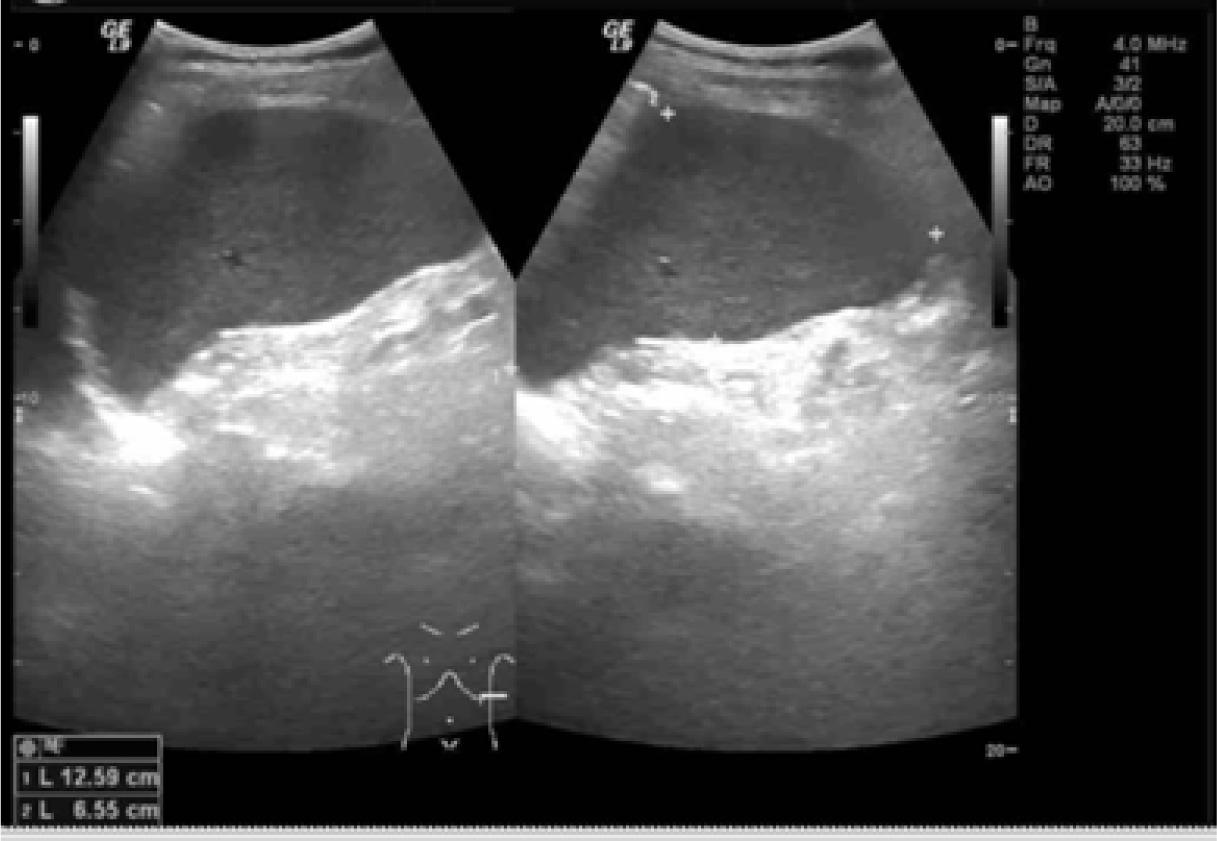
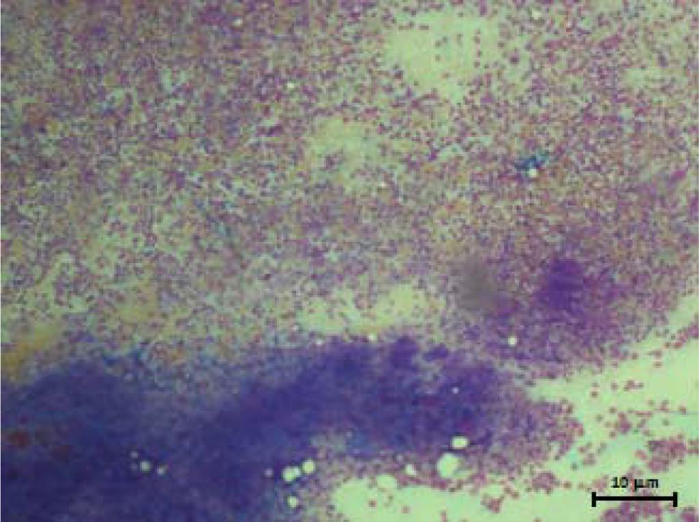
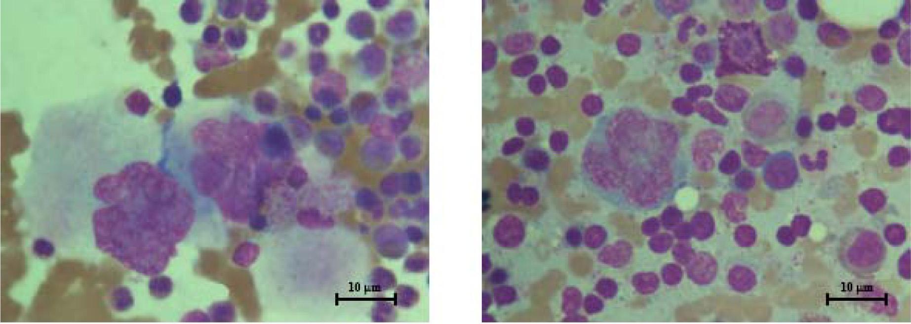
Have a personal or library account? Click to login
Janus kinase 2 negative polycythemia vera Cover

Figures & Tables

Figure 1

Ultrasound images show splenomegaly (size 6.6 cm × 12.6 cm) without a focal mass. No ascites or lymphadenopathies were noted.

Figure 2

Micrograph showing hypercellularity with prominent proliferation of erythroid cells, increased megakaryocytes of varying in shapes and sizes (Wright–Giemsa stain, ×400; bar 60 μm)

Figure 3

left and right: Megakaryocytes from bone marrow aspirates showing megakaryocytes with a nuclear chromatin pattern varying from dispersed, no megakaryocytic clusters were seen, no dysplastic changes and no increase in blasts were noted (Wright–Giemsa stain, ×1,000; bars 10 μm).
DOI: https://doi.org/10.5372/1905-7415.0905.443 | Journal eISSN: 1875-855X | Journal ISSN: 1905-7415
Language: English
Page range: 697 - 700
Published on: Jan 31, 2017
Published by: Chulalongkorn University
In partnership with: Paradigm Publishing Services
Publication frequency: 6 issues per year

© 2017 Pravinwan Thungthong, Supat Chamnanchanunt, Tawatchai Suwanban, Chajchawan Nakhakes, Kunapa Iam-arunthai, published by Chulalongkorn University
This work is licensed under the Creative Commons Attribution-NonCommercial-NoDerivatives 3.0 License.