
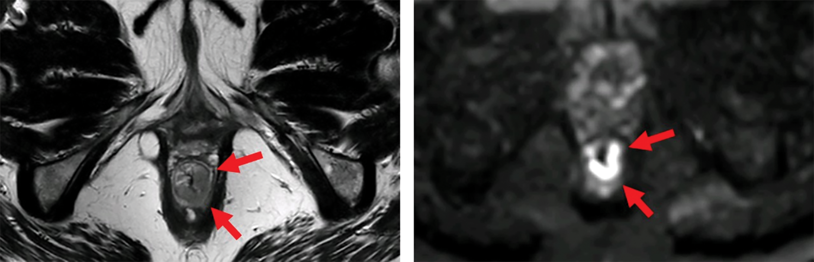
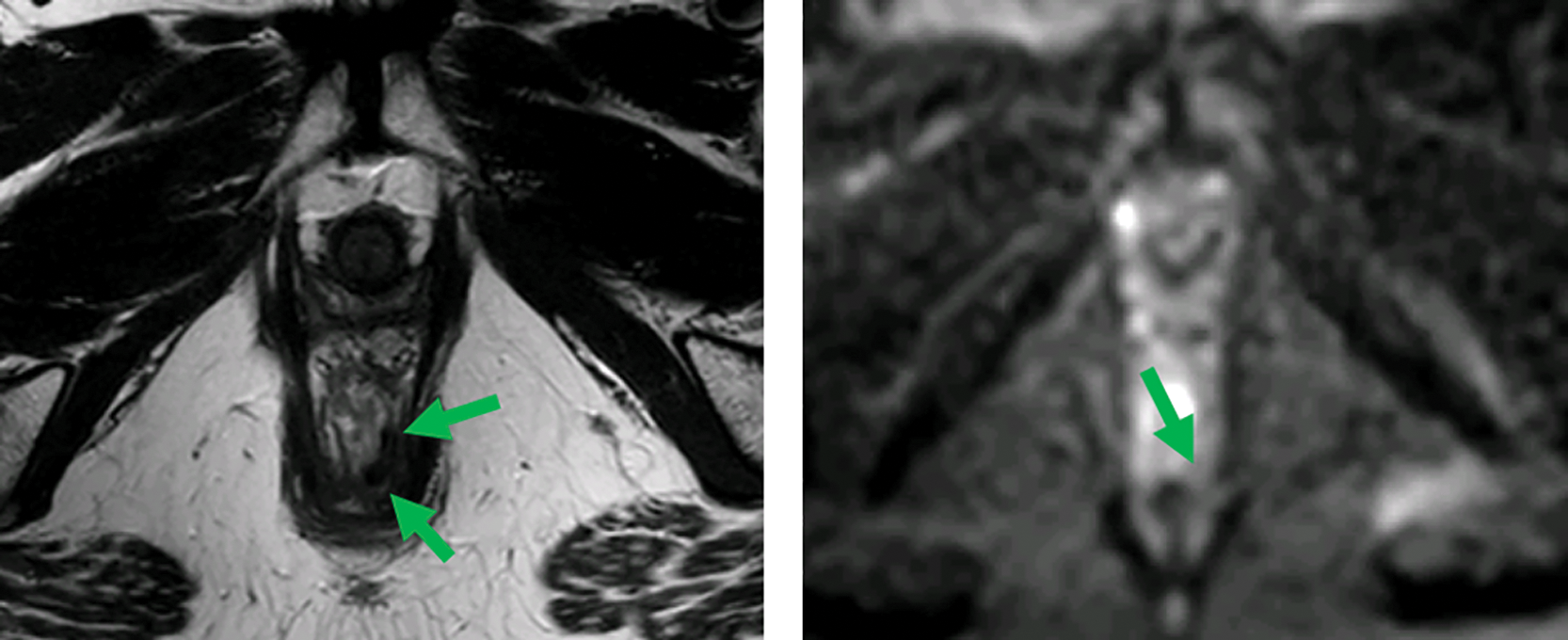
Figure 1
MRI before (top) and after (bottom) neoadjuvant therapy. Axial T2‑weighted and b1000 DWI images show a distal rectal tumor (red arrows). After neoadjuvant therapy, a T2 dark, fibrotic remnant is seen without restricted diffusion (green arrows), indicative of a clinical complete response.