

Figure 1
Axial fat‑suppressed contrast‑enhanced T1‑WI shows an ill‑defined hypovascular pancreatic head mass (arrow) with dilatation of the CBD and pancreatic duct. The suspected diagnosis was PDAC.
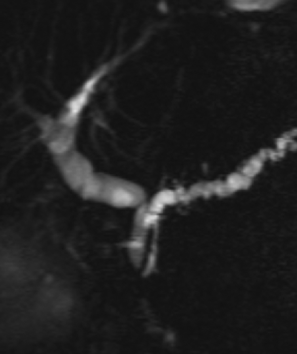
Figure 2
Coronal oblique thick‑slab MRCP image shows 2 MRCP signs that suggest the mass may be inflammatory in nature rather than neoplastic: (a) the “duct penetrating sign” (the pancreatic duct runs through the mass and is not interrupted), and (b) the “attraction sign” (the common bile duct is bent at a 90° angle and attracted to the mass). The final diagnosis was indeed inflammatory pseudomass.