
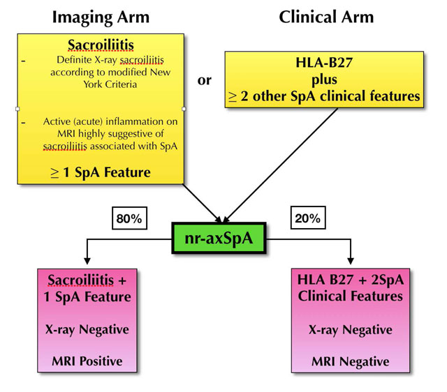
Table 1
Summary of ASAS Criteria and nr-SpA [10].
Table 2
Clinical features as part of ESSG criteria [17].
| Variable | Definition |
|---|---|
| Inflammatory spinal pain* | History or present symptoms of spinal pain in back, dorsal, or cervical region, with at least four of the following: (a) onset before age 45, (b) insidious onset, (c) improved by exercise, (d) associated with morning stiffness, (e) at least three months duration. |
| Synovitis | Past or present asymmetric arthritis or arthritis predominantly in the lower limbs. |
| Family history | Presence in first-degree or second-degree relatives of any of the following: (a) ankylosing spondylitis, (b) psoriasis, (c) acute uveitis, (d) reactive arthritis, (e) inflammatory bowel disease. |
| Psoriasis | Past or present psoriasis diagnosed by a doctor. |
| Inflammatory bowel disease | Past or present Crohn disease or ulcerative colitis diagnosed by a doctor and confirmed by radiographic examination or endoscopy. |
| Alternating buttock pain | Past or present pain alternating between the right and left gluteal regions. |
| Enthesopathy | Past or present spontaneous pain or tenderness at examination at the site of the insertion of the Achilles tendon or plantar fascia. |
| Acute diarrhoea | Episode of diarrhoea occurring within 1 month before arthritis. |
| Urethritis/cervicitis | Non-gonococcal urethritis or cervicitis occurring within one month before arthritis. |
| Sacroiliitis | Bilateral grade 2–4 or unilateral grade 3–4, according to the following radiographic grading system: 0 = normal, 1 = possible, 2 = minimal, 3 = moderate and 4 = ankylosis. |
Table 3
Typical Structural and Inflammatory Lesions on Spinal Imaging.
| Corner Inflammatory Lesion (CIL) | This presents as bone marrow oedema and appears as a triangular or L shape in one quadrant of the vertebra, commonly along the anterior or posterior margin on mid sagittal imaging. Related to the entheses of the anterior and posterior longitudinal ligaments with the annulus fibrosis and the cerebral body. |
| Central Inflammatory Lesion | Andersson lesion, typically appears as a semi-circular area of bone marrow oedema, related to the vertebral end plate adjacent to the intervertebral discs and can be associated with erosions. |
| Costotransverse Joint Inflammation (CTJ) | Adjacent bone marrow oedema on the far lateral sagittal images, related to the junction of the rib and the transverse process of the adjacent thoracic vertebra. Absent at T11 and T12. |
| Costovertebral Joint inflammation (CVJ) | Can affect any joint from T1 to T12. Circular pattern of bone marrow oedema related to the posterior intervertebral disc and middle column of the vertebral body. It can extend to the adjacent soft tissue, rib margin and posterior aspect of vertebral bodies. |
| Enthesitis of spinal ligaments supraspinous ligament and interspinal ligaments | Supraspinous, interspinous ligament inflammation, seen along the spinous processes in the mid sagittal slices, along the posterior elements. |
| Syndesmophytes/ankylosis | Manifests as a linear continuous marrow signal between vertebral bodies on MRI. May occur on para sagittal slices and not on the central sagittal imaging. |
Table 4
Typical Structural and Inflammatory Lesions in the SIJ.
| Erosions | Most varied in presentation. Visible on T1 images as loss of cortical bone associated with adjacent low bone marrow signal intensity. If active, manifests as a hyper intense lesion or with extensive adjacent bone marrow oedema on STIR. (Figure 2) |
| Fat Infiltration | Can be difficult to diagnose in young active adults with patchy marrow fat. If at least two these criteria are followed, may be easier to diagnose accurately. Juxta-articular – in contact with the articular surface. (Figure 1) Geographical – sharp margins Signal – Uniform marrow signal intensity on T1-W images. |
| Sclerosis | Uniform Low signal intensity on T1 and STIR imaging, in the subchondral region. (Figure 3) |
| Ankylosis | Continuous marrow signal intensity across the joint. Can also manifest as marrow across parallel sclerotic tram-track lines believed to be residual joint lines from previous long erosive changes affecting the SIJ. |
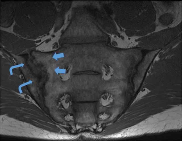
Figure 1
T1-W mid coronal oblique image through the SIJ in a 42-year-old male who presented with long standing low back pain, raised inflammatory markers and a positive HLAB27. Juxta-articular geographical fat infiltration (blue arrows) is noted as structural lesions, along the sacral margin of the SIJ. Pseudowidening of the joint on MRI with large elongated erosions along the iliac aspect and subarticular sclerosis (curved arrowheads) of the right SIJ.
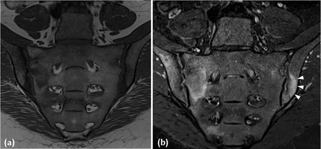
Figure 2
(a) T1-W Mid-coronal MRI in a 38-year-old HLAB27 positive, Female patient who presented with inflammatory back pain and buttock pain. Areas of joint space widening with iliac sided erosions and adjacent marrow low signal intensity are noted in keeping with active inflammatory lesions. (b) Corresponding STIR image in a 38-year-old HLAB27 positive, Female patient who presented with inflammatory back pain and buttock pain, demonstrates extensive high signal intensity (white arrowheads) in the adjacent subchondral region and marrow in keeping with inflammation associated with an active erosion.
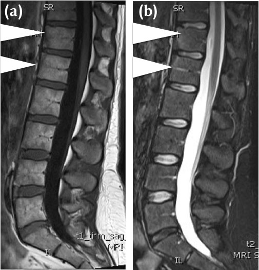
Figure 3
(a) 32-year-old female HLA b27 positive, with chronic low back pain and early morning stiffness. Mid sagittal T1-W MRI demonstrates triangular areas of heterogenous intermediate to low signal intensity to fat (between white arrowheads), vertebral corner lesions at T12 and LI. The features are of sclerosis and a Romanus’ Lesion on MRI. (b) 32-year-old female HLA b27 positive, with chronic low back pain and early morning stiffness. Mid sagittal STIR MRI demonstrates corresponding triangular areas of low signal intensity (between arrowheads) at T12/L1. The features are of sclerosis and a Romanus’ Lesion on MRI.