
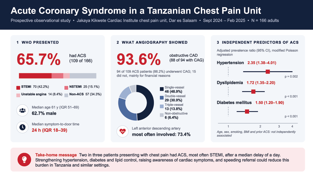
Graphical Abstract
Introduction
Cardiovascular disease (CVD) is the leading global cause of morbidity and mortality (1). In 2023, CVD accounted for 19.2 million deaths and 437 million disability-adjusted life years (DALYs), the majority of which occurred in low- and middle-income countries (LMICs) (2, 3, 4). Among CVDs, acute coronary syndrome (ACS), comprising ST-elevation myocardial infarction (STEMI), non-ST-elevation myocardial infarction (NSTEMI), and unstable angina (UA), is a major contributor to CVD-related mortality, with more than 7 million people diagnosed annually (5). Profound disparities in outcomes from ACS persist worldwide: high-income countries have achieved >50% reductions in age-standardized ACS mortality over the past two decades compared with <15% reductions in LMICs (6), and case fatality rates following cardiovascular events are three to four times higher in low-resource settings (2, 7).
In sub-Saharan Africa (SSA), the epidemiological transition and rising prevalence of cardiovascular risk factors have led to a rapid increase in the burden of ACS (8, 9). Reported ACS prevalence among hospitalized patients with CVD in SSA ranges from 0.2% to 22.3%, with hypertension identified as the leading risk factor (10). In Tanzania, a prospective study of 349 patients with non-traumatic chest pain found ACS in 9.6% of cases (11). In another study from northern Tanzania, 22.3% of 681 patients presenting with chest pain and shortness of breath to a tertiary hospital were diagnosed with acute myocardial infarction (40% STEMI, 60% NSTEMI), with hypertension and tobacco use as the predominant risk factors (12). However, ACS remains under-investigated in the country, and existing data on its prevalence and determinants have not characterized the angiographic profiles of patients undergoing coronary angiography (CAG) or specifically examined those presenting with chest pain to a dedicated chest pain unit.
The rising burden of ACS in Tanzania and across SSA underscores the need to strengthen diagnostic capacity and generate region-specific data to inform clinical care and policy. Although ACS is increasingly recognized, diagnostic limitations and heterogeneous clinical presentations hinder timely recognition and management. In this study, we aim to determine the prevalence, clinical characteristics, angiographic patterns, and factors associated with ACS among patients presenting with chest pain to a dedicated chest pain unit during a six-month prospective study at a large tertiary cardiac center in Dar es Salaam, Tanzania.
Methods
Study setting
This study was conducted in the chest pain unit of Jakaya Kikwete Cardiac Institute (JKCI) in Dar es Salaam, Tanzania. JKCI is a government-owned national specialized and university teaching hospital that provides comprehensive cardiovascular care, as well as training and research services. It serves patients from all regions of the United Republic of Tanzania and receives referrals from neighboring countries. Hypertensive heart disease is the most prevalent cardiac diagnosis among adults undergoing echocardiography at JKCI, affecting 70% of older and 17% of younger patients (13).
As a national referral center, JKCI receives patients referred from regional referral and designated hospitals across Tanzania, and many patients evaluated in the chest pain unit may therefore have undergone an initial assessment at another facility before transfer. The referral source and any prior evaluation of individual participants were not systematically captured in the study dataset. Because JKCI is a national referral center in an urban setting, its chest pain population is expected to be enriched for patients with a higher pretest probability of ACS; we consider the implications of this for generalizability in the Discussion.
The chest pain unit at JKCI was established in 2021 to streamline the evaluation and management of patients presenting with chest pain. On arrival, patients are assessed immediately. A focused history and physical examination are performed, followed by an ECG and measurement of high-sensitivity cardiac troponin. Patients diagnosed with ACS are referred to the catheterization laboratory; CAG and PCI are performed when indicated, in accordance with standard protocols. Specifically, the immediate evaluation consists of triage on arrival, a 12-lead ECG obtained and interpreted immediately on arrival, and venous sampling for hs-cTnI at presentation, with repeat sampling approximately 3–6 hours later when the initial result is non-diagnostic (see Definition of ACS and Components). Patients with STEMI are transferred directly to the catheterization laboratory for primary PCI, whereas patients with NSTE-ACS undergo CAG with timing determined by risk stratification by the treating cardiologist. Additional investigations, such as transthoracic echocardiography and computed tomography pulmonary angiography, are obtained as clinically indicated. Patients with alternative diagnoses are managed by the relevant clinical service or discharged.
Study design and duration
We conducted a prospective observational hospital-based study. The study was carried out over a six-month period, from September 2024 to February 2025. During this time, all eligible patients presenting with chest pain to the JKCI chest pain unit were consecutively screened for participation. The six-month study period was prespecified in the protocol approved by the MUHAS Research and Ethics Committee (MUHAS-REC-07-2024-2386), and enrollment began once ethical clearance and institutional permission had been obtained. Previous reports from JKCI have analyzed retrospective clinical and procedural records (14); in contrast, the present study required prospective enrollment, written informed consent, and standardized interviewer-administered data collection at the time of presentation, which could not be applied retrospectively to patients seen before September 2024.
Study population and eligibility criteria
The study population consisted of adult patients who presented with chest pain as their primary complaint to the JKCI chest pain unit during the study period. We included patients who were 18 years of age or older, who presented with chest pain as the main reason for seeking care, and who provided written informed consent to participate in the study. We excluded patients who had experienced chest trauma within the preceding two weeks or who had undergone chest surgery within the preceding two weeks, in order to avoid misclassifying pain related to recent injury or surgery as possible ACS. Application of these criteria yielded the final study cohort.
Sample size and sampling procedure
The required sample size was estimated using a single-population proportion formula. We assumed a 95% confidence level, a 5% margin of error, and an expected ACS prevalence of 9.6% among chest pain patients, based on prior data from Tanzania. Using these parameters, we calculated a minimum sample size of 148 participants (an unadjusted estimate of 134, inflated by approximately 10% to allow for non-response or incomplete data). During the six-month study period, a consecutive sampling approach was employed. All adult patients presenting to the chest pain unit who met the eligibility criteria were approached and invited to participate throughout the six-month study period.
Data collection
Data were collected prospectively using a structured, interviewer-administered questionnaire developed specifically for this study. The questionnaire captured sociodemographic characteristics, cardiovascular risk factors, details of the presenting complaint, prior medical history, and medication use. Additional clinical information, including vital signs at presentation, anthropometric measurements, ECG findings, laboratory results, and angiographic data, was obtained from the patients’ medical records and from the hospital’s electronic health information system.
The principal investigator and two medical-doctor research assistants carried out all data collection activities. Members of the on-call team in the chest pain unit notified the research team whenever an eligible patient arrived. After eligibility was confirmed and informed consent was obtained, the research team conducted a face-to-face interview with the patient, and when necessary, with a close relative, to collect the required information.
Anthropometric measurements were obtained using standard methods. Body weight was measured to the nearest kilogram using a calibrated scale, and height was measured to the nearest centimeter using a stadiometer. Body mass index (BMI) was calculated as weight in kilograms divided by the square of height in meters. Clinical examination findings and ECG reports were recorded at the time of presentation. Laboratory investigations, including cardiac troponin, lipid profile, random blood glucose, serum creatinine, and blood urea nitrogen, were performed in the hospital laboratory as part of routine care, and the results were documented in the study forms. High-sensitivity cardiac troponin I (hs-cTnI) was measured using an electrochemiluminescence immunoassay (Elecsys Troponin I hs, Roche Diagnostics GmbH, Mannheim, Germany) on the cobas pro e 801 analyzer. For patients who underwent CAG, procedural reports from the catheterization laboratory were reviewed to extract information on the presence and extent of coronary artery disease and the coronary vessels involved.
Variables and definitions
The primary outcome was the presence or absence of ACS among patients presenting with chest pain. Patients were classified as having ACS if they met diagnostic criteria based on clinical presentation, ECG changes, and elevated cardiac biomarkers, as described below. Patients who did not meet these criteria were classified as non-ACS. Coronary angiographic findings were a secondary, descriptive outcome assessed only among participants who underwent CAG during the index presentation. The decision to perform CAG was made by the treating team as part of routine care and was not dictated by the study protocol.
The independent variables included sociodemographic characteristics, cardiovascular risk factors and comorbidities, and clinical characteristics as assessed by the treating physician using prespecified criteria. Hypertension was defined as a documented prior diagnosis of hypertension in the medical record, current use of at least one antihypertensive medication, or elevated blood pressure at enrollment, defined as systolic blood pressure ≥140 mmHg and/or diastolic blood pressure ≥90 mmHg on two measurements taken at least five minutes apart (15). Diabetes mellitus was defined as a documented prior diagnosis of diabetes or current use of glucose-lowering medication, or as hyperglycemia at enrollment, defined as fasting plasma glucose >126 mg/dL (7.0 mmol/L) or random plasma glucose >200 mg/dL (11.1 mmol/L) in the presence of classic symptoms of hyperglycemia, in accordance with American Diabetes Association criteria (16). Dyslipidemia was defined as a documented prior diagnosis of dyslipidemia, current use of lipid-lowering therapy, or abnormal fasting lipid profile at enrollment (total cholesterol ≥5.2 mmol/L, LDL cholesterol ≥3.4 mmol/L, or triglycerides ≥1.7 mmol/L), in line with American College of Cardiology (ACC)/American Heart Association (AHA) guideline thresholds (17). BMI was classified according to World Health Organization criteria as normal weight (18.5–24.9 kg/m²), overweight (25.0–29.9 kg/m²), and obese (≥30.0 kg/m²) (18). Smoking/tobacco use was defined by participant self-report as current or past use of cigarettes or other smoked tobacco products; never-users were classified as non-smokers. Family history of CVD was defined as self-reported history of myocardial infarction, stroke, or sudden cardiac death in a first-degree relative.
Laboratory variables included hs-cTnI, total cholesterol, low-density lipoprotein (LDL) cholesterol, high-density lipoprotein (HDL) cholesterol, random blood glucose, serum creatinine, and blood urea nitrogen. For patients who underwent CAG, angiographic variables included the presence of obstructive coronary artery disease (OCAD), the number of diseased vessels, and the specific coronary arteries involved (left main [LM], left anterior descending [LAD], left circumflex [LCX], and right coronary artery [RCA]).
HEART score
For descriptive risk stratification, we calculated the HEART score (History, ECG, Age, Risk factors, Troponin) for each participant when sufficient data were available (19). Each component was scored from 0 to 2 according to the original HEART score definitions, yielding a total score between 0 and 10. The score was calculated by trained study physicians using prospectively collected clinical and laboratory data; bedside clinicians were not required to compute the score for clinical decision-making. We categorized HEART scores as low risk (0–3), intermediate risk (4–6), and high risk (7–10). The HEART score was used only to describe the risk profile of the study population and was not included as a predictor in the multivariable analysis of factors associated with ACS.
Chest pain classification
At presentation, the treating physician classified chest pain as “cardiac” or “non-cardiac” based on routine clinical assessment. “Cardiac” chest pain was generally characterized as substernal discomfort with a typical quality and duration, often exertional and/or relieved by rest or nitroglycerin. Pain that did not meet these features (e.g., pleuritic, reproducible with palpation, clearly positional) was classified as non-cardiac.
Definition of ACS and components
The diagnosis of ACS was made in accordance with contemporary standard criteria and guided by the 2025 ACC/AHA Guideline for the Management of Patients with Acute Coronary Syndromes (20). The diagnosis integrated three components: symptoms suggestive of myocardial ischemia, ECG changes, and cardiac biomarker results. All clinical determinations were made by the treating cardiologist using prespecified study definitions.
Symptoms of myocardial ischemia were defined as new or worsening chest pain or discomfort with typical cardiac characteristics, including substernal pressure, squeezing, or tightness, with or without radiation to the arm, neck, jaw, or back. Duration of ≥10–20 minutes and provocation by exertion or emotional stress were considered supportive features.
Ischemic ECG changes were defined a priori as any of the following, as interpreted by the treating cardiologist or an on-call cardiology fellow: ST-segment elevation consistent with STEMI (≥1 mm in at least two contiguous limb leads, or ≥2 mm in at least two contiguous precordial leads, with sex- and lead-specific thresholds for V2–V3, or new left bundle branch block); ST-segment depression ≥0.5 mm in at least two contiguous leads; T-wave inversion ≥1 mm in at least two contiguous leads; or other dynamic changes judged to be compatible with acute ischemia.
Cardiac troponin was assessed using hs-cTnI, measured by an electrochemiluminescence immunoassay on the cobas pro e 801 analyzer (Roche Diagnostics). An hs-cTnI value >40 ng/L (the 99th-percentile upper reference limit and lower limit of the assay’s measuring range) was considered elevated. In routine practice, hs-cTnI was measured at presentation and repeated approximately 3–6 hours later when clinically indicated. A dynamic change between serial hs-cTnI measurements was considered supportive of myocardial injury.
Patients were categorized as having:
STEMI if they had persistent ST-segment elevation meeting the above criteria (or new left bundle branch block) together with an elevated hs-cTnI level in an appropriate clinical context.
NSTEMI if they had elevated hs-cTnI without ST-segment elevation on ECG, in the presence of symptoms and/or ECG changes compatible with myocardial ischemia.
Unstable angina if they had ischemic chest pain at rest or with minimal exertion, no elevation in hs-cTnI (all values below the 99th percentile), and either ischemic ECG changes or a high clinical suspicion for coronary ischemia based on symptoms and risk-factor profile.
For descriptive analyses, non-ST-elevation acute coronary syndrome (NSTE-ACS) was defined as the combination of NSTEMI and unstable angina.
In patients with an initially equivocal hs-cTnI (values close to, but not clearly above, the 99th percentile), high clinical suspicion was defined as the combination of typical ischemic chest pain, cardiovascular risk factors, and/or dynamic but non-diagnostic ECG changes; in such cases, repeat hs-cTnI testing was performed at the discretion of the treating cardiologist. Serial hs-cTnI measurements were obtained in these patients, and the final diagnosis of ACS versus non-ACS was based on the full clinical, ECG, and biomarker information.
CAG Definitions
CAG was performed using standard techniques by interventional cardiologists at JKCI. Coronary lesions were assessed visually by the operating cardiologist. OCAD was defined as ≥70% luminal diameter stenosis in any major epicardial coronary artery (LAD, LCX, or RCA) or ≥50% stenosis in the LM. Non-obstructive disease was defined as lesser degrees of stenosis or angiographically normal coronary arteries. The extent of OCAD was categorized as:
Single-vessel disease (SVD): OCAD involving one major epicardial coronary artery,
Double-vessel disease (DVD): OCAD involving two vessels, and
Triple-vessel disease (TVD): OCAD involving all three major epicardial vessels.
The culprit vessel was identified by the interpreting cardiologist based on angiographic appearance in conjunction with the clinical presentation and ECG findings.
Statistical analysis
Data were entered into a secure database and analyzed using IBM SPSS Statistics for Windows, Version 27 (IBM Corp., Armonk, NY, USA). Continuous variables were summarized using medians and interquartile ranges because many variables were not normally distributed. Categorical variables were summarized as frequencies and percentages. Normality of continuous variables was assessed using the Shapiro–Wilk test and visual inspection of histograms. Age is additionally reported as mean ± standard deviation (SD) to facilitate comparison with prior ACS registries.
To identify factors associated with ACS, we used modified Poisson regression with robust standard errors to directly estimate prevalence ratios. We selected this approach rather than logistic regression because ACS was common (>10% prevalence) in our study population, so odds ratios from logistic regression would substantially overestimate the corresponding risk or prevalence ratios. Modified Poisson regression provides unbiased and interpretable estimates of prevalence ratios for binary outcomes in cross-sectional data.
We first performed univariable analyses to examine the crude association between each potential predictor and ACS, and we expressed these associations as crude prevalence ratios with their corresponding 95% CIs. We then constructed a multivariable modified Poisson regression model to identify independent predictors of ACS. Variables were considered for inclusion in the multivariable model if they demonstrated a p-value of less than 0.20 in univariable analysis or if they were established traditional risk factors for ACS based on the literature. The variables included in the final multivariable model were age group, sex, smoking or tobacco use, hypertension, diabetes mellitus, dyslipidemia, BMI category, and previous history of ACS. We reported adjusted prevalence ratios with 95% CIs, and we considered a two-sided p-value of less than 0.05 to indicate statistical significance. Model convergence and the plausibility of parameter estimates were assessed before results were interpreted. Because previous history of ACS was uncommon, this variable was assessed for sparse-data bias (quasi-complete separation), and, as a sensitivity analysis, the multivariable model was re-estimated after excluding it. Adjusted prevalence ratios from the final model were also displayed as a forest plot.
Data quality assurance
Several measures were undertaken to ensure data quality, validity, and reliability. Research assistants received training on study procedures, questionnaire administration, and standardized measurement of clinical and anthropometric parameters. The questionnaire was pre-tested among a small group of chest pain patients at JKCI before the main study, and minor revisions were made to improve clarity and completeness. During data collection, the principal investigator supervised the research process, reviewed completed questionnaires on a daily basis for completeness and consistency, and resolved discrepancies by referring to the original medical records. Data entry was double-checked, and obvious errors were corrected after verification.
Ethical considerations
The study was conducted in accordance with the ethical principles of the Declaration of Helsinki and relevant national regulations. Ethical approval was obtained from the Muhimbili University of Health and Allied Sciences Research and Ethics Committee (Ref. No. DA.282/298/01.C/2386; MUHAS-REC-07-2024-2386). Permission to conduct the study at JKCI was granted by the hospital management. Written informed consent was obtained from all participants prior to enrollment. For participants who were unable to read, the consent form was read aloud in their preferred language, and consent was documented using a thumbprint in the presence of an independent witness. Participants were informed that their decision to participate or not would not affect their care. All data were anonymized and stored securely, and access was restricted to the research team to maintain confidentiality.
Results
Study flow diagram
During the study period, 168 consecutive adults presenting to the JKCI chest pain unit with a primary complaint of chest pain were screened for eligibility. We excluded two patients who had undergone recent chest surgery within the preceding two weeks, leaving 166 participants who met all eligibility criteria and were enrolled in the study. No eligible patients declined to participate. Of the 166 enrolled participants, 109 (65.7%) were ultimately diagnosed with ACS and 57 (34.3%) with non-ACS conditions (Figure 1). All 109 participants with ACS were recommended to undergo CAG, of whom 94 (86.2%) underwent the procedure during the index presentation and 15 (13.8%) did not, primarily because of financial constraints. No participant with a non-ACS diagnosis underwent CAG.
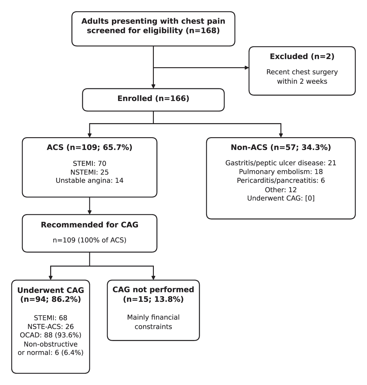
Figure 1
Flow diagram of patient screening, exclusions, final diagnostic categories, and coronary angiography.
Characteristics of patients with chest pain attending JKCI chest pain unit
The median age of participants was 61 years (IQR 51–69; mean 60.1 ± 12.6 years; Shapiro–Wilk p = 0.036), and 45.8% were in the 45–64-year age group; 62.7% were male. Most participants were insured (72.3%), and more than three-quarters (81.9%) had attained at least secondary school education. The most prevalent comorbidity was hypertension (75.9%), followed by diabetes mellitus (48.2%) and dyslipidemia (47.6%). Less than one-third of participants (21.1%) reported a history of current or prior smoking (Table 1).
Table 1
Sociodemographic characteristics of patients with chest pain attending chest pain unit at JKCI, N = 166.
| VARIABLES | n (%) |
|---|---|
| Demographic Characteristics | |
| Age, median (IQR) | 61 (51–69) |
| Age, mean ± SD | 60.1 ± 12.6 |
| Age group in years, n (%) | |
| >65 | 71 (42.8) |
| 45–64 | 76 (45.8) |
| <45 | 19 (11.4) |
| Sex, n (%) | |
| Male | 104 (62.7) |
| Female | 62 (37.3) |
| Insurance, n (%) | |
| Yes | 120 (72.3) |
| No | 46 (27.7) |
| Education level, n (%) | |
| No formal education | 1 (0.6) |
| Primary | 29 (17.5) |
| Secondary | 34 (20.5) |
| Tertiary and above | 102 (61.4) |
| Comorbidities/risk factors | |
| Hypertension | |
| Yes | 126 (75.9) |
| No | 40 (24.1) |
| Diabetes mellitus | |
| Yes | 80 (48.2) |
| No | 86 (51.8) |
| Dyslipidemias | |
| Yes | 79 (47.6) |
| No | 87 (52.4) |
| BMI category | |
| Normal (18.5–24.9 kg/m²) | 58 (34.9) |
| Overweight (25.0–29.9 kg/m²) | 63 (38) |
| Obese (≥30.0 kg/m²) | 45 (27.1) |
| Smoking/tobacco use | |
| Current/past | 35 (21.1) |
| Never | 131 (78.9) |
Clinical and laboratory characteristics of patients with chest pain attending JKCI chest pain unit
Physician-determined cardiac chest pain was present in 95 of 166 participants (57.2%). The median time from onset of chest pain to presentation was 24 hours (IQR 18–39). Only seven patients (4.2%) had a documented previous history of ACS, and 15 (9.0%) reported a family history of CVD. Based on the HEART score, 47 patients (28.3%) were categorized as low risk (0–3), 20 (12.0%) as intermediate risk (4–6), and 99 (59.6%) as high risk (7–10) (Table 2).
Table 2
Clinical and laboratory characteristics of patients with chest pain attending chest pain unit at JKCI, N = 166.
| VARIABLES | |
|---|---|
| Clinical history | n (%) |
| Chest pain characteristic | |
| Cardiac | 95 (57.2) |
| Non-cardiac | 71 (42.8) |
| Onset to presentation in hours, median (IQR) | 24 (18–39) |
| Previous history of ACS, n (%) | |
| Yes | 7 (4.2) |
| No | 159 (95.8) |
| Family history of CVD, n (%) | |
| Yes | 15 (9) |
| No | 151 (91) |
| HEART score, n (%) | |
| 0–3 | 47 (28.3) |
| 4–6 | 20 (12.0) |
| 7–10 | 99 (59.6) |
| Clinical examination | Median (IQR) |
| Systolic blood pressure (mmHg) | 125 (111–145.25) |
| Diastolic blood pressure (mmHg) | 75 (66.75–84) |
| Pulse rate (b/min) | 81 (73–94.25) |
| Anthropometric measurement | Median (IQR) |
| Weight (kg) | 75 (70–80) |
| Height (cm) | 166 (160–170) |
| Laboratory investigation | Median (IQR) |
| hs-cTnI (ng/L)a | 84.7 (6.51–1688.38) |
| Total cholesterol (mmol/l) | 4.5 (3.9–5.87) |
| LDL (mmol/l) | 3.1 (2.4–3.9) |
| HDL (mmol/l) | 1.19 (1.02–1.3) |
| Random blood glucose (mmol/l) | 10.2 (7.6–15.28) |
| Serum creatinine (micromol/l) | 95.32 (77–124.12) |
| BUN (mmol/l) | 4.99 (3.63–7.71) |
[i] a Normal range ≤40 ng/L (99th-percentile upper reference limit).
Prevalence of ACS among patients with chest pain attending chest pain unit at JKCI
Among the 166 patients presenting with chest pain to the JKCI chest pain unit, 109 (65.7%; 95% CI 58.2–72.5%) were diagnosed with ACS and 57 (34.3%) had non-ACS conditions (Table 3). Within the ACS group, STEMI was the most frequent subtype, accounting for 70 patients (42.2% of the total cohort), followed by NSTEMI in 25 patients (15.1%) and unstable angina in 14 patients (8.4%). Among patients without ACS, the most common diagnoses from medical records were acute gastritis or peptic ulcer disease (21 patients, 12.7% of the total cohort), pulmonary embolism (18 patients, 10.8%), and other conditions, including musculoskeletal and non-specific chest pain (12 patients, 7.2%), while pericarditis or pancreatitis was identified in 6 patients (3.6%).
Table 3
Spectrum of diagnoses among patients presenting with chest pain at the JKCI chest pain unit (N = 166).aIncludes musculoskeletal and non-specific chest pain.
| DIAGNOSTIC CATEGORY | n (%) |
|---|---|
| ACS (total) | 109 (65.7) |
| STEMI | 70 (42.2) |
| NSTEMI | 25 (15.1) |
| Unstable angina | 14 (8.4) |
| Non-ACS (total) | 57 (34.3) |
| Pulmonary embolism | 18 (10.8) |
| Gastritis/peptic ulcer disease | 21 (12.7) |
| Pericarditis/pancreatitis | 6 (3.6) |
| Othersa | 12 (7.2) |
Angiographic profiles of patients presenting with chest pain at JKCI who underwent CAG
All 109 patients with ACS were recommended to undergo CAG; 94 (86.2%) underwent the procedure (56.6% of the total cohort), and 15 (13.8%) did not, primarily due to financial constraints. No patient with a non-ACS diagnosis underwent CAG. Among the 94 patients who underwent CAG, OCAD was identified in 88 (93.6%), while 6 (6.4%) had non-obstructive or angiographically normal coronary arteries (Table 4).
Table 4
Angiographic profile of patients presenting with chest pain at JKCI chest pain unit.
| ACS | |||
|---|---|---|---|
| ALL ACS (N = 94) | STEMI (N = 68) | NSTE-ACS (N = 26) | |
| Distribution of the coronary artery involved | |||
| OCAD, n (%)a | 88 (93.6) | 67 (98.5) | 21 (80.8) |
| SVD, n (%) | 46 (48.9) | 34 (50.0) | 12 (46.2) |
| DVD, n (%) | 29 (30.9) | 23 (33.8) | 6 (23.1) |
| TVD, n (%) | 13 (13.8) | 10 (14.7) | 3 (11.5) |
| Non-OCAD, n (%) | 6 (6.4) | 1 (1.5) | 5 (19.2) |
| Type of coronary artery involved | |||
| LM, n (%) | 5 (5.3) | 4 (5.9) | 1 (3.8) |
| LAD, n (%) | 69 (73.4) | 53 (77.9) | 16 (61.5) |
| LCX, n (%) | 28 (29.8) | 21 (30.9) | 7 (26.9) |
| RCA, n (%) | 42 (44.7) | 33 (48.5) | 9 (34.6) |
[i] a≥70% luminal stenosis in a major epicardial coronary artery or ≥50% stenosis in the left main coronary artery.
Among the 94 ACS patients who underwent CAG, SVD was the most frequent angiographic pattern, occurring in 46 patients (48.9%), followed by DVD in 29 patients (30.9%) and TVD in 13 patients (13.8%). Similar proportions were observed when stratified by ACS subtype: in the STEMI group, 50.0% had SVD, 33.8% had DVD, and 14.7% had TVD, whereas in the NSTE-ACS group, 46.2% had SVD, 23.1% had DVD, and 11.5% had TVD. Non-obstructive or angiographically normal coronary arteries were more frequent among patients with NSTE-ACS (5 of 26, 19.2%) than among those with STEMI (1 of 68, 1.5%).
The LAD artery was the most commonly involved vessel among ACS patients who underwent CAG, being affected in 69 of 94 patients (73.4%). The RCA and LCX arteries were involved in 42 (44.7%) and 28 (29.8%) patients, respectively, while LM disease was uncommon and observed in only 5 patients (5.3%). In the STEMI subgroup, LAD involvement was seen in 53 patients (77.9%), RCA in 33 (48.5%), LCX in 21 (30.9%), and LM in 4 (5.9%). A similar pattern was observed in NSTE-ACS, where the LAD was involved in 16 patients (61.5%), the RCA in 9 (34.6%), the LCX in 7 (26.9%), and the LM in 1 (3.8%).
Univariable and multivariable analysis of factors associated with ACS among patients with chest pain who attended the JKCI chest pain unit
In univariable modified Poisson regression, five factors were significantly associated with ACS at p < 0.20: hypertension, smoking or tobacco use, diabetes mellitus, dyslipidemia, and previous history of ACS (Table 5). These variables, together with age group, sex, and BMI category (traditional ACS risk factors), were entered into the multivariable model. After adjustment, three factors remained independently associated with ACS: hypertension (aPR 2.35; 95% CI 1.38–4.01; p = 0.002), diabetes mellitus (aPR 1.50; 95% CI 1.20–1.90; p < 0.001), and dyslipidemia (aPR 1.72; 95% CI 1.35–2.20; p < 0.001) (Table 5). Age, sex, smoking or tobacco use, previous history of ACS, and BMI category were not independently associated with ACS in the adjusted analysis. All seven participants with a previous history of ACS had ACS at the index presentation (quasi-complete separation); the corresponding crude prevalence ratio is therefore determined by the outcome prevalence in the reference group and should be interpreted with caution. In a sensitivity analysis excluding previous history of ACS, the adjusted estimates for the remaining covariates were materially unchanged (hypertension aPR 2.34, 95% CI 1.39–3.94; diabetes mellitus aPR 1.53, 95% CI 1.22–1.93; dyslipidemia aPR 1.70, 95% CI 1.34–2.17). Adjusted prevalence ratios are displayed in Figure 2.
Table 5
Modified Poisson regression analysis for factors associated with ACS among patients with chest pain who attended the JKCI chest pain unit.
| VARIABLES | ACS, n (%) | UNIVARIABLE cPR (95% CI) | p-VALUE | MULTIVARIABLEa aPR (95% CI) | p-VALUEb |
|---|---|---|---|---|---|
| Sex | |||||
| Female | 37 (59.7) | Ref | |||
| Male | 72 (69.2) | 1.16 (0.91–1.48) | 0.23 | 1.11 (0.93–1.32) | 0.27 |
| Age group | |||||
| <45 | 9 (50) | Ref | |||
| 45–64 | 50 (64.1) | 1.28 (0.79–2.1) | 0.32 | 0.84 (0.53–1.34) | 0.47 |
| >65 | 50 (71.4) | 1.43 (0.88–2.32) | 0.15 | 0.85 (0.56–1.30) | 0.45 |
| Hypertension | |||||
| No | 10 (25) | Ref | |||
| Yes | 99 (78.6) | 3.14 (1.82–5.42) | <0.001 | 2.35 (1.38–4.01) | 0.002 |
| Smoking/tobacco use | |||||
| No | 80 (61.1) | Ref | |||
| Current/past | 29 (82.9) | 1.36 (1.11–1.66) | 0.003 | 1.08 (0.87–1.34) | 0.48 |
| Diabetes mellitus | |||||
| No | 36 (41.9) | Ref | |||
| Yes | 73 (91.3) | 2.18 (1.68–2.82) | <0.001 | 1.50 (1.20–1.90) | <0.001 |
| Previous history of ACS | |||||
| No | 102 (64.2) | Ref | |||
| Yes | 7 (100) | 1.56 (1.39–1.75) | <0.001 | 1.28 (0.94–1.73) | 0.11 |
| Dyslipidemia | |||||
| No | 35 (40.2) | Ref | |||
| Yes | 74 (93.7) | 2.33 (1.79–3.03) | <0.001 | 1.72 (1.35–2.20) | <0.001 |
| BMI status | |||||
| Normal | 37 (63.8) | Ref | |||
| Overweight | 43 (68.3) | 1.07 (0.83–1.38) | 0.61 | 0.89 (0.72–1.09) | 0.27 |
| Obese | 29 (64.4) | 1.01 (0.76–1.35) | 0.95 | 0.85 (0.69–1.06) | 0.15 |
[i] aAdjusted for age group, sex, smoking or tobacco use, hypertension, diabetes mellitus, dyslipidemia, BMI category, and previous history of ACS.
bBold values indicate p < 0.05.
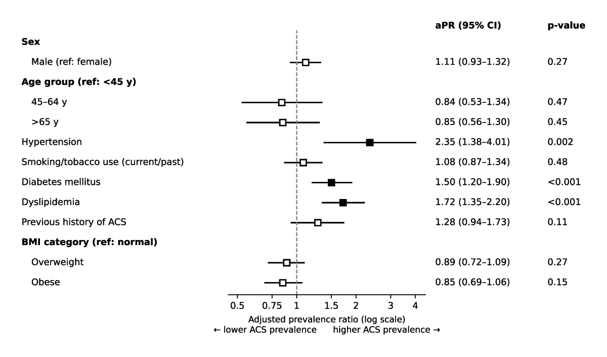
Figure 2
Forest plot of aPR with 95% confidence intervals for factors associated with ACS in the multivariable modified Poisson regression model (N = 166).a, b
aThe dashed vertical line indicates no association (aPR = 1.0); the horizontal axis is log-scaled.
bFilled squares indicate p < 0.05.
Discussion
In this prospective study of 166 patients presenting with chest pain, the median age was 61 years (IQR 51–69). This is similar to prior Tanzanian and regional data, where mean ages around 60 years have been reported (12), and reflects the relatively young age at which ACS occurs in SSA compared with high-income settings (10). In contrast to global data, where most ACS events occur in adults ≥65 years and risk continues to rise into the 70s and 80s (21, 22), our findings suggest earlier onset of coronary artery disease, likely driven by high prevalence of modifiable risk factors and gaps in primary prevention.
Nearly two-thirds of participants were male (62.7%), and the median time from symptom onset to presentation was 24 hours (IQR 18–39). This delay is markedly longer than reported in cohorts from India and the Gulf region, where median times to hospital arrival range from 3.6 to 7.4 hours (23, 24), but is consistent with previous Tanzanian data showing delays measured in days (12). Such late presentation is likely multifactorial, including limited awareness and recognition of cardiac symptoms, poor health-seeking behavior, and referral delays from lower-level facilities, and may contribute to the high burden of STEMI and adverse outcomes (25, 26).
We found the prevalence of ACS to be 65.7%. This markedly high proportion contrasts with estimates from previous studies in Tanzania (11, 12, 21, 27) and elsewhere in SSA (10, 28), most of which reported substantially lower ACS prevalence among patients with chest pain or cardiovascular presentations. This discrepancy likely reflects differences in study populations and settings: prior work has largely been conducted in general emergency departments or mixed medical wards, whereas our study was undertaken at a tertiary-level cardiac referral center and enrolled only patients presenting with chest pain to a dedicated chest pain unit, a context expected to enrich for higher-risk cases. Our study also identified STEMI as the most common ACS subtype, a pattern consistent with several other African series (29, 30) and global data (31). The predominance of STEMI aligns with regional trends in SSA, where delayed healthcare-seeking behavior and limited access to preventive cardiovascular services contribute to a higher burden of acute and severe coronary presentations.
Referral patterns and access also bear on the generalizability of our findings. JKCI receives patients referred from facilities nationwide, many after an initial evaluation elsewhere, and access to a national cardiac center in Dar es Salaam is likely to favor patients who are urban, insured, and of higher socioeconomic status. Consistent with this, 61.4% of our participants had attained tertiary education and 72.3% were insured, in marked contrast to a recent prospective ACS surveillance study of 568 adults with chest pain or shortness of breath in a northern Tanzanian emergency department, which reported an ACS prevalence of 22.7% in a population with considerably lower educational attainment (32). Our estimates of ACS prevalence, risk-factor burden, and angiographic patterns should therefore be regarded as representative of patients who reach a tertiary cardiac referral center, rather than of the broader population of Tanzanians with chest pain, among whom ACS is likely less prevalent and more frequently unrecognized (12, 32).
In our study, the LAD was the predominantly affected vessel among patients with ACS who underwent CAG, accounting for 73.4% of cases. SVD was the most common angiographic pattern, observed in 48.9% of patients. These findings are consistent with a study from Tanzania by Khanbhai et al. (33), in which the LAD was also the most frequently involved vessel (76%) and SVD was a common pattern (43.6%). Similar CAG patterns have been reported in several other regional and global studies (23, 24, 34). The predominance of LAD involvement is likely related to its extensive myocardial territory and its origin as a proximal branch of the left main coronary artery. The LAD supplies a large portion of the myocardium, including the anterior wall, interventricular septum, and apex of the left ventricle—regions that are critical for cardiac output and overall ventricular function—thereby making it particularly susceptible to atherosclerotic plaque formation and rupture (35, 36, 37).
Three cardiometabolic conditions, hypertension, diabetes, and dyslipidemia, were independently associated with ACS in our study, reflecting the growing burden of noncommunicable diseases in Tanzania and underscoring the urgent need for effective, targeted strategies in cardiovascular risk reduction and primary prevention (38, 39, 40, 41, 42). In contrast, other well-established predictors of ACS, such as age, sex, smoking, obesity, and prior ACS, were not independently associated with ACS in the adjusted model. This may be explained by factors such as limited sample size or a relatively homogeneous study population, in which these variables lacked sufficient variability to reach statistical significance. Nonetheless, their clinical importance remains substantial and should not be underestimated in risk stratification and management planning. We also note that South Asian ancestry is a well-recognized independent risk factor for premature coronary artery disease (43), and Dar es Salaam has a long-established community of South Asian descent. Self-reported ethnicity was not collected in our study questionnaire, so we were unable to examine this association; future studies at JKCI should collect self-reported ancestry, given its potential relevance to risk stratification in this urban population.
To our knowledge, this is the first study in Tanzania to prospectively describe the prevalence, clinical characteristics, angiographic patterns, and factors associated with ACS among patients presenting to a dedicated chest pain unit in a specialized cardiac hospital with CAG capability. The use of standardized ECG interpretation, high-sensitivity troponin, and CAG strengthens diagnostic validity and provides granular insight into coronary anatomy in this setting.
This study also has important limitations. Its single-center design at a tertiary cardiac referral hospital restricts generalizability to the broader Tanzanian population with chest pain and precludes causal inference. The cohort was predominantly insured (72.3%) and highly educated (61.4% with tertiary education or higher), which may not reflect the socioeconomic profile of patients accessing lower-level facilities. In addition, 13.8% of ACS patients, mostly clinically stable NSTE-ACS patients with financial constraints, did not undergo CAG, potentially leading to underestimation or mischaracterization of the angiographic profile. Furthermore, we did not systematically apply the recommended 0-hour/1-hour or 0-hour/2-hour hs-troponin rule-in/rule-out algorithms for myocardial infarction (19) because these protocols had not yet been adopted at JKCI and were not feasible given laboratory turnaround times and staffing constraints. Consequently, some patients classified as having unstable angina may have had small NSTEMIs, leading to potential underdiagnosis and misclassification. Other limitations should also be noted. Data on occupation and ethnicity were not collected, precluding assessment of these characteristics, including South Asian ancestry, and limiting further characterization of participants’ socioeconomic status. Because the sample size was calculated assuming an ACS prevalence of 9.6%, the precision of our prevalence estimate (95% CI 58.2–72.5%; margin of error approximately ±7.2 percentage points) was lower than originally planned, and the study had limited power to detect modest independent associations in the multivariable model. The estimate for previous history of ACS was based on only seven participants, all of whom had ACS, and is therefore unstable. The referral source and any prior evaluation at other facilities were not systematically recorded. The six-month enrollment period may not capture seasonal or longer-term variation in presentations, and studies with longer enrollment periods are warranted. Finally, procedural characteristics of CAG, such as vascular access site and radiation dose, were not systematically collected for this study; these will be examined in a separate, dedicated analysis of the JKCI catheterization laboratory experience.
Conclusion
Among patients presenting with chest pain at a tertiary cardiac center in Tanzania, the prevalence of ACS was high. Conventional cardiovascular risk factors were strongly associated with ACS, and there was a notable delay in seeking care after symptom onset. These findings underscore the urgent need for public health initiatives to address modifiable risk factors, raise awareness of CVD, and strengthen systems of care for chest pain management. Future multicenter and longitudinal studies are warranted to improve the generalizability of these results.
List of Abbreviations
ACC – American College of Cardiology
ACS – Acute Coronary Syndrome
AHA – American Heart Association
aPR – Adjusted Prevalence Ratio
BMI – Body Mass Index
BUN – Blood Urea Nitrogen
CAG – Coronary Angiography
CI – Confidence Interval
cPR – Crude Prevalence Ratio
CVD – Cardiovascular Disease
DALY – Disability-Adjusted Life Year
DVD – Double-Vessel Disease
ECG – Electrocardiogram/Electrocardiography
HDL – High-Density Lipoprotein
HEART – History, ECG, Age, Risk Factors, Troponin (risk score)
hs-cTnI – High-Sensitivity Cardiac Troponin I
IBM SPSS – International Business Machines Statistical Package for the Social Sciences
IQR – Interquartile Range
JKCI – Jakaya Kikwete Cardiac Institute
LAD – Left Anterior Descending Coronary Artery
LCX – Left Circumflex Coronary Artery
LDL – Low-Density Lipoprotein
LM – Left Main Coronary Artery
LMIC – Low- and Middle-Income Country
MUHAS – Muhimbili University of Health and Allied Sciences
NSTE-ACS – Non-ST-Elevation Acute Coronary Syndrome
NSTEMI – Non-ST-Elevation Myocardial Infarction
OCAD – Obstructive Coronary Artery Disease
PCI – Percutaneous Coronary Intervention
RCA – Right Coronary Artery
SD – Standard Deviation
SSA – Sub-Saharan Africa
STEMI – ST-Elevation Myocardial Infarction
SVD – Single-Vessel Disease
TVD – Triple-Vessel Disease
UA – Unstable Angina
Ethics and Consent
Written informed consent was obtained from all study participants. Ethical approval was granted by the Muhimbili University of Health and Allied Sciences Research and Ethics Committee (Ref. No. DA.282/298/01.C/2386; MUHAS-REC-07-2024-2386), and permission to conduct the study was obtained from the management of Jakaya Kikwete Cardiac Institute. All procedures were performed in accordance with the Declaration of Helsinki and applicable national ethical guidelines and regulations.
Acknowledgements
The authors would like to thank all the institutions, health staff, and study participants involved in the study.
Data Accessibility Statement
The datasets generated and/or analyzed during the current study are available from the corresponding author upon reasonable request.
Author Contributions
RS, FOR, DP, JM, KK: conceptualized, data collection, data analysis, drafted the initial manuscript. All authors critically reviewed the manuscript and agreed on the final version.