
Figure 1
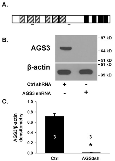
Genetic modification of NRK epithelial cells using lentiviral vectors to reduce endogenous levels of AGS3. (A) Schematic of AGS3 protein structure. Hatched bars = TPR motifs; solid black = G-protein regulatory (GPR) domains; solid lines = AGS3 shRNA target sites. (B) Protein lysates from NRK-52E cells transduced with lentiviral vectors expressing either control (NRK-Ctrl) or two distinct AGS3-specific shRNA (NRK-AGS3sh) were isolated for immunoblot analysis using a polyclonal AGS3 antibody. β-actin was shown as a loading control. Protein standards are shown on the right (in kD) for each blot image. (C) Graphical analysis of the AGS3 band intensity. Three different samples were used in each group, and were shown in the bars. * P < 0.001 significant difference between NRK cells expressing Ctrl versus AGS3-specific shRNA.
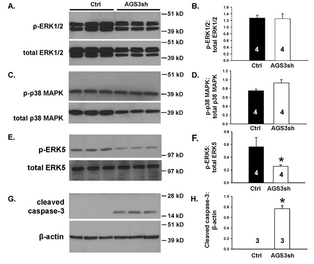
Figure 2
Reduction in endogenous AGS3 protein by short hairpin RNA technology decreases ERK5 activation and promotes cleavage of caspase-3. NRK cells transduced with lentiviral vectors expressing either control (Ctrl) or two distinct AGS3-specific shRNA (AGS3sh) were plated and collected after 2–3 days in culture for protein lysate isolation. Phospho- and total (A) ERK1/2, (C) p38 MAPK, (E) ERK5, and (G) cleaved caspase-3 were detected by Western blot analysis. β-actin was used as a loading control in (G). Protein standards are shown on the right (in kD) for each blot image. Densitometry of the bands was determined for each of the MAPK and cleaved caspase-3 using NIH ImageJ software. Ratios of the phospho-to-total bands for the MAPK (B, D, F) or cleaved caspase-3:β-actin (H) were calculated and graphed. Sample numbers were shown in the bars.
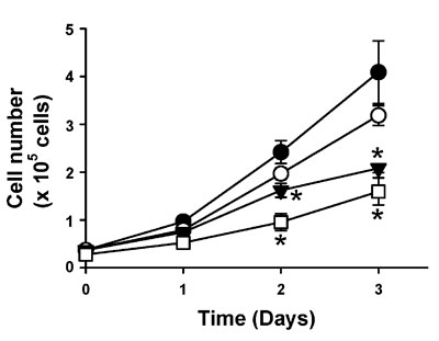
Figure 3
ERK5 inhibition reduces renal epithelial cell number. NRK cells transduced with lentiviral vectors expressing either control (Ctrl) or two distinct AGS3-specific shRNA (AGS3sh) were plated. NRK-Ctrl cells were counted in the presence of either vehicle (DMSO; ●), or two different doses of a selective ERK5/BMK1 inhibitor, BIX02189, at 3 (○) and 10 µM (▼). As a comparison, cell counts were determined for the NRK-cells expressing the AGS3 shRNA (NRK-AGS3sh; □). *P < 0.05 significant difference between vehicle (DMSO)-treated group. n = 3–6 experiments per time point.
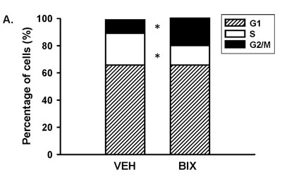
Figure 4
Inhibition of ERK5 signaling blocks the transition from G2 to M phase of the cell cycle. NRK-Ctrl cells were incubated with vehicle (DMSO) or BIX-02189 (10 µM) for 16 hours, and cells were fixed for propidium iodide (PI) staining to detect cell cycle progression by flow cytometry. The percentage of cells in each stage of the cell cycle (G1, S, and G2/M) was graphed. n = 4 different experiments. * P < 0.05 significant difference of each specific cell cycle stage between vehicle- versus BIX-02189 treated cells.
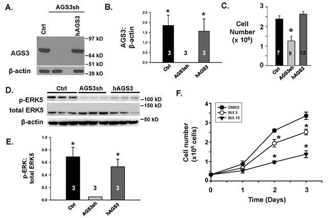
Figure 5
mTOR phosphorylation status following a reduction in endogenous levels of AGS3. (A) NRK cells were transduced with the following lentiviral vectors: 1) control (Ctrl); 2) two distinct AGS3-specific shRNA (AGS3sh); or a combination of the AGS3-specific shRNA with a human AGS3cDNA (hAGS3). A representation Western blot is shown to demonstrate AGS3 protein levels. β-actin was used as a loading control. n = 3 different transduced NRK cell lines were generated to perform the cell culture studies in Figures 5 and 6. (B) Densitometry of the AGS3 bands was graphed relative to β-actin. * P < 0.01 significant difference between the NRK-AGS3sh cells. (C) NRK-Ctrl, NRK-AHS3sh, and hAGS3 cells were counted by hemocytometry after 48 hours. n = number of cell counting experiments. * P < 0.05 significant difference between NRK-AGS3sh versus the other two groups. (D) Western blots were performed for p-ERK5 and total ERK5 in NRK-Ctrl and NRK-AGS3sh cells to confirm that recovery of ERK5 activity could be detected following over-expression of human AGS3 in the NRK-AGSsh cells. β-actin was used as a loading control. n = 3 different cell lines tested per genetic modification. Protein standards are shown on the right (in kD) for each blot image. (E) Densitometry of the p-ERK5 and total ERK5 bands were measured and graphed as a ratio. * P < 0.05 significant difference between the NRK-AGS3sh cells. (F) NRK-hAGS3 cells were counted in the presence of either vehicle (DMSO; ●), or two different doses of a selective ERK5/BMK1 inhibitor, BIX02189, at 3 (¡) and 10 µM (▼). *P < 0.05 significant difference between vehicle (DMSO)-treated group. n = 3 experiments per time point.
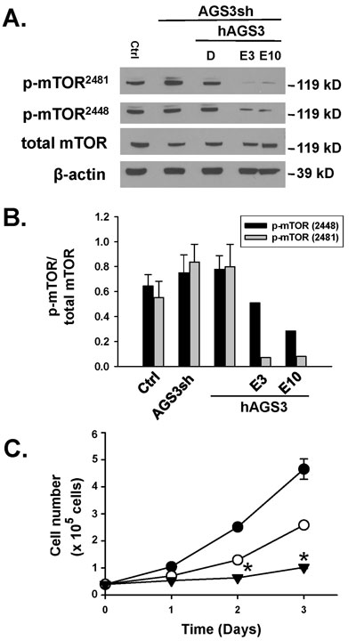
Figure 6
Lack of a change in mTOR phosphorylation following a reduction in endogenous levels of AGS3. (A) The same genetically modified cells described in Figure 5 were used to detect the phosphorylation state of mTOR at Ser2448 or Ser2481 by Western blot analysis. In two separate groups of cells, everolimus at 3 and 10 nM was applied for a 24 hr period to confirm as a positive control that mTOR phosphorylation is reduced. β-actin was used as a loading control. Protein standards are shown on the right (in kD) for each blot image. (B) Densitometry of the bands was determined using NIH ImageJ software, and ratios of the phospho-to-total bands were calculated and graphed. (C) NRK-hAGS3 cells were counted by hemocytometry over a 72 hr period in the presence of vehicle (DMSO; ●) or two different doses of a selective mTOR inhibitor, everolimus, at 3 (○) and 10 nM (▼). n = 2–3 experiments per time point. * P < 0.05 significant difference between control versus Everolimus treatment.