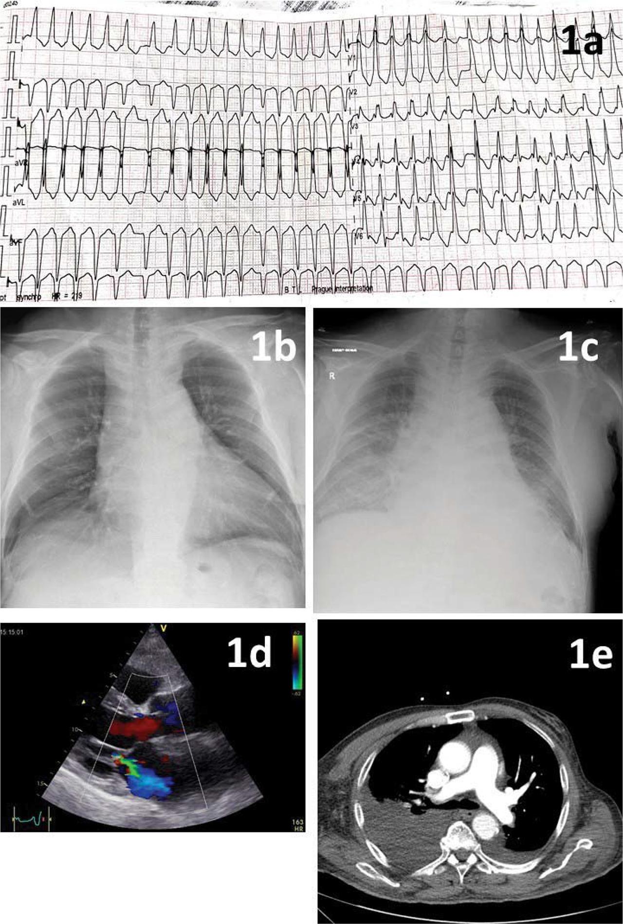
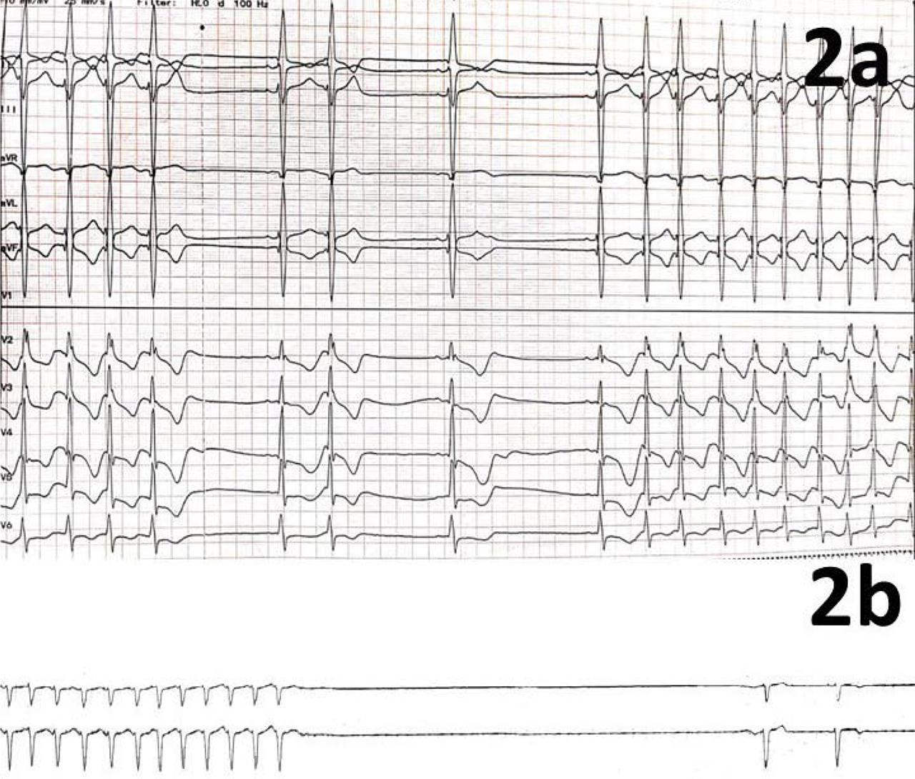
Have a personal or library account? Click to login
Thyrotoxicosis and Heart Failure – a Case Report Cover

Figures & Tables

Figure 1

ECG and imaging. 1a – ECG: irregular rhythm (AF), HR ~ 180 bpm, borderline QRS length (120 ms), RBBB pattern, left anterior fascicular block, ST-T segment depression in the anterolateral leads. 1b – CXR 6 months prior – increased cardiac index, no congestion. 1c – CXR at admission – bilateral pulmonary congestion, apical vascular redistribution, and right pleural effusion, 1d – transthoracic echocardiography – moderate functional mitral regurgitation, dilated LA (50 ml/m2), no LVH, mild LV dilation (EDLVD 54 mm), 1e – CT pulmonary angiography – excludes PE, confirms the presence of moderate pleural effusion. Legend: ECG = electrocardiogram, AF = atrial fibrillation, HR = heart rate, RBBB = right bundle branch block, CXR = chest X-ray, LA = left atrium, LVH = left ventricular hypertrophy, LV = left ventricle, CT = computed tomography, PE = pulmonary embolism.

Figure 2

2a - ECG – day 7 – intermittent conversion to SR with a slow HR (40–50 bpm). 2b - ECG Holter – day 8–9 – alternating moderate / high-rate AF and sinus rhythm, several 5–8 second post conversion symptomatic sinusal pauses (7 second pause pictured). Legend: ECG = electrocardiogram, SR = sinus rhythm, HR = heart rate, AF = atrial fibrillation.
DOI: https://doi.org/10.47803/rjc.2021.31.2.385 | Journal eISSN: 2734-6382 | Journal ISSN: 1220-658X
Language: English
Page range: 385 - 390
Published on: May 3, 2022
Published by: Romanian Society of Cardiology
In partnership with: Paradigm Publishing Services
Publication frequency: 4 issues per year

© 2022 Alexandru-George Cotoban, Vlad Damian Vintila, Cristina Constantinescu, Antonia Nica, Berenice Maria Claudia Suran, Dragos Vinereanu, published by Romanian Society of Cardiology
This work is licensed under the Creative Commons Attribution 4.0 License.