
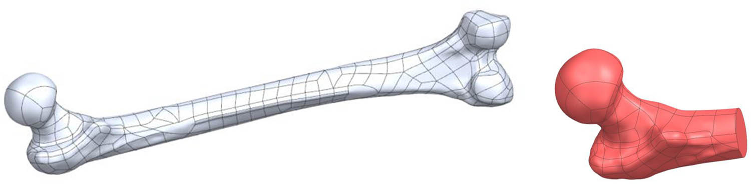
Figure 1
(a) Input geometry – full femoral model; (b) working geometry – proximal femur.
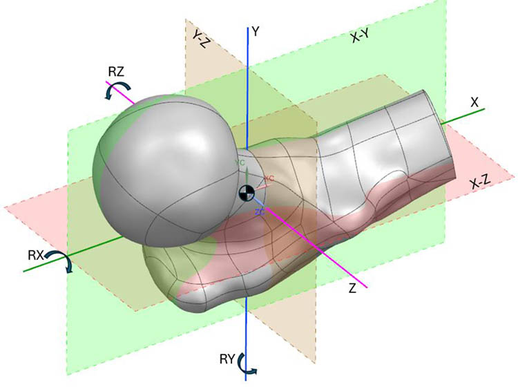
Figure 2
Coordinate system and principal working planes in the Siemens NX environment with rotation axes (RX, RY, RZ).
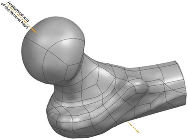
Figure 3
Algorithmically determined head-neck axis of the proximal femur.
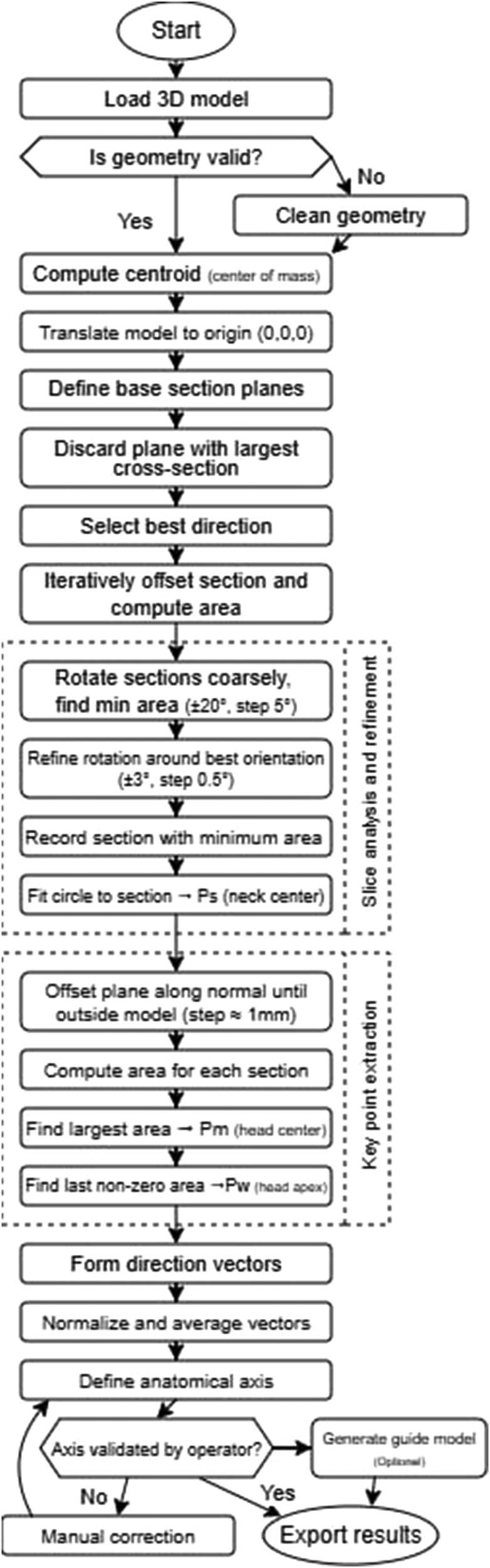
Figure 4
Flowchart of the algorithm for determining topological parameters of the proximal femur.
Table 1
Algorithm validation results across four femoral models
| Parameter | Model 1 | Model 2 | Model 3 | Model 4 |
|---|---|---|---|---|
| Model cleanup | 5 | 5 | 5 | 5 |
| Initial orientation robustness (five positions) | 4/5 pos. score 5, 1 pos. score 4 | 4/5 pos. score 4, 1 pos. score 2 | 3/5 pos. score 5, 2 pos. score 4 | 4/5 pos. score 5, 1 pos. score 4 |
| Multi-surface handling | 5 | 1 (error; after manual fix: 5) | 4 | 5 |
| Head direction identification | 5 | 2 | 4 | 4 |
| Neck isthmus point (Ps) accuracy | 5 | 2 | 3 | 5 |
| Head center point (Pm) accuracy | 5 | 2 | 4 | 5 |
| Head apex point (Pw) accuracy | 5 | 2 | 4 | 5 |
| Anatomical axis accuracy | 5 | 2 | 4 | 4 |
| Mean score | 4.9 | 2.5 | 4.0 | 4.6 |
Scoring: 1 = program failure; 2 = runs but incorrect; 3 = partially correct; 4 = nearly correct with minor deviations; 5 = fully correct. Overall mean: 4.1/5.0 (82%).
Table 2
Comparison of femoral component positioning accuracy across methods in BHR. Clinical methods report accuracy (deviation from intended or reference axis); the proposed algorithm reports repeatability
| Method | Study | Setting | Angular error metric | Notes |
|---|---|---|---|---|
| Conventional mechanical jig | Davis et al. [11] | Cadaver | 7.3° mean error (SSA target 135°) | n = 6 paired limbs |
| Gravius et al. [24] | In vitro | 6.5 ± 7.8° mean error | Conventional instruments | |
| Wirth and Gossé [25] | Clinical | 7 ± 5.7° mean deviation | Manufacturer centering device | |
| Morison et al. [29] | Clinical | 5.6° mean deviation (pre-nav. exp.); 2.2° (post-nav. exp.) | 213 consecutive HRAs | |
| Imageless navigation | Davis et al. [11] | Cadaver | 1.7° mean error | n = 6 paired limbs |
| Olsen et al. [19] | Clinical | 2.8 ± 2.0° mean deviation from plan | 100 consecutive BHRs; 86% within ±5° | |
| Ganapathi et al. [26] | Clinical | 0% outliers > 5° (vs 38% without) | 51 navigated vs 88 non-navigated | |
| Fluoroscopy-based navigation | Gravius et al. [24] | In vitro | 0 ± 0.7° mean error | Cadaver: 1 ± 1.4° |
| Chiron et al. [27] | Clinical | AP < 1.41°; profile < 0.80° | 160 consecutive HRAs | |
| Wirth and Gossé [25] | Clinical | 3 ± 3.4° mean deviation | Central drilling under fluoroscopy | |
| Patient-specific template | Kitada et al. [28] | Synthetic | RMSE 0.954–2.969° | vs navigation RMSE 1.468–3.213° |
| Kunz et al. [18] | Clinical | 1.14° mean deviation | 3D-printed drill templates | |
| Proposed algorithm* | Present study | Computational (pilot) | ± 0.5° repeatability* | 4 models, 5 orientations each |
[i] *The value reported for the proposed algorithm represents intrinsic computational repeatability (precision), not accuracy against an independent reference axis. It is therefore not directly comparable with the clinical accuracy values listed for other methods. The algorithm determines the axis on a digital model prior to surgery; additional error sources – including CT reconstruction, registration, surgical execution, and cementation – would contribute to the total clinical error chain. The comparison is provided to contextualize the magnitude of the algorithmic variability relative to known clinical positioning errors, not to claim equivalence of the measured quantities.