Figure 1.
Figure 2.
Figure 3.
Figure 4.
Figure 5.
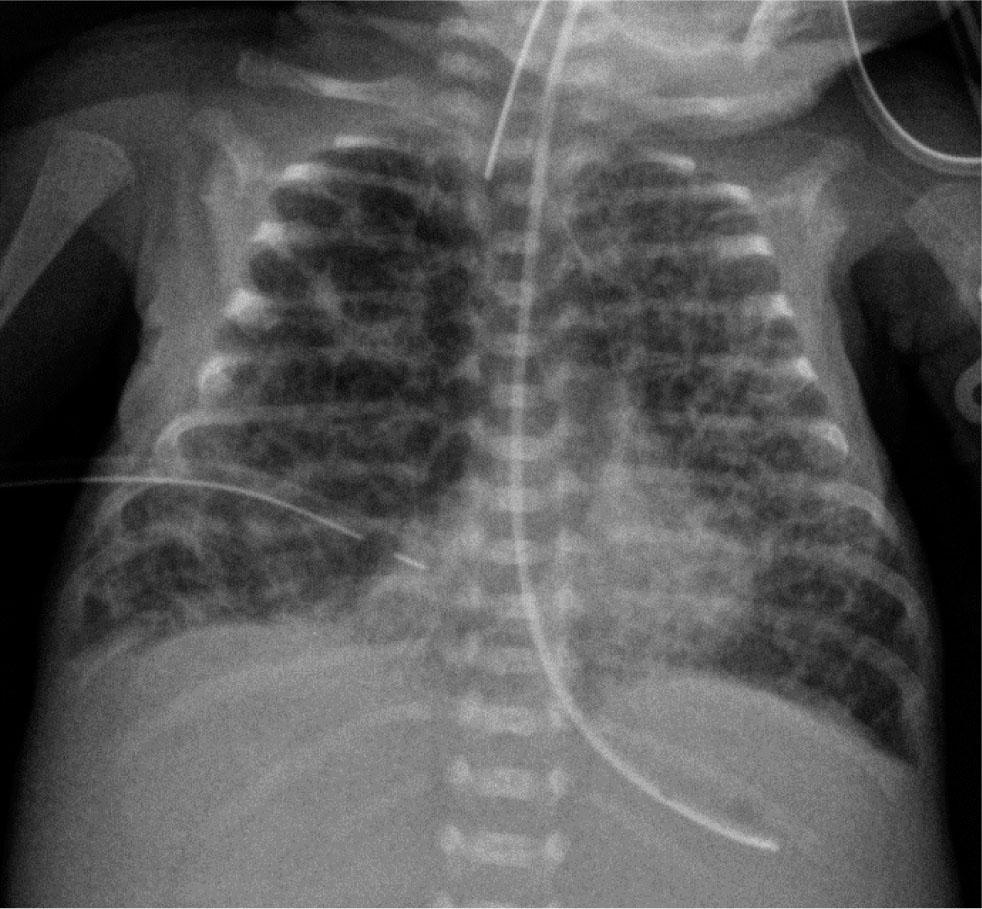
Figure 6.
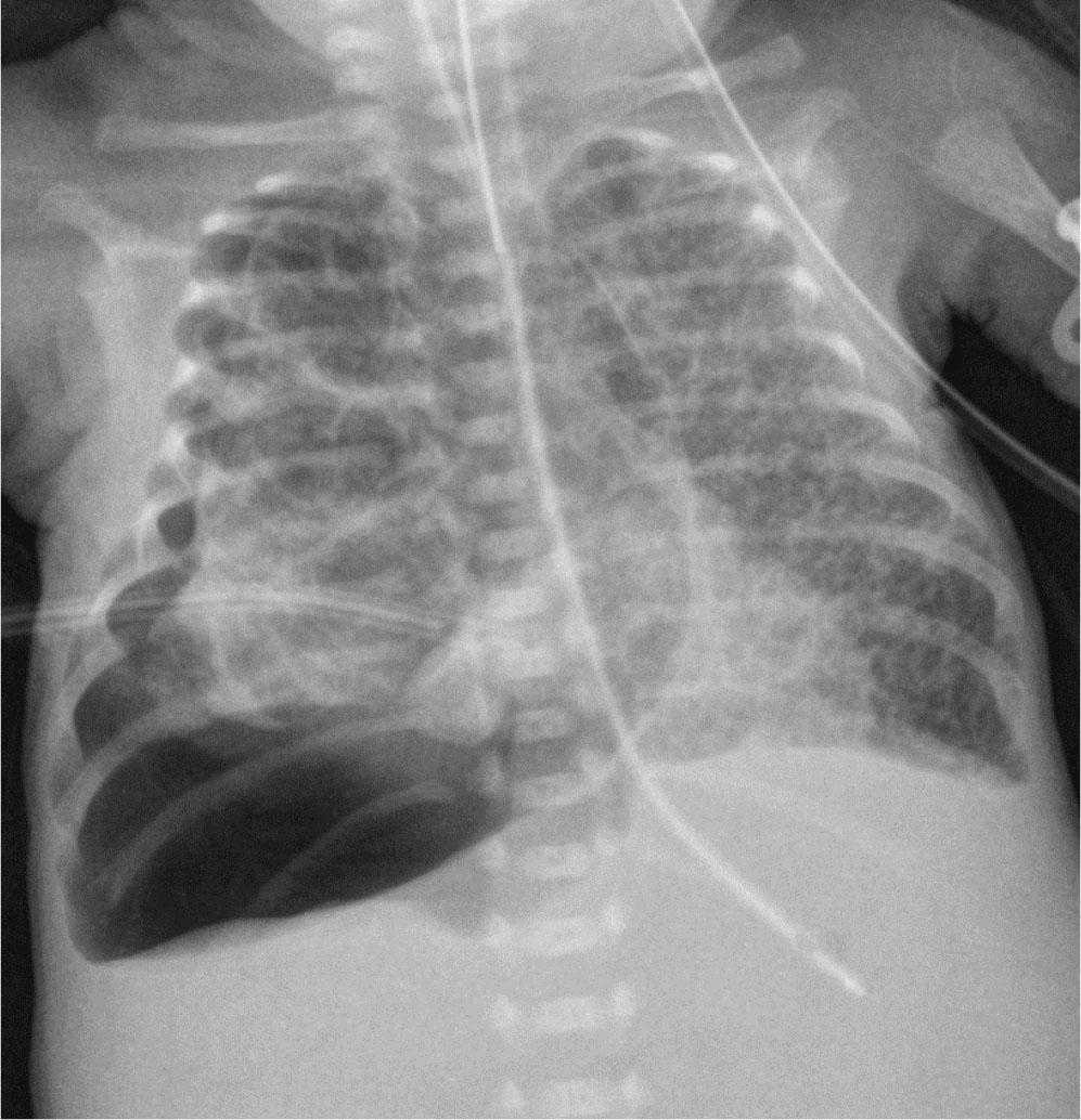
Figure 7.
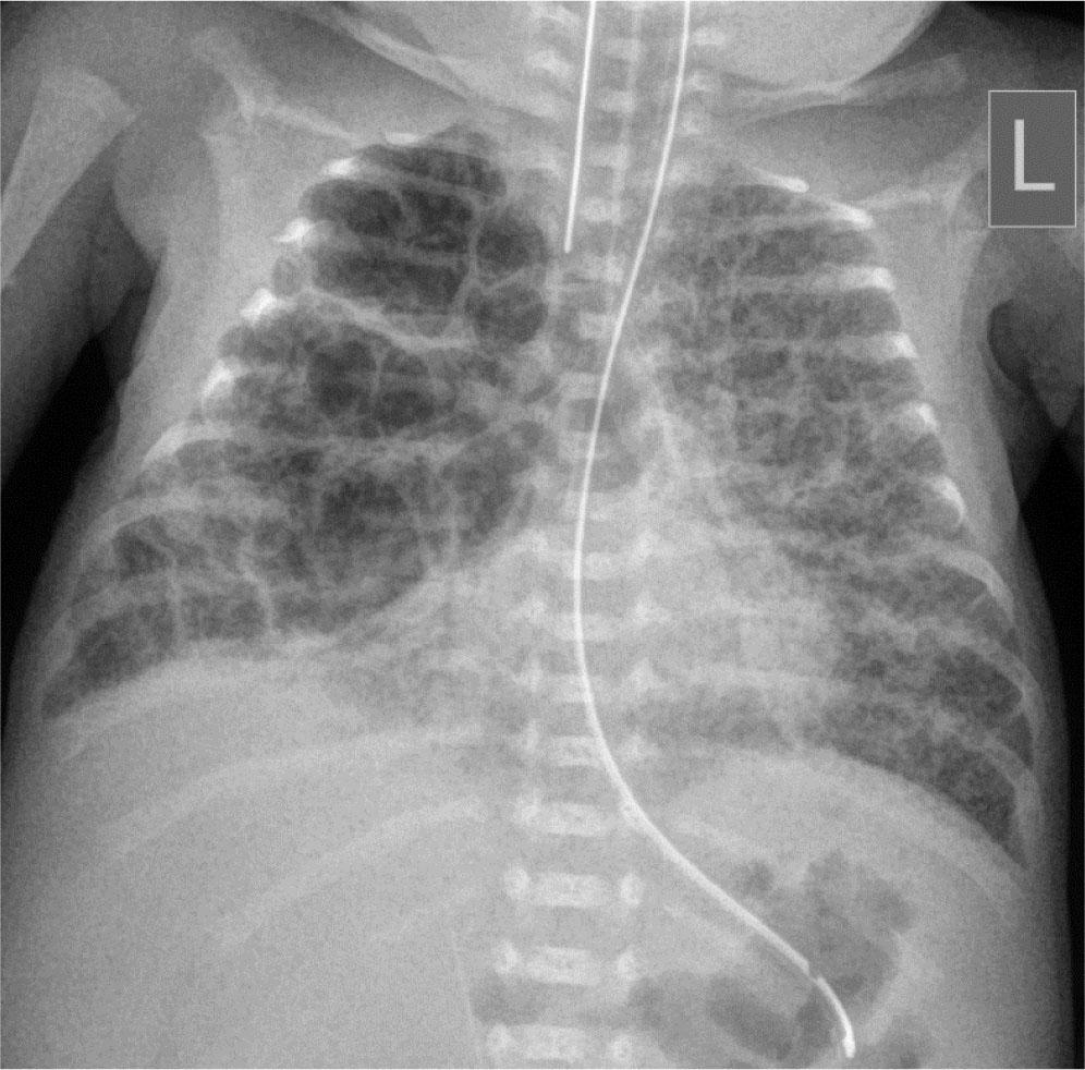
Figure 8.
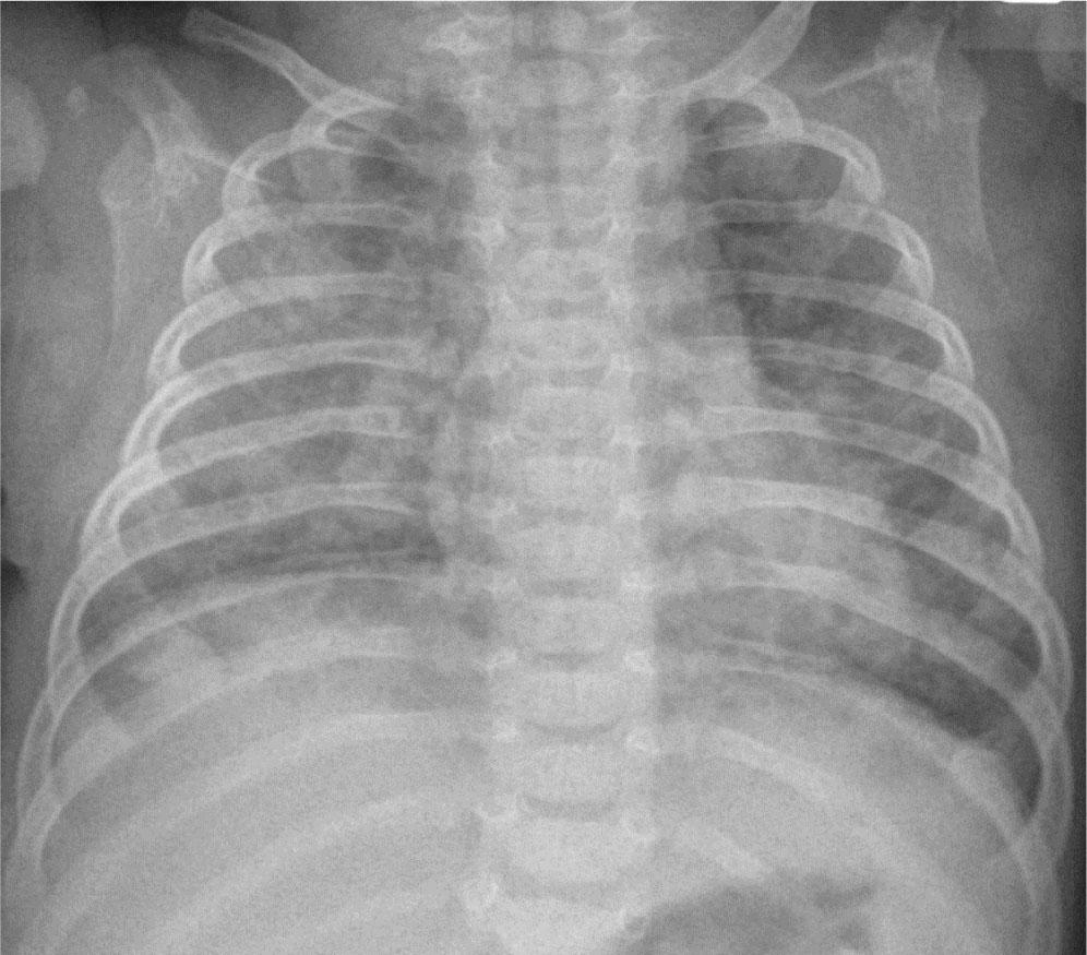
Figure 9.
Figure 10.

© 2023 Magdalena Rutkowska, Martyna Woynarowska, Iwona Terczyńska, Małgorzata Seroczyńska, Dariusz Mydlak, Jarosław Mądzik, Ewa Nowakowska, Katarzyna Niepokój, Sławomir Szczepaniak, Krystyna Polak, published by Institute of Mother and Child
This work is licensed under the Creative Commons Attribution 4.0 License.