
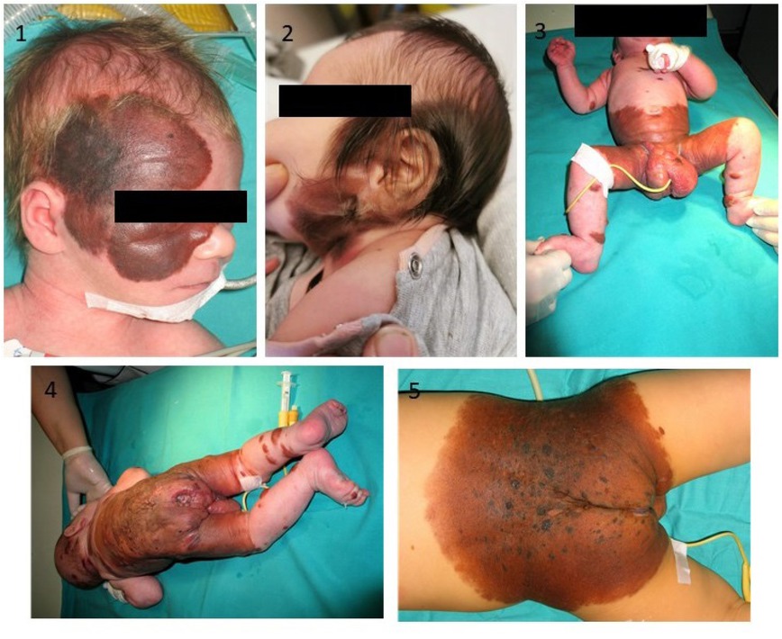
Figure 1
Examples of CMN: head localisation of the nevi (1, 2), anterior and posteriori view of neonate with bathing trunk localisation of the nevus before first operation (3, 4), neonate with back localisation of main nevus (5). Photographed by Marcin R. Szulżycki.
Table 1
Characteristics of patients and results of molecular investigation
| Patient No | Year of birth/sex | Localization of nevus | Melanoma arising in the nevus | Neurocutaneous melanosis (NCM) | Age during first operation | Number of operations | Treatment completed | CMN size | Satellite nevi | BRAF variants detected | NRAS variants detected |
|---|---|---|---|---|---|---|---|---|---|---|---|
| 1 | 2011/m | B | 6/12 | 5 | + | >20 cm | + | none | p.Gln61Lys | ||
| 2 | 2012/m | H | + | 7/12 | 4 | > 0,5% PAS | 0 | none | none | ||
| 3 | 2012/m | H | 1/12 | 2 | + | 0,5 % PAS | 0 | none | p.Gln61Lys | ||
| 4 | 2009/f | BT | 4y | 6 | > 20 cm | ++ | none | none | |||
| 5 | 1989/f | N | 15y | 4 | + | > 20 cm | 0 | none | none | ||
| 6 | 2003/f | B | 1/12 | 5 | + | > 20 cm | + | none | none | ||
| 7 | 2007/f | BT | + | 1/12 | 14 | > 20 cm | 0 | none | none | ||
| 8 | 2011/m | B | 3/12 | 2 | + | > 20 cm | 0 | none | p.Gln61Lys | ||
| 9 | 2009/f | B | 4/12 | 8 | + | > 20 cm | + | none | none | ||
| 10 | 2007/f | H | 2y | 8 | > 0,5% PAS | + | none | p.Gln61Lys | |||
| 11 | 2011/f | B | 3/12 | 5 | + | > 20 cm | + | none | p.Gly12Asp | ||
| 12 | 2006/m | BT | + | 4/12 | 21 | > 20 cm | nd | none | none | ||
| 13 | 2009/m | B | + | 2y | 5 | > 20 cm | ++ | none | p.Gln61Lys | ||
| 14 | 2002/m | BT | 2y | 10 | + | not assessed | 0 | none | p.Gln61Arg | ||
| 15 | 2012/m | HN | 3/12 | 2 | > 0,5% PAS | 0 | none | none | |||
| 16 | 2010/f | B | + | 6/12 | 4 | + | > 20 cm | + | none | none | |
| 17 | 2006/f | BT | + | + | 6/12 | 13 | + | > 20 cm | nd | none | none |
| 18 | 2008/m | H | + | + | 1/12 | 3 | + | > 0,5% PAS | - | none | none |