
Introduction
Listeria monocytogenes is an important foodborne pathogen and the causative agent of listeriosis, a disease acquired mainly through consumption of contaminated foods. Clinical manifestations range from mild gastroenteritis to invasive disease, including septicemia and central nervous system infections, with particularly serious consequences for pregnant individuals, neonates, older adults, and immunocompromised patients. Although listeriosis occurs less frequently than many other foodborne infections, invasive disease has a comparatively high case-fatality rate, making L. monocytogenes an important concern for food safety and public health (Vázquez-Boland et al. 2001; Şanlıbaba et al. 2018; Zhang et al. 2021).
A major challenge in controlling L. monocytogenes is its capacity to persist in food-associated environments. The organism can tolerate a range of stresses encountered during food production and storage and can colonize food-contact surfaces through biofilm formation. Biofilms may protect bacterial cells from environmental stresses and facilitate persistence on processing surfaces, thereby increasing opportunities for recurrent contamination of food products (Osek et al. 2022). Contamination may occur at different stages of the food chain, including production, processing, transportation, storage, handling, and retail display. Consequently, foods of both animal and plant origin can serve as potential vehicles for L. monocytogenes. Meat and poultry products are of particular concern because contamination may occur during processing and handling, whereas fresh vegetables may become contaminated before or after harvest and are sometimes consumed with minimal processing. Examining a range of commonly marketed animal- and plant-derived foods can therefore provide useful information on the local occurrence of L. monocytogenes and potential routes of consumer exposure (Osek et al. 2022; Zhang et al. 2021).
In addition to determining its occurrence in foods, characterization of antimicrobial susceptibility and biofilm-forming capacity provides further information on the phenotypic properties of food-derived L. monocytogenes. Although L. monocytogenes generally remains susceptible to important therapeutic antimicrobials, susceptibility varies among food isolates, supporting continued surveillance of antimicrobial susceptibility patterns (Şanlıbaba et al. 2018). Biofilm formation is also an important persistence-associated phenotype because it can facilitate attachment to abiotic surfaces and survival under adverse environmental conditions. Biofilm formation may vary among isolates, highlighting the importance of characterizing food-derived strains rather than assuming uniform behavior across the species.
Interconnected regulatory systems control biofilm development, environmental adaptation, and pathogenicity in L. monocytogenes. The Agr system mediates cell-to-cell signaling and regulates population-dependent behaviors. Within this system, agrB contributes to processing and exporting the signaling peptide, whereas agrA encodes a response regulator involved in transcriptional responses after signal detection (Miller and Bassler 2001; Lemon et al. 2010; Vázquez-Armenta et al. 2020). Other important regulatory elements include prfA, which encodes the major transcriptional regulator controlling multiple genes required for host-cell invasion, intracellular survival, and dissemination, and sigB, which encodes an alternative sigma factor involved in adaptation to environmental stresses such as acid, osmotic, oxidative, and temperature stress (Kazmierczak et al. 2006; Palmer et al. 2011). Because these genes are involved in bacterial communication, stress response, and virulence, changes in their expression can help show how L. monocytogenes responds to antimicrobial treatments.
Plant-derived compounds have received increasing attention as potential complementary approaches for limiting the growth, persistence, and biofilm formation of foodborne microorganisms. Among these compounds, clove essential oil and quercetin are chemically distinct natural products with reported antimicrobial and antibiofilm activities. Clove oil is particularly rich in eugenol, a phenolic compound associated with disruption of bacterial membrane integrity and inhibition of biofilm-associated processes (Wongsawan et al. 2019; Bai et al. 2023; Silva et al. 2024). Previous studies have also demonstrated inhibitory effects of clove oil and eugenol against foodborne bacteria, including L. monocytogenes. Quercetin is a naturally occurring flavonoid with reported antimicrobial and antibiofilm properties. In L. monocytogenes, quercetin has been shown to reduce bacterial adhesion and biofilm development and to influence the expression of stress- and virulence-associated genes, including sigB and prfA (Vázquez-Armenta et al. 2018; Vázquez-Armenta et al. 2020). These findings support evaluating clove oil and quercetin at concentrations below their minimum inhibitory concentrations (sub-MICs) to determine whether they affect biofilm formation and gene expression without completely inhibiting bacterial growth.
Despite evidence for the antimicrobial and antibiofilm activities of clove oil and quercetin, limited information remains on how these compounds affect genes involved in bacterial communication, stress response, and virulence in food-derived L. monocytogenes. In particular, the responses of agrA, agrB, sigB, and prfA to sub-inhibitory concentrations of these compounds have not been extensively characterized together. Furthermore, information on the occurrence, antimicrobial susceptibility, and biofilm-forming characteristics of L. monocytogenes recovered from diverse retail foods in Erbil remains limited. Therefore, this study aimed to determine the occurrence and antimicrobial susceptibility profiles of L. monocytogenes isolated from selected retail foods in Erbil, Iraq; characterize the biofilm-forming capacity of the recovered isolates; and evaluate the effects of sub-MIC concentrations of clove oil and quercetin on biofilm formation and the expression of agrA, agrB, sigB, and prfA in two selected strong biofilm-producing isolates.
Experimental
Materials and Methods
Sample collection and study design
A total of 778 food samples representing 11 food categories of animal and plant origin were collected from selected retail markets in Erbil, Iraq, between 20 August 2024 and March 2025. The selected foods are commonly consumed in the region and may be exposed to L. monocytogenes contamination at different stages of production, transportation, handling, storage, or retail display. The samples comprised local chicken meat (n = 70), frozen chicken meat (n = 88), minced meat (n = 80), local cheese (n = 30), leek (n = 80), lettuce (n = 77), beetroot (n = 87), celery (n = 84), radish (n = 76), watercress (n = 66), and garden cress (n = 40).
Isolation of L. monocytogenes
Isolation of L. monocytogenes was performed according to ISO 11290-1:2017 (International Organization for Standardization, 2017). Briefly, 25 g of each food sample was homogenized with 225 mL of Half-Fraser broth (Hopebio, China) and incubated at 30°C for 24 h. Subsequently, 0.1 mL of the primary enrichment was transferred into 10 mL of Fraser broth and incubated at 37°C for 24 h. The secondary enrichment was streaked onto Listeria CHROMagar and PALCAM agar and incubated at 37°C for 24–48 h. Presumptive colonies were characterized by Gram staining, catalase and CAMP tests, motility, and carbohydrate fermentation tests using rhamnose, xylose, and mannitol. Presumptive isolates were subsequently confirmed as L. monocytogenes by PCR targeting the hly and iap genes, as described below.
Molecular confirmation of L. monocytogenes
Presumptive L. monocytogenes isolates were confirmed by PCR targeting the hly and iap genes using previously published primer sequences (Cao et al. 2018). Genomic DNA was extracted using the AddPrep Genomic DNA Extraction Kit according to the manufacturer’s instructions. Table I presents the primer sequences and expected amplicon sizes. The PCR amplification conditions consisted of an initial denaturation at 94°C for 4 min, followed by 35 cycles of denaturation at 94°C for 30 s, annealing at 54°C for 30 s, and extension at 72°C for 1 min, with a final extension at 72°C for 10 min. Ethidium bromide was used to stain the PCR products after they were separated on a 1.5% agarose gel for 75 minutes at 80V.
Table I
Primer sequences and expected amplicon sizes used for molecular confirmation of Listeria monocytogenes.
| Gene | Primer | Size | Reference |
|---|---|---|---|
| hly | F: ACGCAGTAAATACATTAGTG | 372 | Cao et al. 2018 |
| R: AATAAACTTGACGGCCATAC | |||
| iap | F: TTTGCTAAAGCGGGAATCTC | 205 | Cao et al. 2018 |
| R: AGCCGTGGATGTTATCGTAT |
Antimicrobial susceptibility testing
Antimicrobial susceptibility testing of 29 L. monocytogenes isolates was performed by disk diffusion according to CLSI M45 guidelines. The following antibiotics (TM MEDIA / India) were used: ampicillin (10µg), chloramphenicol (C- 30 μg), imipenem 10µg, tetracycline (30 µg), erythromycin (15 µg), ciprofloxacin (CIP-5μg), gentamicin (10 µg), vancomycin (30µg), penicillin (P-10 μg) and Co-trimoxazole (25 µg). Bacterial suspensions were adjusted to a 0.5 McFarland standard and inoculated onto Mueller–Hinton agar supplemented with 5% defibrinated sheep blood. Antimicrobial disks were applied, and plates were incubated under CLSI-recommended conditions. Inhibition-zone diameters were measured in millimeters and interpreted using L. monocytogenes-specific CLSI criteria. Antimicrobials lacking applicable interpretive criteria were excluded from susceptibility categorization and multidrug-resistance analysis (CLSI 2016).
Assessment of biofilm-forming capacity of L. monocytogenes
Biofilm formation was assessed using a modified crystal violet microtiter plate assay. Bacterial suspensions (5 × 10⁶ CFU/ml) were prepared in Mueller–Hinton broth (MHB) supplemented with 1% glucose, and 20 µl was added to 180 µL of the same medium in 96-well polystyrene plates to obtain a final inoculum of approximately 5 × 10⁵ CFU/ml. Following static incubation at 37°C for 24 h, the wells were washed twice with PBS, stained with 200 µl crystal violet for 15 min, washed, dried, and the bound stain was solubilized with 200 µl of 70% ethanol. Biofilm biomass was quantified at 600 nm (OD600). Uninoculated medium served as the negative control. The cutoff OD (ODc) was calculated as the mean OD of negative controls + 3 SD, and isolates were classified as non-producers (OD ≤ ODc), weak (ODc < OD ≤ 2ODc), moderate (2ODc < OD ≤ 4ODc), or strong biofilm producers (OD > 4ODc). The assay was performed in triplicate (Stepanović et al. 2007).
Minimum inhibitory concentration (MIC) determination of quercetin
The minimum inhibitory concentration (MIC) of quercetin against two selected L. monocytogenes isolates was determined using the broth microdilution method. The isolates were selected based on their antimicrobial-resistance profiles and strong biofilm-forming capacity. A quercetin stock solution (30 mg/ml) was prepared in dimethyl sulfoxide (DMSO), then serially diluted twofold in the appropriate test broth in sterile 96-well microtiter plates. A standardized bacterial suspension was added to each well to achieve the required final inoculum density. Growth-control wells containing inoculated medium without quercetin, sterility-control wells containing uninoculated medium, and vehicle-control wells containing DMSO at the highest final concentration used in the assay were included. The plates were incubated at 37°C for 24 h, and the MIC was defined as the lowest quercetin concentration that completely inhibited visible bacterial growth. The assay was performed in three independent biological replicates. One-half of the experimentally determined MIC (½ × MIC) was subsequently used as the subinhibitory concentration for biofilm-inhibition and gene-expression experiments (Roy et al. 2023).
Collection and extraction of clove oil
Clove buds (Syzygium aromaticum) were obtained from a local market in Erbil, Iraq, and authenticated by the Herbarium of the Biology Department, College of Science, Salahaddin University–Erbil. Clove oil was obtained using a modified Soxhlet extraction procedure based on Chaieb et al. (2007). Briefly, 50 g of powdered cloves was extracted with 250–300 ml of methylene chloride for 6–8 h. Following extraction, the solvent was completely removed under reduced pressure using a rotary evaporator at 40–45°C, and the recovered clove oil was stored in dark glass containers at 4°C until use. For antibacterial assays, the clove oil was dispersed in an aqueous Tween 80 solution at the required concentration, with the corresponding Tween 80 solution included as a vehicle control. Methylene chloride was used only during extraction and was completely removed before biological testing; therefore, viable bacterial cells were not exposed to methylene chloride (Chaieb et al. 2007)
Gas chromatography–mass spectrometry (GC–MS) analysis
The chemical composition of the extracted clove oil was characterized by gas chromatography–mass spectrometry (GC–MS) using established analytical procedures (Adams 2017; Sparkman et al. 2011). The analysis was performed using a Shimadzu GC–MS system equipped with GCMS solution software. Prior to analysis, 1 ml of the extracted clove oil was diluted with 10 ml of hexane, and an aliquot of the diluted sample was transferred to a 1.5-ml GC vial for analysis. The sample was injected in split mode. Chromatographic separation was performed using a nonpolar capillary column with a 5% phenyl methyl polysiloxane stationary phase, with helium as the carrier gas under constant-flow conditions. The oven temperature was held at 70°C for 2 min, then increased at 10°C/min to 250°C. The total analytical run time was 60 min, and the detected compounds eluted within a retention-time range of 19.9–27.3 min. Mass spectra were acquired in electron ionization (EI) mode at 70 eV over a mass-to-charge (m/z) range of 50–360. Compounds were identified by comparing their acquired mass spectra with reference spectra available in the NIST mass spectral library (NIST 2020).
Preparation of clove oil stock solution and determination of MIC and sub-MIC
A clove oil stock solution was prepared by mixing 0.5 ml of clove oil with 4.5 ml of sterile distilled water and 0.05 ml of Tween 80 as an emulsifying agent, according to Costa et al. (2022). The minimum inhibitory concentration (MIC) of clove oil against selected L. monocytogenes isolates was determined using a broth microdilution assay. Serial twofold dilutions of clove oil were prepared to obtain concentrations of 104.5, 52.25, 26.13, 13.06, 6.53, 3.27, 1.63, 0.82, and 0.41 mg/ml, with further twofold dilutions prepared as required. Bacterial suspensions were adjusted to a 0.5 McFarland standard, and 10 µl of the standardized suspension was inoculated into wells containing Mueller–Hinton broth and 100 µl of the corresponding clove oil dilution. The plates were incubated at 37°C for 24 h. The MIC was defined as the lowest clove oil concentration that completely inhibited visible bacterial growth. An inoculated medium without clove oil served as a growth control, while uninoculated sterile medium served as a sterility control. A corresponding Tween 80 vehicle control was also included. The assay was performed in triplicate. One-half of the experimentally determined MIC (½ × MIC) was subsequently used as the subinhibitory concentration for biofilm and gene-expression experiments.
Growth curve
Growth curves were prepared for two selected, more antibiotic-resistant, biofilm-producing L. monocytogenes isolates, with and without the addition of ½ MIC clove oil and quercetin, to assess growth phases over time. First, a few colonies from each selected isolate were subcultured in 10 ml of brain heart infusion (BHI) broth and kept at 37°C for 24 h. At the end of incubation, 1 ml of the bacterial culture was transferred into 100 ml of fresh BHI broth, and the mixture was incubated at 37°C while being constantly shaken at 150 rpm. The bacterial growth was monitored over an 18 h period. To measure growth, 1 ml of the bacterial suspension was aseptically collected at hourly intervals, and the absorbance was measured at 600 nm (Cockerill 2012).
Real-time PCR. RNA extraction
For gene-expression analysis, selected L. monocytogenes isolates were cultured with or without ½ × MIC clove oil or quercetin. Based on the previously established growth curves for each treatment condition, bacterial cells were harvested at their respective mid-exponential growth phases, ensuring physiologically comparable populations. Total RNA was extracted using the AddPrep Total RNA Extraction Kit (Keara) according to the manufacturer’s instructions and treated with RNase-free DNase I to remove residual genomic DNA. RNA concentration and purity were assessed spectrophotometrically using an IMPLEN NanoPhotometer, based on the A260/A280 ratio (Desjardins and Conklin, 2010). The specificity of primers for the target and reference genes was initially confirmed by conventional PCR.
Relative gene expression analysis by RT-qPCR
Relative gene expression was determined using the SuPrimeScript One-Step RT-PCR Kit with SYBR Green I (GeNet Bio, Korea) in a 20-µL reaction volume on a PCRmax Eco 48 Real-Time PCR System. Reverse transcription was performed at 50°C for 20 min, followed by initial denaturation at 95 °C for 10 min and 40 amplification cycles at 95°C for 15 s and 60°C for 60 s. Table II lists the gene-specific primers. Three independent biological experiments were performed for each experimental condition. Amplification specificity was verified by melt-curve analysis. Primer amplification efficiencies were determined from standard curves and were within the acceptable range of 90–110%, with R2 ≥ 0.99. The amplification efficiencies of the target and reference assays were sufficiently comparable for use of the comparative Ct method. No-template controls (NTCs) and no-reverse-transcription (No-RT) controls were included to assess reagent contamination and residual genomic DNA, respectively. We normalized target-gene expression to the 16S rRNA reference gene and calculated it using the 2−ΔΔCt method, with untreated cultures as calibrators.
Table II
Target and housekeeping gene primers and their product sizes used in gene expression experiments.
| Gene | Primer sequence | Size | Reference |
|---|---|---|---|
| sigB | F: GCCGCTTACCAAGAAAATGG R: AATATTTTCGGGCGATGGAC | 91 | Poimenidou et al. 2023 |
| agrA | F: GCAGCCGGACATGAATGG R: AACCACGCGGATCAAACTTC | 62 | Liu et al. 2021 |
| agrB | F: CGGCAGACACAGAAAGTTTG R: TGCGAATGGTATTAGCAACG | 116 | Wiktorczyk-Kapischke et al. 2023 |
| prfA | F: CAATGGGATCCACAAGAATAT R: GATAAAGCCAGACATTATAACGAAAGC | 132 | Kazmierczak et al. 2006 |
| 16S rRNA | F: GGAGCATGTGGTTTAATTCG R: CCAACTAAATGCTGGCAACT | 199 | Roy et al. 2023 |
Statistical analysis
The association between sample origin (animal or plant) and the detection of L. monocytogenes was evaluated using Fisher’s exact test. Gene expression was calculated using the comparative Ct (2−ΔΔCt) method after normalization to the 16S rRNA reference gene. Three independent biological experiments were performed, and results are presented as mean ± standard deviation (SD). Statistical comparisons between each treatment (quercetin or clove oil) and the corresponding untreated control were performed on ΔCt values using two-tailed Student’s t-tests. A p-value < 0.05 was considered statistically significant. Statistical analyses were performed using GraphPad Prism version 10 (GraphPad Software, Boston, MA, USA).
Results and Discussion
Isolation and identification of the isolates
The bacteria were isolated and identified based on colony morphology on Listeria CHROMagar, and finally, characteristic colonies were confirmed by Gram stain and biochemical tests, including the catalase test. Presumptive colonies were confirmed as L. monocytogenes using PCR targeting the hly and iap genes, as shown in Fig. 1.
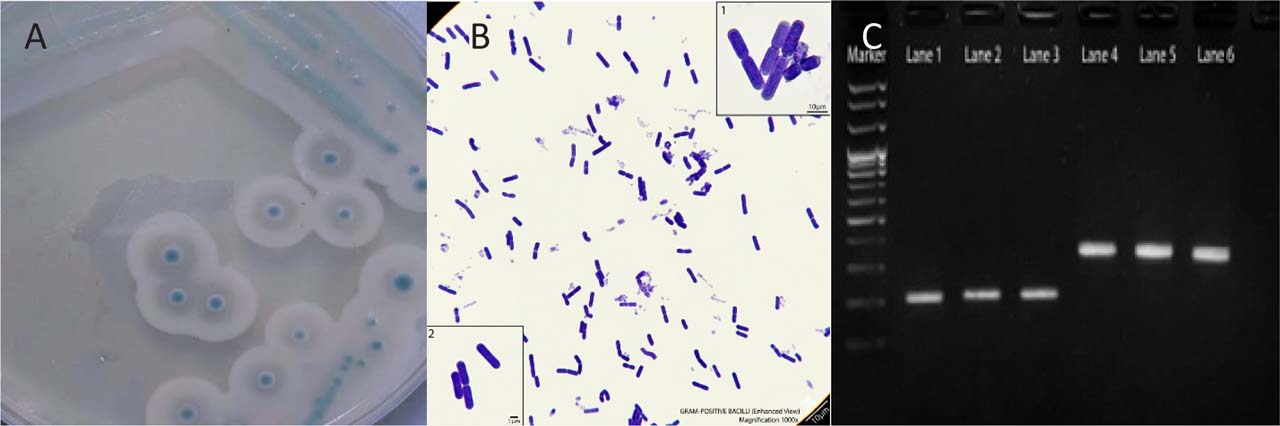
Fig. 1.
Phenotypic and molecular identification of Listeria monocytogenes. (A) Typical L. monocytogenes colonies on Listeria CHROMagar, appearing as small blue colonies surrounded by a white halo. (B) Microscopic examination showing Gram-positive coccobacilli. (C) PCR amplification of L. monocytogenes: lane M, molecular size marker; lanes 1–3, hly gene (372 bp); and lanes 4–6, iap gene (205 bp). PCR products were separated on a 1.5% agarose gel.
Distribution of L. monocytogenes in various foods
L. monocytogenes was detected in 29 of 778 examined samples, corresponding to an overall prevalence of 3.73%. The prevalence was 2.99% in animal-origin samples and 4.12% in plant-origin samples. However, Fisher’s exact test showed no significant association between sample origin and L. monocytogenes detection (p = 0.551)
Table III presents the distribution of L. monocytogenes in different food samples. Overall, L. monocytogenes prevalence was 3.7% among all samples examined. No significant difference was observed between foods of plant and animal origin. However, contamination was relatively higher in frozen chicken meat and certain vegetables, particularly beetroot, lettuce, and leeks. In contrast, local chicken meat, watercress, and local cheese samples showed low or no contamination. Recent studies similarly report increased L. monocytogenes recovery from poultry meat due to cross-contamination during processing and the organism’s psychrotrophic ability, which allows it to survive in frozen environments (EFSA 2020). Fresh vegetables also showed considerable positivity, consistent with recent findings highlighting contamination through soil, irrigation water, and post-harvest handling (Smith et al. 2021). Leafy vegetables such as lettuce and garden cress demonstrated notable levels, supporting reports that rough surfaces promote bacterial adherence (Zhang & Wiedmann, 2020). This study detected no L. monocytogenes in the local cheese samples examined, consistent with reports of low or undetectable prevalence in some dairy products (Carvalho et al. 2022).
Table III
Distribution of Listeria monocytogenes in different local food samples.
| Sample type | No. of samples | No. of positive samples for L. monocytogenes | % of positive samples for L. monocytogenes |
|---|---|---|---|
| Local chicken meat | 70 | 1 | 1.4 |
| Frozen chicken meat | 88 | 5 | 5.68 |
| Minced meat | 80 | 2 | 2.5 |
| Lettuce | 77 | 4 | 5.1 |
| Root beet | 87 | 5 | 5.7 |
| Celery | 84 | 2 | 2.38 |
| Local cheese | 30 | 0 | 0 |
| Radish | 76 | 3 | 3.95 |
| Leeks | 80 | 4 | 5 |
| Watercress | 66 | 1 | 1.5 |
| Garden cress | 40 | 2 | 5 |
| Total | 778 | 29 | 3.7 |
Evaluation of antimicrobial susceptibility of L. monocytogenes isolates
Table IV summarizes the antibiotic resistance profiles of the 29 Listeria monocytogenes isolates. Overall, the isolates were highly susceptible to several commonly used antibiotics, including β-lactams and other key therapeutic agents. In contrast, notable resistance was observed to several antibiotics, particularly moderate resistance to erythromycin. Only minimal resistance was found to trimethoprim–sulfamethoxazole. The antimicrobial susceptibility pattern observed in this study differs from those reported in previous investigations, suggesting that geographic location and local antimicrobial practices influence resistance in L. monocytogenes. Reports of resistance to commonly used antibiotics, including β-lactams and aminoglycosides (Zhang et al. 2019), as well as the high prevalence of multidrug resistance described in Iraqi isolates (Al-Brefkani 2023), may reflect extensive or uncontrolled antibiotic usage in clinical and agricultural settings. In contrast, the susceptibility observed here may indicate reduced selective pressure or differences in isolate sources, such as food-related versus clinical environments. The variability reported in more recent European studies (Nikolaou et al. 2025) further supports the idea that resistance trends are dynamic and may change over time due to improved antimicrobial stewardship and regulatory measures.
Table IV
Antibiotic susce ptibility test of 29 Listeria monocytogenes isolates.
| Antibiotics | Dose µg/disk | Sensitivity of the isolates (No.) | Resistant strains (%) | ||
|---|---|---|---|---|---|
| Sensitive | Intermediate | Resistant | |||
| Ampicillin | 10 | 29 | 0 | 0 | 0% |
| Gentamicin | 10 | 27 | 2 | 0 | 0% |
| Tetracycline | 30 | 14 | 6 | 9 | 31% |
| Co-trimoxazole | 25 | 27 | 0 | 2 | 6.9% |
| Vancomycin | 30 | 29 | 0 | 0 | 0% |
| Ciprofloxacin | 5 | 24 | 1 | 4 | 13.8% |
| Penicillin | 10 | 29 | 0 | 0 | 0% |
| Erythromycin | 15 | 16 | 1 | 12 | 41.4% |
| Imipenem | 10 | 29 | 0 | 0 | 0% |
| Chloramphenicol | 30 | 28 | 1 | 0 | 0% |
Biofilm formation
The biofilm-forming capability of the 29 L. monocytogenes isolates was estimated using the microtiter plate assay, as presented in Table V. Overall, most isolates exhibited moderate biofilm-forming capacity, followed by strong producers, while fewer isolates were grouped as weak biofilm producers. The predominance of moderate biofilm-forming isolates observed in this study may reflect an adaptive balance between energy expenditure and persistence in food-related environments. Moderate biofilm production can provide sufficient protection against environmental stressors such as cleaning procedures, temperature fluctuations, and nutrient limitation while avoiding the higher metabolic cost associated with strong biofilm formation. Studies of isolates from food-processing environments have suggested this adaptive strategy (Bonsaglia 2014). In contrast, the variability reported in clinical isolates, where weaker biofilm formation is more common (Doijad 2015), may indicate that biofilm formation is less critical for survival in host-associated conditions than in external environments. These differences suggest that ecological niche and selective pressures influence biofilm-forming capacity. L. monocytogenes’ ability to form biofilms at varying levels highlights its persistence in food-processing settings and underscores the importance of effective sanitation strategies to control contamination and reduce potential public health risks.
GC mass analysis of clove oil
GC mass analysis of clove oil shows that Eugenol accounted for 94.97% of the total detected peak area in the extracted oil as it is clear from Table VI and Fig. 2.
Table VI
Chemical composition of clove oil extract determined by GC–MS
| NO. | Retention Time | Area (%) | Name |
|---|---|---|---|
| 1 | 20.167 | 94.97 | Eugenol |
| 2 | 22.217 | 0.56 | Caryophyllene |
| 3 | 23.050 | 0.05 | Isoeugenol, TMS derivative |
| 4 | 23.175 | 0.11 | Alpha -Caryophyllene |
| 5 | 24.475 | 3.89 | Phenol, 2-methoxy-4-(1-propenyl)- |
| 6 | 27.375 | 0.41 | Caryophyllene oxide |
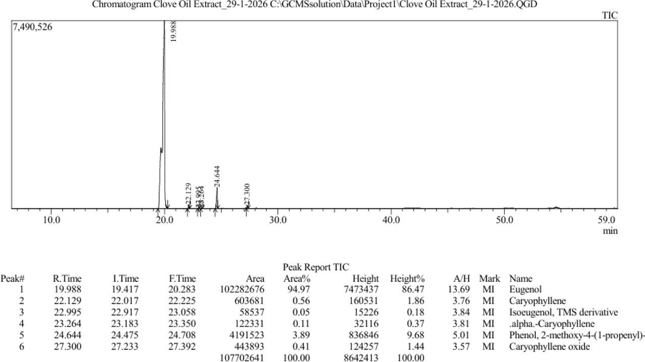
Fig. 2.
Analytical parameters, identification, and quantification of clove oil extract by GC mass.
Eugenol is the major constituent of clove oil, generally accounting for 55–90% of its composition, and exhibits in vitro antibacterial, antifungal, and antiviral activities (Marchese, Barbieri et al. 2017). Studies have shown that clove oil and its major component, eugenol, strongly inhibit a wide range of foodborne microorganisms. These effects are linked to their ability to impair microbial adhesion and motility, suppress biofilm formation, and reduce the production of key virulence factors. These effects are also associated with its ability to disrupt microbial cell membranes. Moreover, in vivo studies indicate that both clove oil and eugenol are generally recognized as safe for use (Hu, Zhou et al. 2018). Clove oil’s impact on L. monocytogenes cell membrane permeability was demonstrated in another investigation (Cui, Zhang et al. 2018). The results showed that when bacterial isolates were treated with clove oil, significant macromolecules like protein, ATP, and DNA leaked out, while the concentration of intracellular enzymes like β-galactosidase and AKP dropped. Using the inhibition rate and superposition rate tests, the same study demonstrated its impact on the TCA pathway in the respiratory system. Three crucial enzymes in the TCA pathway, isocitrate dehydrogenase, citrate synthase, and α-ketoglutarate dehydrogenase can have their activity reduced following treatment with clove oil.
MIC determination and planktonic growth analysis of L. monocytogenes isolates
In the evaluation of antibacterial activity of quercetin and clove oil against two isolates of multidrug-resistant and biofilm-producing L. monocytogenes, the quercetin MIC value was 0.45 mg/ml, as shown in Table VII. This concentration is moderately high compared with other published reports. In research performed on a mixed culture of L. monocytogenes, the MIC value of quercetin was found to be 0.25mg/ml (Roy et al. 2023). Previous research on Gram-positive bacteria has shown that quercetin exhibits a wide range of MICs, generally between 125 and 500 µg/ml, varying by organism and strain. For instance, the reported MIC levels ranged from 250 to 500 µg/ml when testing different Staphylococcus species and similar Gram-positive microbes (da Costa Júnior, de Oliveira Santos et al. 2018). In contrast, the antibacterial activity of clove oil against the same two isolates of L. monocytogenes was 0.4 mg/ml. This MIC value is in the same range as several studies report that show clove oil inhibits L. monocytogenes. In a study, they reported an MIC of ~0.5 mg/ml (MBC 1.0 mg/ml) for clove oil against L. monocytogenes, which is slightly higher (Cui et al. 2018). Meanwhile, much lower MICs (0.05 mg/ml) were reported by Somrani et al. (2022). These variations may result from differences in the tested bacterial isolates, chemical composition of the compounds, extraction procedures, solvent systems, inoculum concentration, growth conditions, and the method used to determine the MIC endpoint. More recently, Fidelis et al. (2025) reported that food-plant essential oils, including clove oil, may inhibit biofilm formation and disrupt quorum sensing in foodborne pathogens. However, they emphasized the need to validate these effects in food matrices and to consider toxicity, allergenicity, formulation, and possible bacterial adaptation (dos Santos Ferreira, de Aguiar Bernardo et al. 2025).
Growth curve
Growth curves were constructed for the two selected isolates by monitoring bacterial growth for 18 h in the presence and absence of sub-MIC concentrations of clove oil and quercetin (Fig. 3). Both isolates reached the stationary phase approximately 12–14 h after inoculation. Because bacterial gene expression is influenced by growth phase and growth rate (Klumpp and Hwa, 2014), these growth curves were used to identify the respective mid-exponential phase for RNA collection and subsequent RT-qPCR analysis.
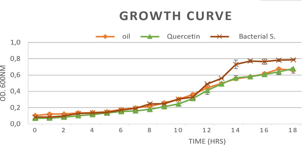
Fig. 3.
Growth curves of Listeria monocytogenes isolates cultured with or without sub-MIC concentrations of clove oil and quercetin for 18 h. Data points represent the means of three biological replicates.
Repression of regulatory genes by clove oil in selected L. monocytogenes isolates
Clove oil showed a marked anti-virulence effect against L. monocytogenes at a subinhibitory concentration, as shown by significant repression of key regulatory genes involved in quorum sensing, stress adaptation, and virulence control (Fig. 4A and Fig. 4B; Table VIII). In both LM1 and LM2 isolates, treatment with clove oil resulted in downregulation of agrA and agrB, indicating interference with the agr quorum-sensing system. Since agrB is required for the processing and export of the autoinducing peptide, whereas agrA functions as the response regulator controlling downstream transcription, the simultaneous suppression of these genes suggests disruption of both signal production and signal transduction. This finding is mechanistically relevant, as the agr system plays an important role in coordinating population-density-dependent behaviors associated with virulence, biofilm formation, and adaptation to adverse environmental conditions (Autret et al. 2003; Zetzmann et al. 2019; Mao et al. 2023).
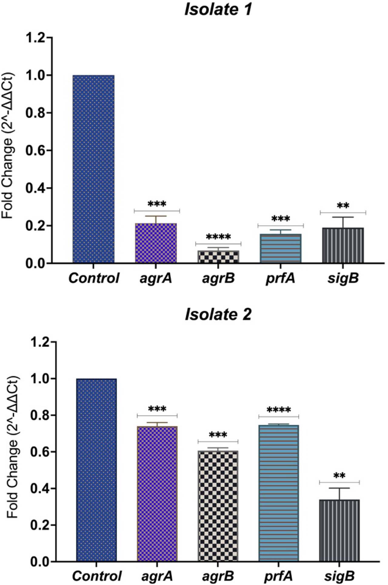
Fig. 4.
Effect of clove oil on the relative expression of agrA, agrB, prfA, and sigB in Listeria monocytogenes isolates LM1 (A) and LM2 (B). Gene expression was normalized to the 16S rRNA reference gene and calculated using the 2−ΔΔCt method relative to the untreated control. Bars represent the mean ± SD of three biological replicates.
Table VIII
Relative gene expression (agrA, agrB, prfA, and sigB) in Listeria monocytogenes isolates treated with sub-MIC clove oil.
| Isolate No. | Gene | Fold change | Regulation |
|---|---|---|---|
| LM1 | agrA | 0.21 | down-regulated |
| agrB | 0.07 | down-regulated | |
| prfA | 0.15 | down-regulated | |
| sigB | 0.19 | down-regulated | |
| LM2 | agrA | 0.74 | down-regulated |
| agrB | 0.61 | down-regulated | |
| prfA | 0.74 | down-regulated | |
| sigB | 0.37 | down-regulated |
The greater reduction in agrA and agrB expression observed in LM1 compared with LM2 may reflect isolate-dependent differences in susceptibility to clove oil, possibly related to differences in regulatory background or physiological state. Nevertheless, both isolates showed the same overall transcriptional pattern, supporting the conclusion that clove oil consistently interferes with agr-mediated signaling. Because agr downregulation has been associated with reduced secretion of virulence-associated proteins, weakened quorum-sensing regulation, and attenuation of infection-related traits, the present findings provide molecular support for the quorum-quenching activity of clove oil against L. monocytogenes (Autret et al. 2003; Zetzmann et al. 2019).
In addition to its effect on quorum sensing, clove oil significantly repressed prfA, the master transcriptional regulator of virulence in L. monocytogenes. This observation is particularly important because PrfA controls the expression of major pathogenicity determinants, including genes involved in host-cell invasion, intracellular survival, and cell-to-cell spread. Therefore, reduced prfA transcription strongly suggests attenuation of the organism’s global virulence potential. The more pronounced suppression in LM1 and the moderate but clear reduction in LM2 indicate that clove oil can impair this central regulatory pathway across different isolates. From an antivirulence perspective, repression of prfA is especially significant because targeting a master regulator may reduce pathogenicity without directly affecting bacterial viability, thereby potentially lowering selective pressure for resistance development (de las Heras et al. 2011).
A similarly important finding was the downregulation of sigB, which indicates that clove oil may also weaken the bacterial stress-response network. Sigma B is a global alternative sigma factor that regulates many genes associated with tolerance to acid, osmotic, oxidative, and other environmental or host-related stresses. It also contributes to the regulation of virulence-associated functions. Consequently, reduced sigB expression suggests impaired adaptive capacity and decreased stress tolerance in treated cells. This effect may compromise L. monocytogenes’ ability to persist under food-processing conditions and within the host environment, further reducing its pathogenic fitness (Palmer et al. 2011).
Taken together, the simultaneous downregulation of agrA, agrB, sigB, and prfA indicates that clove oil exerts a multi-target regulatory effect on L. monocytogenes. Rather than acting on a single virulence determinant, clove oil appears to interfere with interconnected pathways governing cell-to-cell communication, environmental adaptation, and pathogenicity. In this context, inhibiting the agr system may impair quorum-sensing-dependent coordination of virulence traits, repressing sigB may reduce stress resilience, and downregulating prfA may suppress expression of major pathogenicity genes. Together, these effects likely weaken the bacterium’s ability to survive, adapt, and establish infection.
The present findings are in agreement with previous reports demonstrating that disruption of agr-mediated signaling weakens Listeria pathogenicity and affects biofilm-associated behavior (Autret et al. 2003; Zetzmann et al. 2019). They are also consistent with the established functions of SigB as a key stress-response regulator and PrfA as the master activator of the LIPI-1 virulence program (Palmer et al. 2011; de las Heras et al. 2011). Furthermore, the present results generally support the observations of Zhang et al. (2020), who reported that clove oil downregulated agrA and prfA, thereby inhibiting biofilm formation. Collectively, these data suggest that clove oil acts not only as an antimicrobial compound but also as a quorum-sensing inhibitory and antivirulence agent capable of reducing the pathogenic potential of L. monocytogenes.
Repression of regulatory genes by quercetin in selected L. monocytogenes isolates
Quercetin treatment significantly downregulated all examined regulatory genes in both L. monocytogenes isolates (Fig. 5A, B and Table IX). The most pronounced effect was observed for the quorum-sensing regulators agrA and agrB, particularly in isolate LM1, indicating marked suppression of the agr signaling system. In addition, substantial repression of the master virulence regulator prfA and the general stress-response regulator sigB was detected in both isolates. Collectively, these findings indicate that quercetin broadly affects regulatory pathways associated with quorum sensing, virulence, and stress adaptation.
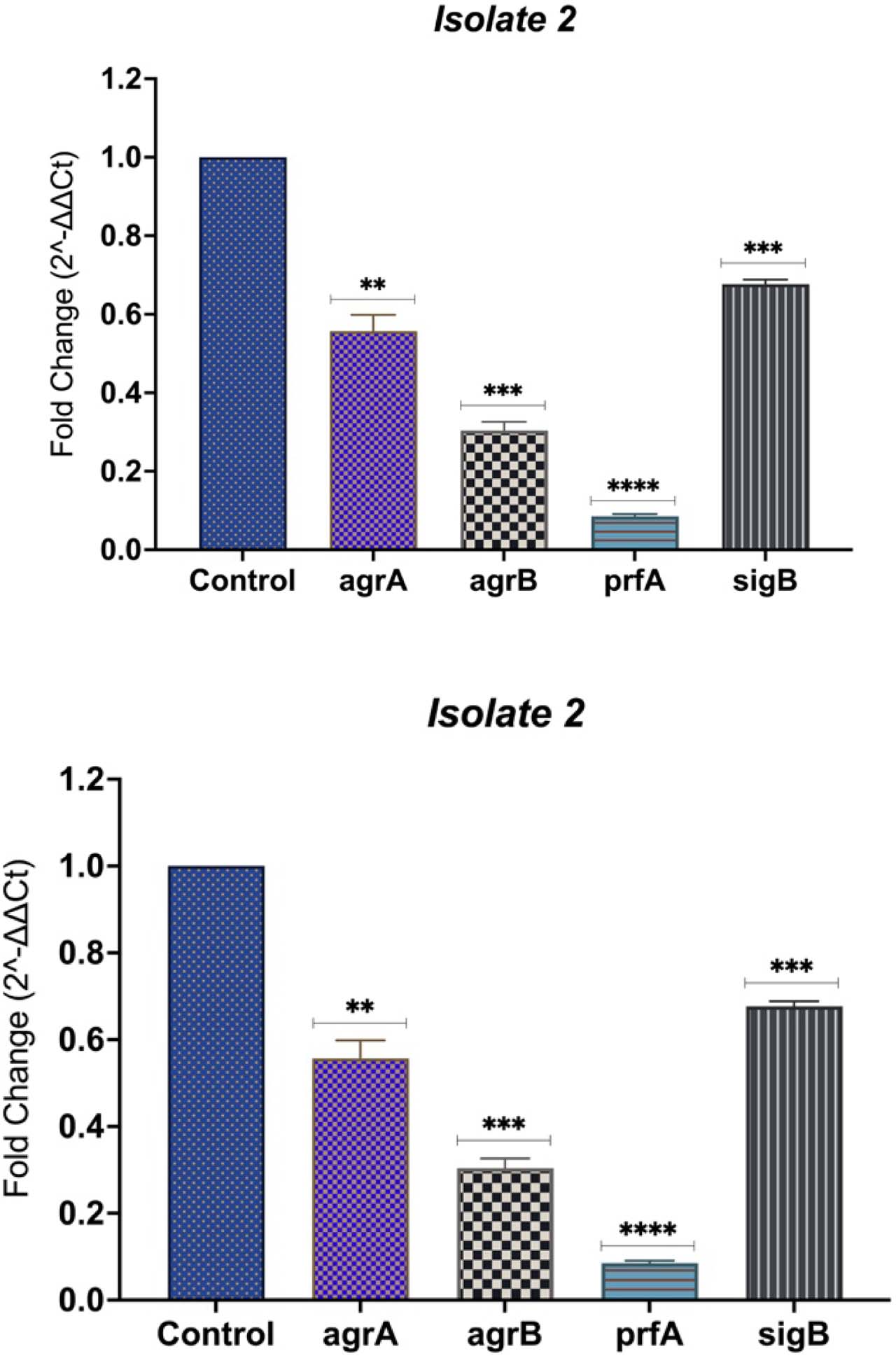
Fig. 5.
Effect of quercetin on regulatory gene expression in Listeria monocytogenes isolates. Relative expression levels of agrA, agrB, prfA, and sigB following quercetin treatment are shown for (A) isolate 1 and (B) isolate 2. Gene expression was normalized to the 16S rRNA reference gene and calculated using the 2−ΔΔCt method, relative to the corresponding untreated control. Bars represent the mean ± SD of three independent biological replicates. Asterisks indicate statistically significant differences compared with the untreated control (p < 0.05, *p < 0.01, **p < 0.001, ***p < 0.0001).
Table IX
Relative gene expression (agrA, agrB, prfA, and sigB) in Listeria monocytogenes isolates treated with sub-MIC quercetin.
| Isolate No. | Gene | Fold Change | Regulation |
|---|---|---|---|
| LM1 | agrA | 0.07 | downregulated |
| agrB | 0.08 | downregulated | |
| prfA | 0.16 | downregulated | |
| sigB | 0.24 | downregulated | |
| LM2 | agrA | 0.3 | downregulated |
| agrB | 0.56 | downregulated | |
| prfA | 0.06 | downregulated | |
| sigB | 0.24 | downregulated |
These findings indicate that quercetin acts at multiple regulatory levels in L. monocytogenes, suggesting a broader antivirulence mechanism than inhibition of a single target. The downregulation of agrA and agrB supports the idea that quercetin interferes with the agr quorum-sensing system, a regulatory circuit associated with virulence and adaptive behavior in Listeria. This interpretation agrees with Autret et al. (2003), who identified the agr locus and showed its role in bacterial virulence, and with later work showing that impaired Agr peptide sensing alters the biofilm phenotype of L. monocytogenes. Therefore, these findings provide evidence that quercetin suppresses this signaling pathway at the regulatory-gene level, which strengthens the argument that its activity includes quorum-sensing inhibition rather than only general growth suppression. These results also agree with previous studies on quercetin. Roy et al. (2022b) showed that quercetin reduced L. monocytogenes biofilm formation and repressed virulence-related genes, including sigB and prfA, in mixed cultures on food-contact surfaces. The current findings extend that observation by showing that quercetin also suppresses agrA and agrB, suggesting that its activity may begin upstream at the level of quorum-sensing regulation, with downstream effects on stress response and virulence control. This broader mechanistic interpretation helps explain why quercetin has both antibiofilm and antivirulence activity and supports its potential use as a natural control compound against L. monocytogenes.
The potential application of clove oil and quercetin in food preservation has gained attention due to their antimicrobial and antibiofilm properties. However, their effectiveness may vary in real food systems depending on food composition and processing conditions (Hyldgaard et al. 2012). Therefore, further studies using food-matrix models, sensory evaluation, and safety assessments are required before potential industrial application.
Study limitations
The findings of this study should be interpreted cautiously because food samples were obtained from selected retail markets in Erbil using non-random sampling, and the number of samples varied across food categories, which may limit comparisons and broader generalization. Therefore, the prevalence results mainly reflect the sampled foods and markets. In addition, molecular analyses and gene-expression evaluations were performed on only two selected strong biofilm-producing L. monocytogenes isolates, which may limit the applicability of these findings to the wider population of isolates. Nevertheless, the large sample size and standardized identification methods support the reliability of the findings within the studied population. Further studies involving broader sampling, larger isolate collections, and food-matrix models are required to validate the antibiofilm effects of clove oil and quercetin under realistic food conditions.
Conclusions
In conclusion, the findings of this study suggest that sub-MIC concentrations of clove oil and quercetin can reduce biofilm formation and downregulate selected quorum-sensing, stress-response, and virulence-associated genes, including agrA, agrB, sigB, and prfA, in two selected multidrug-resistant, strong biofilm-producing L. monocytogenes isolates. These results indicate that both compounds may have potential antibiofilm and antivirulence activity. However, because gene-expression analysis was performed on only two selected isolates under in vitro conditions, the findings should be interpreted cautiously. Further studies involving a larger number of isolates, food-matrix models, sensory evaluation, toxicity assessment, and in vivo validation are required before these compounds can be recommended for practical application in food preservation.
Notes
[1] Ethical statement
The Ethics Committee of the College of Dentistry, Hawler Medical University, reviewed and approved the research proposal. Because the study involved isolating bacteria from food samples and did not include human participants or experimental animals, informed consent was not applicable.
[2] Contributed by Author contributions
Shean Ismail Salih, the PhD student, conducted the experimental work, contributed to the methodology and investigation, collected and analysed the data, and prepared the initial draft of the manuscript. Ahang Hasan Mawlood supervised the study, contributed to the study design and interpretation of the results, and critically reviewed and edited the manuscript. Both authors read and approved the final version of the manuscript.