
Introduction
Cancer remains one of the most significant public health burdens worldwide. In 2022, an estimated 20 million new cases were diagnosed globally, with 9.7 million associated deaths recorded during the same period (WHO 2024). At the population level, approximately one in five individuals will develop cancer over their lifetime, and the disease is responsible for death in one in nine men and one in twelve women (WHO 2024).
Cancer patients, particularly those undergoing chemotherapy or other immunosuppressive treatments, are highly susceptible to bacterial infections (Alevizakos et al. 2016). Consequently, infections in cancer patients are associated with a substantially higher mortality rate, with studies reporting up to a threefold increase in the risk of death compared with infected individuals without cancer (Nanayakkara et al. 2021). This elevated risk is partly attributable to the growing prevalence of multidrug-resistant (MDR) bacteria in clinical, animal, and environmental settings, posing significant epidemiological and therapeutic challenges, especially in oncology healthcare facilities (Naghavi et al. 2024).
Current strategies to combat MDR include the development of new antibiotics, improved infection prevention measures, optimized antibiotic use, and the application of nanoparticles (NPs) (Khan et al. 2019). NPs are particularly promising because they can simultaneously target multiple microbial structures and pathways, thereby limiting the ability of microorganisms to develop antibiotic resistance (Baptista et al. 2018). Moreover, several metallic nanoparticles have been reported to exhibit acceptable biocompatibility at low concentrations (Alamdari et al. 2020). While NPs can be synthesized using physical or chemical methods, biosynthesis using plants or microorganisms offers an environmentally friendly and cost-effective alternative, utilizing naturally occurring biomolecules as reducing and stabilizing agents (Koul et al. 2021; Alloosh et al. 2021).
Among metal-based nanoparticles, zinc oxide nanoparticles (ZnO-NPs) are distinguished by several advantageous characteristics, including the relatively low cost of zinc as a raw material, favorable optical and electrical properties, and broad regulatory acceptance (Liao et al. 2020). Zinc is also recognized as a safe element by the U.S. Food and Drug Administration (FDA), with a recommended daily intake of 10–15 mg (Rahman et al. 2018). These characteristics make ZnO-NPs suitable for a wide range of biomedical applications, including antimicrobial therapy (Abdulsattar et al. 2018).
In this study, ZnO-NPs were biosynthesized using an aqueous leaf extract of ARVR and comprehensively characterized. The phytochemical composition of the extract was further analyzed, and the antibacterial, antibiofilm, and cytotoxic activities of the synthesized nanoparticles were systematically evaluated against MDR bacterial strains isolated from cancer patients. To the best of our knowledge, this represents the first reported investigation into ARVR-mediated ZnO-NPs as multifunctional agents with potential activity against clinically relevant microbial pathogens, alongside an assessment of their mammalian cell safety profile.
Experimental
Materials and Methods
Bacterial strain isolation and identification
Bacterial isolates were collected in 2024 from the laboratory unit of Hiwa Hospital, Sulaymaniyah, Iraq. Species identification and antimicrobial susceptibility testing (AST) were conducted using the BD Phoenix™ automated system (BD Diagnostic Systems).
Molecular identification was carried out by PCR following previously described protocols (Mohamedsalih and Sabir 2020). Genomic DNA was extracted using the Presto™ Mini gDNA Bacteria Kit (Geneaid) in accordance with the manufacturer’s instructions. Amplification of the bacterial 16S rRNA gene was performed in a 50 μl reaction volume containing Prime Taq Premix (2×) (GeNet Bio) and 0.5 μM of each primer: forward (5′-AGAGTTTGATYMTGGCTCAG-3′) and reverse (5′-ACGGYTACCTTGTTACGACTT-3′) (Satokari, Vaughan, Akkermans, Saarela, & de Vos, 2001). Thermal cycling parameters comprised an initial denaturation step at 95°C for 30 s, followed by 30 cycles of denaturation at 95°C for 30 s, annealing at 58°C for 30 s, and extension at 72°C for 2 min, with a final extension at 72°C for 5 min. PCR products were subjected to Sanger sequencing at Macrogen (Republic of Korea). Sequence quality was assessed and edited using Chromas software (Technelysium, v.2.6.6).
Construction of the phylogenetic tree
The partial 16S rRNA gene sequences of the isolates were submitted to the NCBI database via the WebSub platform to obtain accession numbers. Taxonomic identification of the bacterial isolates was carried out using BLAST searches of the partial 16S rRNA gene sequences against the NCBI nucleotide database. Multiple sequence alignment of these 16S rRNA sequences with closely related reference sequences was performed using ClustalX version 2.1 with default parameters (Larkin et al. 2007). Phylogenetic analysis was performed in MEGA version 11 (Kumar et al. 2016) using the Neighbor-Joining method with 1000 bootstrap replicates, and tree visualization was also carried out in MEGA.
Collection and molecular identification of plant
Specimens of ARVR were collected from Betwata Mountain, Ranya, Kurdistan Region, Iraq (36°19′18.43″ N, 44°56′48.61″ E) (Fig. 1). Taxonomic identification was performed using both morphological and molecular approaches. Morphological identification was carried out by experts at the College of Agricultural Herbarium, University of Sulaimani, Iraq, while molecular identification followed the protocols of Khal et al. (2023b). Briefly, genomic DNA was extracted from 300 mg of powdered leaf tissue. The internal transcribed spacer (ITS) region was amplified using primers ITS1 (5′-TCCGTAGGTGAACCTGCGG-3′) and ITS4 (5′-TCCTCCGCTTATTGATATGC-3′) (Hsiao et al. 1995) in a 26 μl reaction mixture comprising 5 μl template DNA, 10 μl Taq Master Mix, 2 μl of each primer, and 7 μl nuclease-free water. Cycling conditions consisted of an initial denaturation at 94°C for 10 min, followed by 35 cycles of 94°C for 1 min, 55°C for 1 min, and 72°C for 2 min, with a final extension at 72°C for 10 min. Amplicons were visualized on 1.5% agarose gels, purified using a Gel Extraction Kit, and sequenced at Macrogen Inc. (Seoul, Republic of Korea). The sequence was aligned and compared with NCBI reference sequences for species identification, and accession numbers were obtained by submitting the sequences via the NCBI WebSub platform.
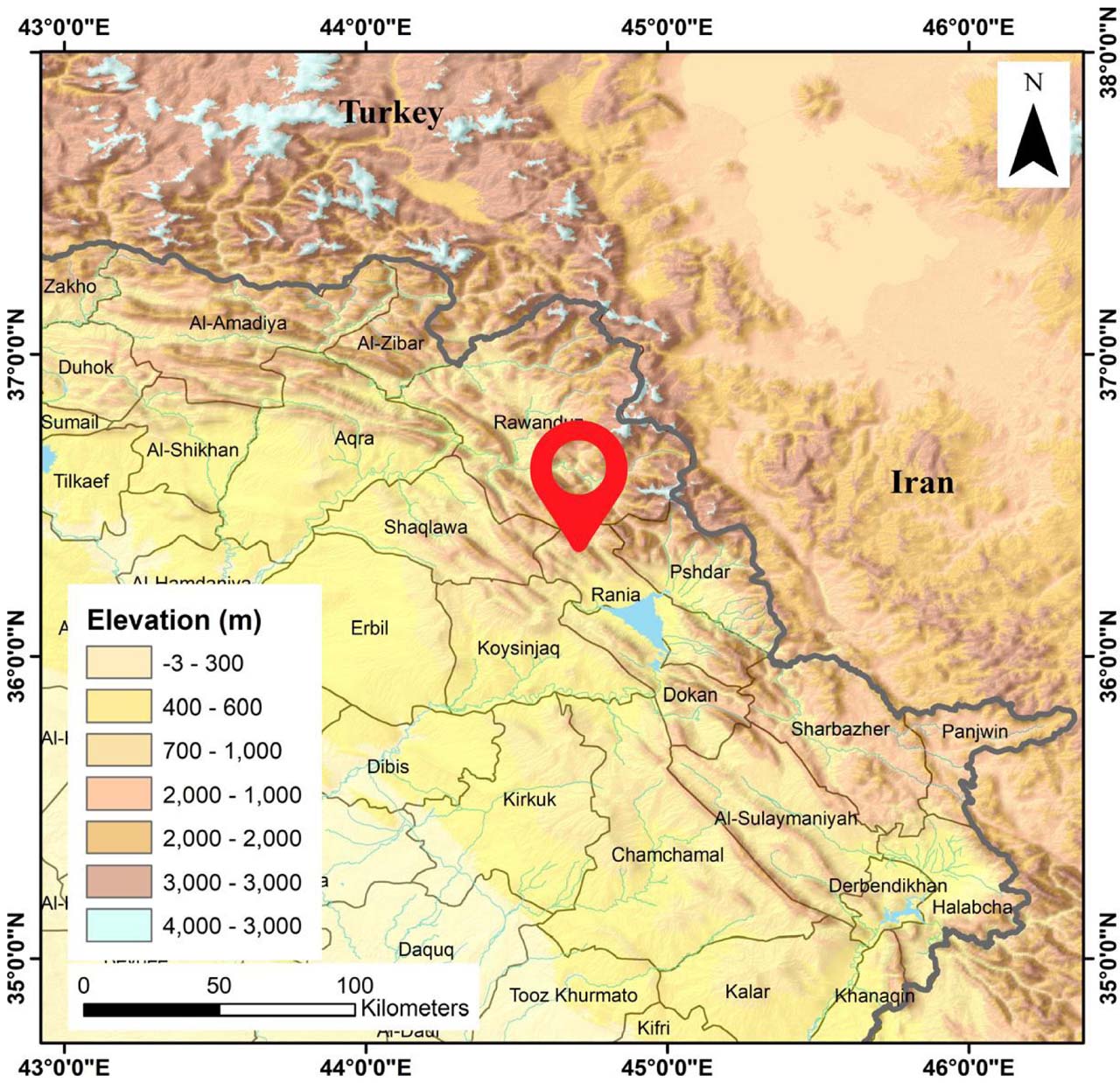
Fig. 1.
Collection site of ARVR on Betwata Mountain near Ranya, Kurdistan Region, Iraq. The red marker indicates the sampling location (36°19′18.43″ N, 44°56′48.61″ E). Elevation is shown in meters, with darker shades representing higher altitudes.
GC–MS chemical profiling and phytochemical analysis of the plant extract
The chemical composition of ARVR leaf extract was analyzed using gas chromatography–mass spectrometry (GC–MS). GC–MS analysis was performed on an Agilent 7890B gas chromatograph coupled to an Agilent 5977 mass selective detector (Agilent Technologies, USA) and fitted with an HP-5MS UI capillary column (30 m × 0.25 mm i.e., 0.25 μm film thickness), operated in spitless injection mode. The oven temperature was held at 40°C for 1 min and then increased to 300°C at a rate of 10°C min−1. Helium was used as the carrier gas at a constant flow rate of 1 ml min−1. The injector temperature was set to 290°C, and 1 μl of sample was injected. Mass spectra were recorded over an m/z range of 35–650, with ion source, quadrupole, and transfer line temperatures maintained at 230, 150, and 290°C, respectively. A solvent delay of 35–40 min was applied. Chemical constituents were identified by comparing the acquired mass spectra with those in standard mass spectral libraries (Arunachalam et al. 2012; Khal et al. 2023a).
Phytochemical analyses were conducted to evaluate the major bioactive constituents of the extract. Total phenolic content was determined using the Folin–Ciocalteu method (Singleton and Rossi 1965), which is based on the reduction of the phosphomolybdic–phosphotungstic reagent by phenolic compounds. Total flavonoid content was quantified using the aluminum chloride colorimetric method (Shraim et al. 2021), which involves the formation of a stable colored complex between flavonoids and Al3+. All the experiments were performed in triplicate, and results were expressed as milligrams of gallic acid equivalents per gram of extract (mg GAE g−1).
Biosynthesis of ZnO-NPs
ARVR leaves were used for the biosynthesis of ZnO-NPs following a previously described protocol (Salih et al. 2020). Ten grams of shade-dried, crushed leaves were extracted in 100 ml of distilled water by sonication for 15–20 min and heating at 40°C for 2 h. The extract was filtered through Whatman filter paper and stored overnight in an airtight bottle.
For NPs synthesis, 50 ml of 0.1 M zinc nitrate solution (Zn(NO3)2·6H2O; Sigma-Aldrich) was prepared and stirred at 60 ± 3°C for 10 min. An equal volume of the plant aqueous extract was then added (1:1 ratio) and pH was adjusted to 8.0 with 2 M NaOH, stirred for 2 h until the solution turned pale yellow (Fig. 2). The product was collected by centrifugation at 10,000 rpm for 20 min.
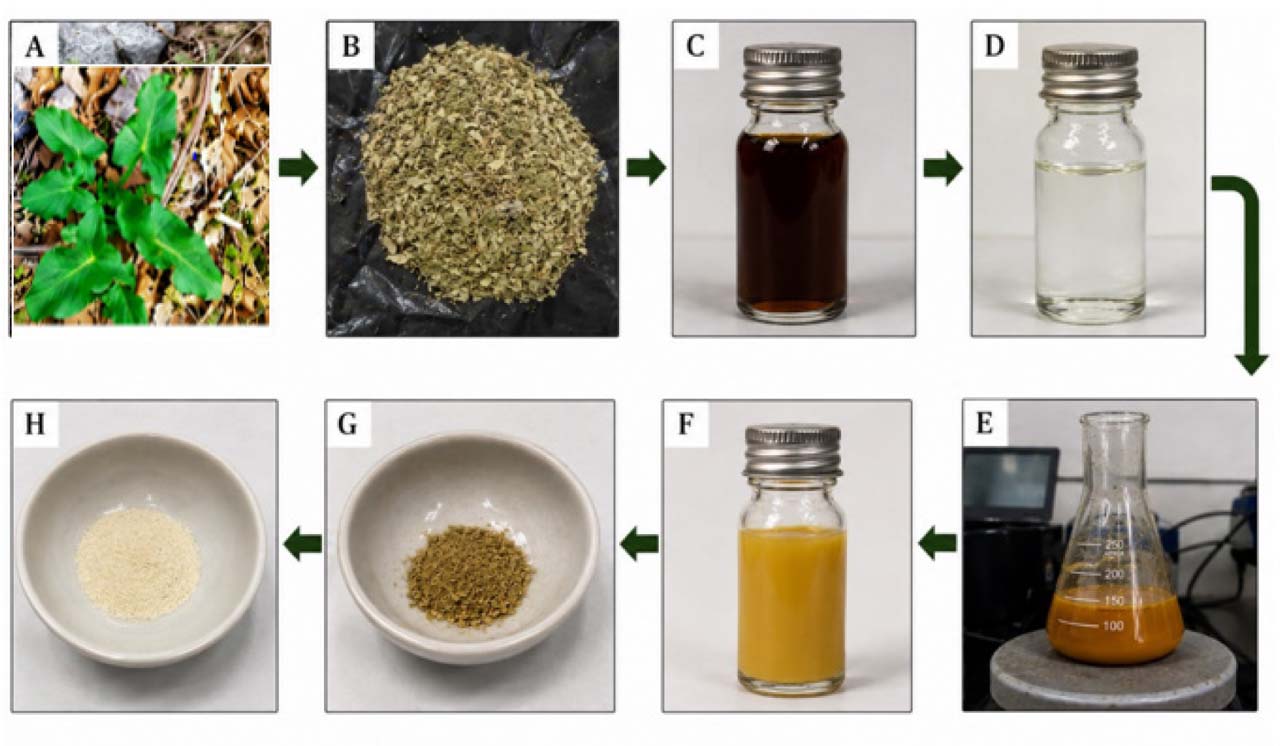
Fig. 2.
Diagrammatic illustration of the biofabrication process of ZnO-NPs. (A) ARVR specimen. (B) Dried leaf powder. (C) Aqueous extract. (D) Zn(NO3)2·6H2O solution. (E and F) ZnO-NPs after 2 h of stirring and heating at 60 ± 3°C with plant extract. (G) ZnO-NPs before calcination. (H) ZnO-NPs after calcination in a muffle furnace.
The precipitate was washed three times with distilled water, dried in a hot air oven at 80°C for 24 h, and calcined in a muffle furnace at 430°C for 2 h to improve crystallinity and remove residual organics. The resulting fine, cream-colored ZnO-NPs powder was used for physicochemical characterization and for the evaluation of antimicrobial and antibiofilm activities (Fig. 2).
Characterization of biosynthesized ZnO-NPs
The biosynthesized ZnO-NPs were comprehensively characterized using complementary physicochemical techniques to assess their optical properties, crystalline structure, surface chemistry, elemental composition, size distribution, colloidal stability, and morphology.
UV–visible spectroscopic analysis was first performed to confirm nanoparticle formation and evaluate optical characteristics. Measurements were conducted using a UV–Vis spectrophotometer (UviLine 9400, SI Analytics, EU) with a spectral resolution of 2 nm over the wavelength range of 200–800 nm, employing freshly prepared aqueous nanoparticle suspensions (Karanpal et al. 2019).
The crystalline structure and average crystallite size of the ZnO-NPs were subsequently analyzed by X-ray diffraction (XRD) using an X’Pert Pro diffractometer (PANalytical, Almelo, Netherlands) equipped with Cu-Kα radiation (λ = 1.5406 Å), operated at 45 kV and 40 mA. Diffraction patterns were recorded over a 2θ range of 10°–70° with a step size of 0.02° and a scan rate of 1°/min using powdered, oven-dried samples. The average crystallite size was estimated using the Debye–Scherrer equation (Holzwarth and Gibson 2011):
where D is the crystallite size (nm), K is the shape factor (0.90), λ is the X-ray wavelength, β is the full width at half maximum (FWHM) of the most intense peak, and θ is the Bragg angle.To investigate surface functional groups associated with nanoparticle reduction and stabilization, Fourier transform infrared (FTIR) spectroscopy was performed. Spectra were recorded in the range of 4000–400 cm−1 using an FTIR spectrometer (Nicolet iS50, Thermo Fisher Scientific) with a spectral resolution of 4 cm−1 and 32 scans per sample.
Elemental composition and purity were analyzed by energy-dispersive X-ray spectroscopy (EDS) coupled with a JEOL JSM-7610F scanning electron microscope (JEOL, Tokyo, Japan). Measurements were carried out in low-vacuum mode at an accelerating voltage of 15 kV, with a spot size of 3 and a working distance of 10.2 mm. Both point analysis and elemental mapping were conducted to confirm elemental distribution.
The hydrodynamic size distribution and dispersion behavior of the ZnO-NPs in aqueous media were evaluated using dynamic light scattering (DLS) with a HORIBA DLS instrument (HORIBA, Kyoto, Japan). Samples were dispersed in deionized water at a concentration of 0.1 mg·ml−1, sonicated for 10 min, equilibrated at 25°C, and analyzed in triplicate. The Z-average diameter and polydispersity index (PDI) were determined using cumulant analysis. In parallel, colloidal stability was assessed by zeta potential measurements using a HORIBA Zetasizer instrument, with analyses performed in deionized water at pH 7.0 and a controlled temperature of 25°C.
Surface morphology, particle shape, and aggregation state were examined using scanning electron microscopy (SEM) with a CamScan 3200 LV instrument operated at an accelerating voltage of 20 kV. Prior to imaging, samples were coated with a thin gold layer to minimize charging effects. Finally, Transmission electron microscopy (TEM) was employed to obtain high-resolution information on nanoparticle morphology, size distribution, and crystallinity using a Hitachi HF-3300 microscope operated at 300 kV. For TEM analysis, samples were ultrasonicated and deposited onto carbon-coated copper grids before imaging.
Antibacterial activity of ZnO-NPs
The antimicrobial activity of ZnO-NPs was assessed against clinically isolated bacteria using minimum inhibitory concentration (MIC) and minimum bactericidal concentration (MBC) assays. A 5 mg/ml stock solution was prepared by dissolving 0.025 g of ZnO-NPs in 1% dimethyl sulfoxide (DMSO) with distilled water, followed by sonication for 15 min at 30°C (Abomuti et al. 2021). MIC was determined according to CLSI guidelines (CLSI M100, 34th edition) (Clinical & Laboratory Standards, 2024). Overnight cultures of bacterial isolates were adjusted to approximately 108 CFU/ml. In sterile 96-well microtiter plates, 10 μl of bacterial suspension and 100 μl of ZnO-NPs (19–1250 μg/ml) were added to 90 μl of Mueller-Hinton broth (MHB). Blank wells containing ZnO-NPs in MH broth without bacteria were included to correct for background absorbance. Positive controls contained bacterial culture with MH broth, and negative controls contained MH broth only. Plates were incubated at 37°C for 20 h with shaking, and absorbance was measured at 600 nm using a microplate reader. For MBC determination, 5 μl from each well was spotted onto Mueller-Hinton agar (MHA) plates and incubated at 37°C for 24 h.
Biofilm formation assay
Biofilm formation was evaluated using the crystal violet microtiter plate assay, following a previously described method with minor modifications (Mousa et al. 2018). Briefly, bacterial isolates were cultured in Tryptic Soy Broth (TSB) at 37°C for 24 h with shaking. Cultures were adjusted to approximately 108 CFU/ml, and 200 μl of each bacterial suspension was dispensed into sterile 96-well flat-bottom polystyrene microtiter plates. Plates were incubated at 37°C for 24 h under gentle agitation using a PST-60 HL Plus microplate incubator-shaker (BOECO, Germany) to allow biofilm development.
Following incubation, planktonic cells were carefully removed, and the wells were washed three times with 200 μl of phosphate-buffered saline (PBS, pH 7.2) to remove non-adherent bacteria. The plates were then air-dried, and the adherent biofilms were stained with 0.1% (w/v) crystal violet for 30 min. Excess stain was removed by washing the wells three times with distilled water, followed by air-drying at room temperature. The bound dye was solubilized with 200 μl of 95% ethanol, and biofilm biomass was quantified by measuring absorbance at 595 nm using a Biotech Quant microplate spectrophotometer (USA).
Depending on their optical density (OD), bacterial isolates were classified as non-biofilm producers (OD ≤ ODc), weak biofilm producers (ODc < OD ≤ 2×ODc), moderate biofilm producers (2×ODc < OD ≤ 4×ODc), or strong biofilm producers (OD > 4×ODc), where ODc represents the optical density cut-off value calculated from the negative control (Mousa et al. 2018).
Biofilm inhibition assay using ZnO-NPs
The antibiofilm activity of ZnO-NPs was assessed using the crystal violet assay described above, with the inclusion of ZnO-NPs at sub-inhibitory concentrations, following a protocol adapted from Kıvanç et al. (2018). To initiate biofilm formation, bacterial isolates were cultured in tryptic soy broth (TSB) at 37°C for 24 h.
Subsequently, in sterile 96-well flat-bottom microtiter plates, 10 μl of bacterial suspension (108 CFU/ml) was combined with 100 μl of ZnO-NP suspensions at concentrations ranging from 4.8 to 156 μg/ml, and the volume was adjusted with TSB to a final volume of 200 μl per well. Wells containing TSB and bacterial suspension without ZnO-NPs served as untreated biofilm controls.
Plates were incubated at 37°C for 24 h to allow biofilm formation in the presence of ZnO-NPs. After incubation, wells were washed three times with PBS (pH 7.2), air-dried, and stained with 0.1% crystal violet for 30 min. Excess stain was removed by washing with distilled water, and the bound dye was solubilized with 200 μl of 95% ethanol. Absorbance was measured at 595 nm using a microplate reader to quantify biofilm biomass.
The percentage of biofilm inhibition was calculated using the following equation (Ozeki et al. 2014):
MTT Assay for cytotoxicity assessment of ZnO-NPs
The cytotoxic potential of ZnO-NPs was evaluated in human embryonic kidney (HEK-293) cells using the MTT assay. HEK-293 cells, obtained from the Pasteur Institute (Tehran, Iran), were cultured in Dulbecco’s Modified Eagle Medium (DMEM) supplemented with 10% fetal bovine serum (FBS) and 1% penicillin–streptomycin at 37°C in a humidified atmosphere with 5% CO2. ZnO-NPs were sterilized under ultraviolet (UV) irradiation for 72 h, dispersed in culture medium, and sonicated to ensure a homogeneous suspension. Cells were seeded into 96-well plates, allowed to adhere overnight, and exposed to ZnO-NPs at concentrations of 0, 10, 25, 50, 100, 250, 500, and 1000 μg/ml for 24 h. Following treatment, 20 μl of MTT solution (5 mg/ml) was added to each well and incubated for 4 h. The resulting formazan crystals were solubilized in dimethyl sulfoxide (DMSO), and absorbance was measured at 570 nm. Cell viability (%) was calculated relative to untreated controls, and the half-maximal inhibitory concentration (IC50) values were determined using a nonlinear regression model. Morphological changes of HEK-293 cells after ZnO-NP exposure were documented using an inverted light microscope.
Ethics approval
The study protocol was reviewed and approved by the Ethics Committee of the College of Science, Charmo University (Approval No. A23/2024). The microbial isolates used in this study were obtained from a diagnostic laboratory as part of routine clinical testing. All samples were anonymized, and no identifiable patient information was accessed.
Statistical analysis
All antibacterial and antibiofilm activity data are presented as mean ± standard deviation (SD) from three independent biological replicates. Statistical analyses were performed using GraphPad Prism (GraphPad Software Inc.). Differences among groups were evaluated by one-way analysis of variance (ANOVA), followed by Tukey’s post hoc multiple-comparison test. Statistical significance was defined as p < 0.05.
Results and Discussion
MDR bacteria present a major therapeutic challenge, particularly in immunocompromised patients such as those with cancer (Nanayakkara et al. 2021). In this study, ZnO-NPs were biosynthesized using ARVR leaf extract and characterized for their physicochemical properties. The antibacterial and antibiofilm activities of these nanoparticles were evaluated against clinically isolated MDR bacterial strains, and their cytotoxic potential was assessed in mammalian cells.
Phytochemical profile of the plant extract
Biosynthesis of ZnO-NPs depends on plant phytochemicals that naturally act as reducing and capping agents. In this study, an aqueous leaf extract of ARVR was used to reduce and stabilize Zn2+ during nanoparticle formation. The plant was collected from Betwata Mountain, Ranya, Kurdistan Region, Iraq (36°19′18.43″ N, 44°56′48.61″ E), authenticated at the College of Agricultural Herbarium, University of Sulaimani (voucher no. 5051), and plant identification further confirmed by ITS sequencing (GenBank: PX259642).
Although ZnO-NPs have been synthesized using several other plant species, including Nyctanthes arbortristis (Jamdagni et al. 2018), Trifolium pratense (Dobrucka and Długaszewska 2016), and Passiflora caerulea (Kumar et al. 2017), this study represents, to the best of our knowledge, the first report employing ARVR. This locally abundant species has been suggested to contain bioactive phytochemicals such as phenolics and flavonoids, which are known to facilitate nanoparticle reduction and stabilization (Kozuharova et al. 2024). To explore this potential, ARVR was subjected to detailed phytochemical characterization, including GC–MS analysis and quantitative assessment of total phenolics, flavonoids, and antioxidant capacity, to identify key constituents involved in Zn2+ reduction and nanoparticle stabilization.
GC–MS analysis revealed a chemically diverse extract containing fatty acids, phenolic derivatives, and nitrogen-containing compounds, supporting both its biological relevance and suitability for green ZnO-NP synthesis. A total of 56 compounds were detected (Fig. 3, Table I). Unsaturated fatty acids and their methyl esters were the dominant fraction, including 9,12,15-octadecatrienoic acid (16.44%), 9-octadecenoic acid methyl ester (11.78%), n-hexadecenoic acid (9.67%), and hexadecenoic acid methyl ester (4.37%). These molecules contain carboxyl and ester functional groups capable of electron donation and surface binding to Zn2+ ions, thereby facilitating nanoparticle nucleation and acting as effective capping agents, as previously reported for bio-mediated ZnO and metal nanoparticle synthesis (Ahmed and Othman 2024; Bien et al. 2024).
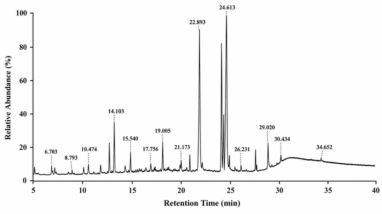
Fig. 3.
Gas chromatography–mass spectrometry (GC–MS) chromatogram of the aqueous leaf extract of Arum rupicola var. rupicola (ARVR). The chromatogram shows the distribution of metabolites detected in the extract, including phenolic compounds, oxygenated metabolites, fatty acids, fatty acid esters, and other phytochemical constituents. A total of 56 compounds were identified based on retention time and mass spectral matching.
Table I
List of chemical compounds identified in the plant extract by GC–MS analysis, including compound names, retention times, and relative peak area percentages.
| No. | Chemical compounds | Area % | R.T. |
|---|---|---|---|
| 1 | Methyl 2-hydroxy-2-methoxyacetate | 1.1038 | 4.315 |
| 2 | Butanal, 3-methyl- | 0.7377 | 4.716 |
| 3 | 1,2-Pentanediol | 0.564 | 4.951 |
| 4 | 2-Propanamine, N-methyl-N-nitroso- | 0.2778 | 5.572 |
| 5 | Pyrazine, methyl- | 0.7731 | 6.507 |
| 6 | Dimethyl Sulfoxide | 3.3008 | 6.703 |
| 7 | Dimethyl Sulfoxide | 0.271 | 7.426 |
| 8 | 1,5-Pentanedithiol | 0.2515 | 7.819 |
| 9 | Acetaldehyde, ethylidenehydrazone | 0.6276 | 7.999 |
| 10 | Pyrazine, 2,5-dimethyl- | 1.2808 | 8.486 |
| 11 | 2-Dodecylcyclobutanone | 0.56 | 8.793 |
| 12 | N-(2-Methylbutylidene)isobutylami | 0.6201 | 9.005 |
| 13 | 2-Furanmethanol, 5-methyl- | 0.1926 | 9.476 |
| 14 | 1,7-Diazabicyclo[2.2.0]heptane | 0.1744 | 9.712 |
| 15 | 2,4-Dihydroxy-2,5-dimethyl-3(2H)-furan-3-one | 1.0664 | 9.94 |
| 16 | Decane | 1.2514 | 10.183 |
| 17 | Propanenitrile, 3,3’-iminobis- | 1.9255 | 10.474 |
| 18 | 2-[(2-Oxocycloheptyl)methyl]cycloheptan-1-one (isomer 2) | 0.2715 | 10.694 |
| 19 | 2-Butenal, 3-methyl- | 0.6502 | 10.914 |
| 20 | Benzeneacetaldehyde | 0.3978 | 11.251 |
| 21 | Furaneol | 0.5105 | 11.487 |
| 22 | 1-Ethyl-2-pyrrolidinone | 0.9112 | 11.77 |
| 23 | But-3-enyl (E)-2-methylbut-2-enoate | 2.1565 | 12.155 |
| 24 | cis-1-Butene,1-butoxy- | 0.2587 | 12.65 |
| 25 | 4H-Pyran-4-one, 2,3-dihydro-3,5-dihydroxy-6-methyl- | 3.3002 | 12.972 |
| 26 | Benzoic acid | 0.2148 | 13.364 |
| 27 | 2-Pyrrolidinone, 5-(hydroxymethyl)- | 3.2193 | 13.561 |
| 28 | Benzofuran, 2,3-dihydro- | 2.3003 | 14.103 |
| 29 | Decanoic acid, 3-hydroxy-, methyl ester | 1.1422 | 14.456 |
| 30 | 2,4-Difluoroanisole | 1.7553 | 15.053 |
| 31 | 2-Methoxy-4-vinylphenol | 1.0778 | 15.54 |
| 32 | Butanoic acid, 3-methyl-, hexyl ester | 0.3884 | 15.807 |
| 33 | DL-Proline, 5-oxo-, methyl ester | 1.1106 | 16.365 |
| 34 | 1,2,4-Benzenetriol | 0.5489 | 16.695 |
| 35 | trans-Cinnamic acid | 0.5023 | 16.986 |
| 36 | 4-Vinylbenzene-1,2-diol | 1.3339 | 17.221 |
| 37 | N,N-Diamylmethylamine | 0.37 | 17.677 |
| 38 | 2-Hydroxy-1-(1’-pyrrolidiyl)-1-buten-3-one | 1.5931 | 18.211 |
| 39 | 2(4H)-Benzofuranone, 5,6,7,7a-tetrahydro-4,4,7a-trimethyl-, (R)- | 0.4428 | 18.502 |
| 40 | Phenol, 4-ethenyl-2,6-dimethoxy- | 1.1138 | 18.769 |
| 41 | 3-Methyl-4-phenyl-1H-pyrrole | 1.7471 | 19.005 |
| 42 | Octahydropyrrolo[1,2-a]pyrazine | 0.3566 | 19.256 |
| 43 | 1,3-Benzenediol, 4,5-dimethyl- | 0.3926 | 20.206 |
| 44 | Tetradecanoic acid | 0.3686 | 20.819 |
| 45 | 2(1H)-Pyridinone, 1,3,6-trimethyl- | 0.9166 | 21 |
| 46 | Phthalic acid, isobutyl undecyl ester | 0.4412 | 22.068 |
| 47 | Hexadecanoic acid, methyl ester | 4.3661 | 22.539 |
| 48 | n-Hexadecanoic acid | 9.6708 | 22.893 |
| 49 | 9-Octadecenoic acid, methyl ester, (E)- | 11.7767 | 24.228 |
| 50 | Methyl stearate | 4.0117 | 24.44 |
| 51 | 9,12,15-Octadecatrienoic acid, (Z,Z,Z)- | 16.4382 | 24.613 |
| 52 | Methyl 9-cis,11-trans-octadecadienoate | 1.963 | 24.998 |
| 53 | 3’-O-Acetylthymidine | 0.5502 | 26.483 |
| 54 | 3-Eicosene, (E)- | 1.5561 | 27.496 |
| 55 | Bis(2-ethylhexyl) phthalate | 1.087 | 28.015 |
| 56 | 1-Tetracosene | 3.809 | 29.02 |
Phenolic compounds, including 2-methoxy-4-vinylphenol, dihydroxy benzene derivatives, and 4H-pyran-4-one derivatives (3.3 %), contribute to Zn2+ reduction and nanoparticle stabilization via surface adsorption and chelation (Cueva et al. 2012). Nitrogen-containing heterocycles, such as pyrrolidinone and pyrazine derivatives, may further stabilize NPs through coordination interactions (Balouiri et al. 2016). Overall, the predominance of fatty acids, phenolics, and heterocycles provides a strong chemical basis for the extract’s dual function as a reducing and capping agent.
Quantitative phytochemical analysis of the ARVR leaf extract revealed a high total phenolic content (508.32 mg/g FW), along with strong antioxidant capacity (438.50 mg/g FW) and a substantial total flavonoid content (229.17 mg/g FW) (Fig. 4). These values are higher than those reported for several plant and algal extracts previously used in green ZnO-NPs synthesis, including Spirulina platensis, Eclipta prostrata, and Piper longum, where total phenolic contents generally fall within the low to mid hundreds of mg/g extract (Ahmed and Othman 2024; Tran et al. 2024). The predominance of phenolic compounds in ARVR is consistent with the pronounced antioxidant activity observed and supports their central role in supplying the reducing power required for Zn2+ reduction and nanoparticle stabilization. Similar associations between phenolic richness and redox activity have been reported in recent plant-mediated ZnO-NPs synthesis studies, while flavonoids typically contribute to a lesser extent to metal ion reduction and capping processes (Ahmed and Othman 2024; Tran et al. 2024). Variations in phytochemical levels among studies are likely attributable to differences in plant species, extraction methods, solvent polarity, and environmental conditions influencing secondary metabolite accumulation (Khal 2024).
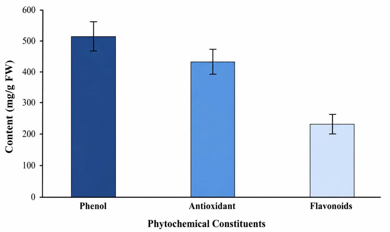
Fig. 4.
Quantitative phytochemical analysis of ARVR leaf extract showing total phenolic content, antioxidant capacity, and total flavonoid content, expressed as mg/g fresh weight (FW). Data are presented as mean ± standard deviation (SD) from three independent biological replicates.
3.2. Bacterial isolation and identification
In this study, four bacterial strains were isolated from cancer patients at Hiwa Hospital in the Kurdistan Region of Iraq.
Initial identification and the AST profile of the isolates were performed using the BD Phoenix system, and results were interpreted according to CLSI breakpoints (CLSI M100, 34th edition, 2024) (Clinical & Laboratory Standards 2024). The isolates comprised two Gram-positive bacteria, S. aureus and S. epidermidis, and two Gram-negative bacteria, E. coli and K. aerogenes. Based on CLSI criteria, all isolates were classified as MDR, exhibiting resistance to at least one agent in three or more antibiotic classes (Magiorakos et al. 2012) (Table II). The Gram-positive isolates were found resistant to several antibiotics in the class of β-lactams, aminoglycosides, macrolides, lincosamides, and fluoroquinolones. The Gram-negative isolates showed broad resistance to aminoglycosides, β-lactams, fluoroquinolones, and sulfonamides. Notably, the E. coli isolate was resistant to carbapenems, a clinically significant finding, as carbapenem-resistant E. coli is recognized by the ECDC (2025) as a major threat to patients and healthcare systems due to rising incidence and limited therapeutic options.
Table II
MDR profiles of the studied bacterial isolates. Antibiotic susceptibility was determined using the BD Phoenix system and interpreted according to CLSI M100 (34th edition, 2024) (Clinical & Laboratory Standards 2024).
| Bacterial strain | Resistant antibiotics (class) | Number of antibiotics classes |
|---|---|---|
| Staphylococcus aureus | Gentamicin (Aminoglycoside); Penicillin, Oxacillin, Cefoxitin (β-lactams); Erythromycin (Macrolide); Clindamycin (Lincosamide); Ciprofloxacin, Levofloxacin (Fluoroquinolones) | 5 |
| Staphylococcus epidermidis | Gentamicin (Aminoglycoside); Penicillin, Oxacillin, Cefoxitin (β-lactams); Erythromycin (Macrolide); Clindamycin (Lincosamide); Ciprofloxacin, Levofloxacin (Fluoroquinolones) | 5 |
| Escherichia coli | Gentamicin, Amikacin (Aminoglycosides); Ampicillin, Cefotaxime, Ceftazidime, Cefazolin (β-lactams); Imipenem, Ertapenem, Meropenem (Carbapenems); Ciprofloxacin, Levofloxacin (Fluoroquinolones); Trimethoprim–sulfamethoxazole (Sulfonamide) | 5 |
| Klebsiella aerogenes | Gentamicin, Amikacin (Aminoglycosides); Ampicillin, Cefazolin, Cefuroxime, Ceftazidime (β-lactams); Ciprofloxacin, Levofloxacin (Fluoroquinolones); Trimethoprim–sulfamethoxazole (Sulfonamide) | 4 |
Confirmation of bacterial identity was achieved through partial sequencing of the 16S rRNA gene. Sequence homology searches and phylogenetic analysis confirmed the isolates as S. epidermidis, S. aureus, E. coli, and K. aerogenes (Fig. 5). The corresponding sequences were deposited in GenBank under the accession numbers S. epidermidis Charmo3 (PQ787848.1), S. aureus Charmo4 (PQ787849.1), E. coli Charmo5 (PQ787850.1), and K. aerogenes Charmo28 (PQ787851.1). Phylogenetic analysis also showed that all isolates clustered closely with reference strains, supported by high bootstrap values, indicating accurate species-level identification. For instance, S. aureus Charmo4 grouped monophyletically with S. aureus SC01 (FJ899095.1), DSM 20231 (MN650918.1), and IIF6SW-P2 (KY218882.1) (Fig. 5).
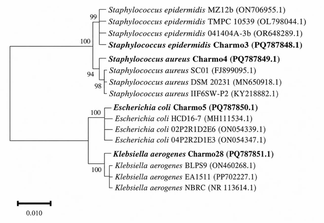
Fig. 5.
Phylogenetic tree of S. epidermidis Charmo3, S. aureus Charmo4, E. coli Charmo5, and K. aerogenes Charmo28 based on 16S rRNA gene sequences. DNA sequences were aligned using ClustalX, and the tree was constructed in MEGA 11 with the neighbor-joining method and 1,000 bootstrap replicates. Bootstrap values are shown at branch nodes.
The scale bar represents 0.01 nucleotide substitutions per site.
Biosynthesis and characterization of the ZnO-NPs
ZnO-NPs were biosynthesized using ARVR extract as a reducing and capping agent. Upon mixing the plant extract with zinc nitrate hexahydrate (Zn(NO3)2·6H2O), the reaction mixture gradually developed a pale-yellow coloration accompanied by precipitate formation, suggesting the initiation of ZnO-NPs formation. This visual change has been widely reported as an initial indicator of ZnO-NPs synthesis in plant-mediated systems (Al-Askar et al. 2023). Because pH plays a crucial role in nanoparticle nucleation, growth, and biomolecule adsorption, the synthesis was conducted at pH 8.0. Near-neutral to mildly alkaline conditions have been reported to favor ZnO crystallinity and yield while preserving the capping efficiency of plant-derived biomolecules (Gherbi et al. 2022).
UV–Vis spectrophotometric analysis revealed a distinct absorption peak at 383 nm (Fig. 6), corresponding to the characteristic band-gap excitation of ZnO-NPs (Gharpure et al. 2022). Compared with bulk ZnO, nanoscale ZnO typically exhibits shifts in the absorption edge due to reduced particle size, surface states, and quantum confinement effects (García 2011). The sharp and well-defined nature of the absorption peak observed here suggests effective stabilization of ZnO-NPs by ARVR-derived phytochemicals. Absorption maxima reported for plant-mediated ZnO-NPs generally range between 310 and 386 nm (EL-Ghwas 2022); and the value obtained in this study falls well within this range, further confirming the successful green synthesis of ZnO-NPs using ARVR extract.
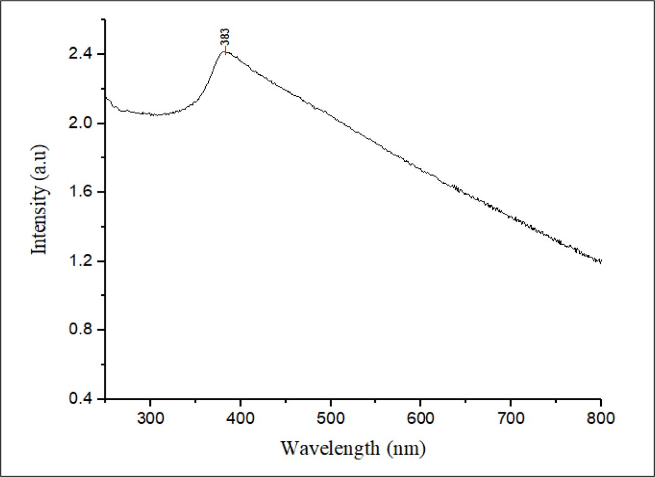
Fig. 6.
UV–Vis absorption spectrum of biosynthesized ZnO-NPs using ARVR leaf extract, showing a characteristic band-edge peak at 383 nm.
Consistent with the UV–Vis results, XRD analysis confirmed the successful formation of crystalline ZnO-NPs (Fig. 7). The diffraction peaks at 2θ values of 31.8°, 34.5°, 36.2°, 47.6°, 56.6°, 62.9°, and 68.2° correspond to the (100), (002), (101), (102), (110), (103), and (200) planes of hexagonal wurtzite ZnO, in good agreement with JCPDS card no. 04-003-2106 (Chopra et al. 2022). Minor intensity variations likely arise from lattice strain or interactions with ARVR-derived phytochemicals, as commonly observed in green synthesis routes (Ismail, 2018). The average crystallite size calculated using the Scherrer equation was approximately 10 nm. This value represents the coherent diffraction domain rather than the actual particle diameter but nonetheless reflects the high crystallinity achieved under mild, plant-mediated synthesis conditions.
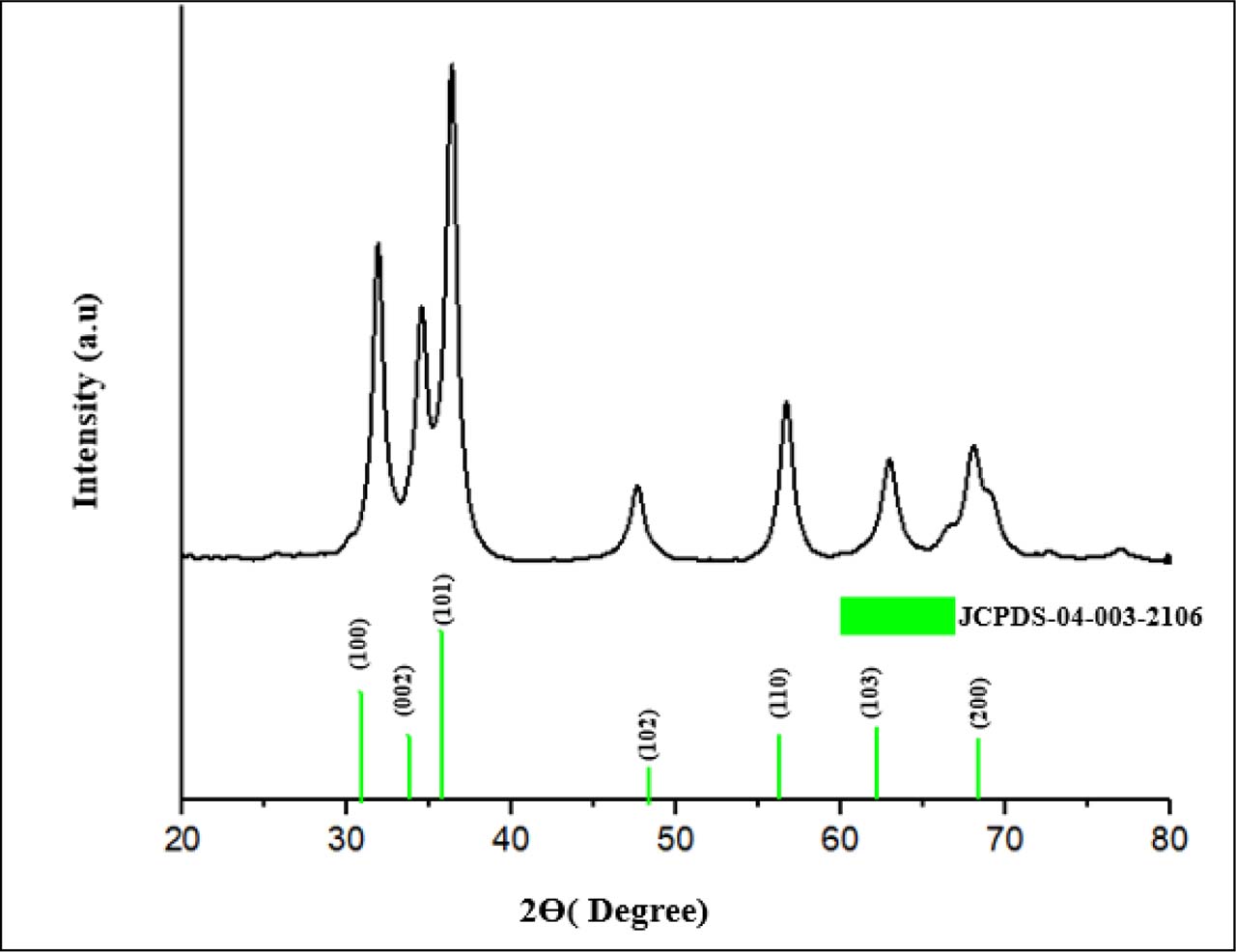
Fig. 7.
X-ray diffraction (XRD) pattern of biosynthesized ZnO-NPs (pH 8.0, calcined at 430°C for 2 h). The diffraction peaks at 2θ = 31.8°, 34.5°, 36.2°, 47.6°, 56.6°, 62.9°, and 68.2° correspond to the (100), (002), (101), (102), (110), (103), and (200) planes of hexagonal wurtzite ZnO, in agreement with the JCPDS standard (card no. 04-003-2106) (Kisi & Elcombe, 1989), confirming high crystallinity and phase purity.
Following confirmation of the crystalline structure of the ZnO-NPs, FTIR analysis was used to identify the functional groups involved in their formation and surface stabilization (Fig. 8). A broad band around 3418.33 cm−1 is attributed to O–H stretching vibrations, arising from surface hydroxyl groups and adsorbed water molecules. The absorption near 2929 cm−1 corresponds to aliphatic C–H stretching, which is consistent with GC–MS results indicating the presence of aldehydes and fatty acid derivatives in the ARVR extract (Suresh et al. 2018). Absorption bands observed between 1100 and 1400 cm−1 are associated with C–O and C–N stretching, indicating the involvement of phenolics, flavonoids, and nitrogen-containing metabolites. The peak at 1626.85 cm−1 corresponds to aromatic ring stretching, while the band at 879.7 cm−1 reflects C–H bending vibrations. Importantly, the strong band at ~526 cm−1 is characteristic of Zn–O stretching, confirming ZnO-NP formation (Tilahun et al. 2023). Overall, the FTIR findings complement the XRD and GC–MS results, demonstrating that ARVR-derived phytochemicals not only facilitated the reduction of Zn2+ ions but also remained associated with the NPs surface, contributing to effective capping and long-term stabilization.
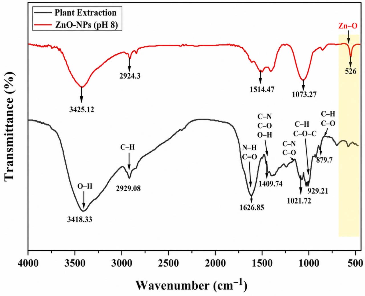
Fig. 8.
FTIR spectra of the ARVR plant leaf extract and the biosynthesized ZnO nanoparticles (ZnO-NPs). The spectra display characteristic absorption bands corresponding to O–H, C–H, C=O, N–H, C–N, and C–O functional groups, highlighting the role of phytochemicals in ZnO-NP formation and stabilization. The distinct band observed at ~526 cm−1 confirms Zn–O stretching vibrations.
Elemental composition was verified by EDS (Fig. 9), which showed strong signals for zinc (1 keV and 8–9 keV) and oxygen (~0.5 keV), with weight percentages of 62.12% Zn and 28.64% O, consistent with ZnO formation. Similar low-intensity bioorganic peaks have been reported in other plant-mediated ZnO-NPs synthesized using Salvia officinalis, lemon juice, and Punica granatum extract (Alvarado et al. 2025). These results validated the purity of the ZnO-NPs.
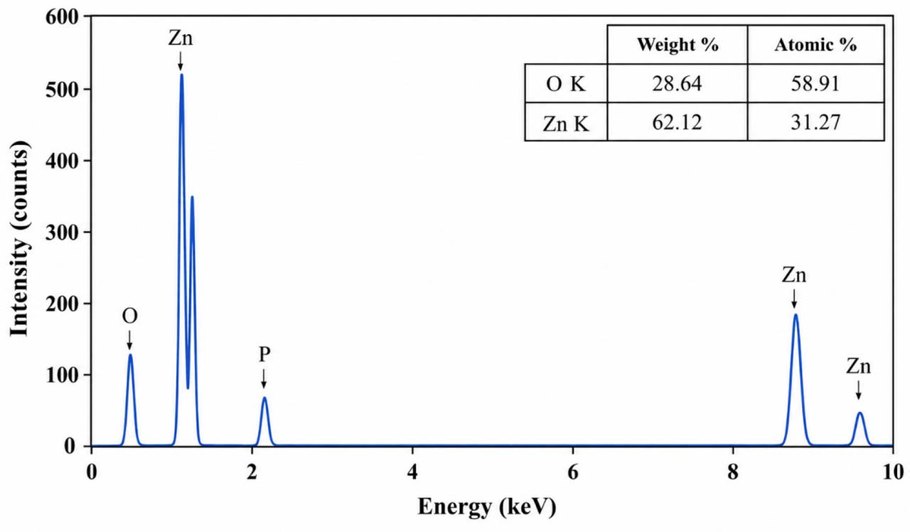
Fig. 9.
Energy-dispersive X-ray spectroscopy (EDS) spectrum of biosynthesized ZnO-NPs. Strong peaks for zinc (Zn) and oxygen (O) confirm ZnO formation, with Zn (62.12 wt%) as the dominant element and O (28.64 wt%).
A minor phosphorus (P) peak was also detected, likely originating from phytochemicals in the plant extract.
To assess the behavior of the ZnO-NPs in suspension, DLS was performed (Fig. 10A). The Z-average hydrodynamic diameter was approximately 80 nm, with a polydispersity index (PDI) of 0.36, indicating reasonably uniform dispersion. For metal oxide nanoparticles, PDI values below 0.5 are generally considered indicative of acceptable colloidal stability (Bhattacharjee, 2016). Comparable hydrodynamic sizes have been reported for ZnO-NPs, with Raghavendra et al. (2020) documenting diameters in the range of 90–140 nm depending on dispersion conditions and surface chemistry (Raghavendra et al. 2022). The values reported in this study fall well within the range commonly described for well-dispersed ZnO-NPs, including hydrodynamic sizes of 30–50 nm with a PDI of 0.28 (Singh et al. 2019) and Z-average diameters of 45–70 nm with PDI values below 0.4 (Ramesh et al. 2015).
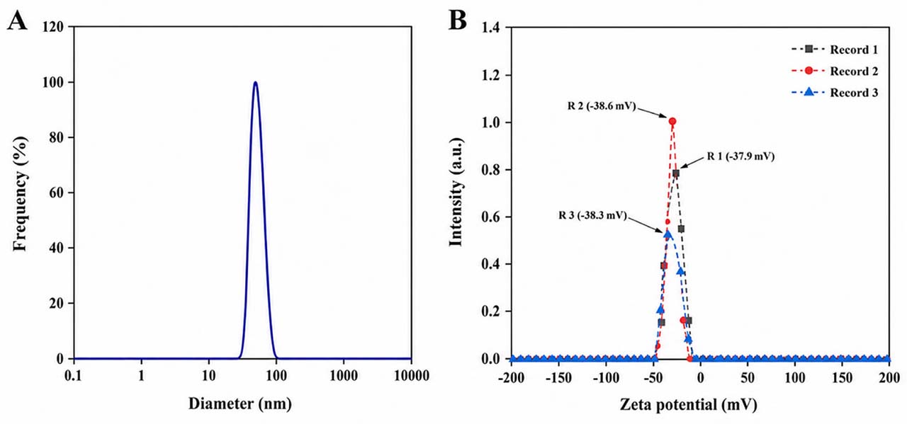
Fig. 10.
(A) DLS analysis showing a Z-average hydrodynamic diameter of ~80 nm and a moderate polydispersity index (PDI) of 0.36, measured in triplicate. (B) Zeta potential of ZnO-NPs in aqueous solution at 25 °C, showing a negative charge of −28.3± 0.286 mV (triplicate readings), indicating good colloidal stability.
Zeta potential measurements were performed to further evaluate surface charge and colloidal stability (Fig. 10B). The ARVR-mediated ZnO-NPs exhibited zeta potential values ranging from −27.9 to −28.6 mV, with an average of −28.3 ± 0.286 mV. Zeta potential magnitudes greater than ±25 mV are generally indicative of good electrostatic stabilization (Kayani et al. 2015). The negative surface charge is attributed to ionized surface hydroxyl groups, while the adsorbed phytochemicals likely contribute additional steric stabilization. These zeta potential values indicate good colloidal stability, suggesting that the ZnO-NPs are likely to remain well-dispersed in aqueous media; an essential property for biological and catalytic applications. These results are consistent with previously reported chemically and biologically synthesized ZnO-NPs, including −29.8 mV, −27.4 mV (Kayani et al. 2015), and −18 to −32 mV (Sirelkhatim et al. 2015). The presence of capping phytochemicals, as indicated by FTIR and GC–MS analysis, likely enhances stabilization by providing additional surface charge and steric hindrance. Overall, the zeta potential confirms that ARVR-mediated ZnO-NPs exhibit physicochemical behavior similar to previously reported ZnO-NPs and fall within the expected stability range for effective dispersion (Raghavendra et al. 2022).
SEM was conducted to examine the surface morphology and overall shape of the biosynthesized ZnO-NPs (Fig. 11). The NPs were found predominantly spherical, with occasional flower-like aggregates, a morphology commonly reported for plant-mediated ZnO-NPs synthesis (Azizi et al. 2013). The prevalence of spherical particles suggests controlled nucleation and growth, while the flower-like structures are likely due to localized aggregation influenced by phytochemicals acting as reducing and stabilizing agents (Ramesh et al. 2015).
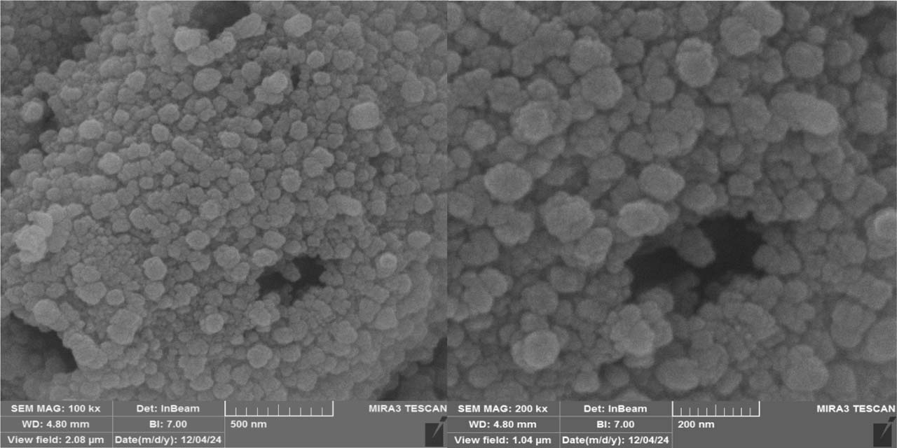
Fig. 11.
SEM images of biosynthesized ZnO-NPs showing predominantly spherical particles with occasional flower-like aggregates. Scale bars: 500 nm (A) and 200 nm (B).
TEM provided a higher resolution view of the particle morphology and primary size (Fig. 12A). The NPs remained predominantly spherical, with size ranging from 20 to 35 nm, with an average diameter of approximately 27.7 ± 3.9 nm, as determined from Gaussian fitting of the size distribution histogram (Fig. 12B). The particles appeared relatively uniform with minimal aggregation, indicating that ARVR-derived phytochemicals effectively acted as capping and stabilizing agents during synthesis.
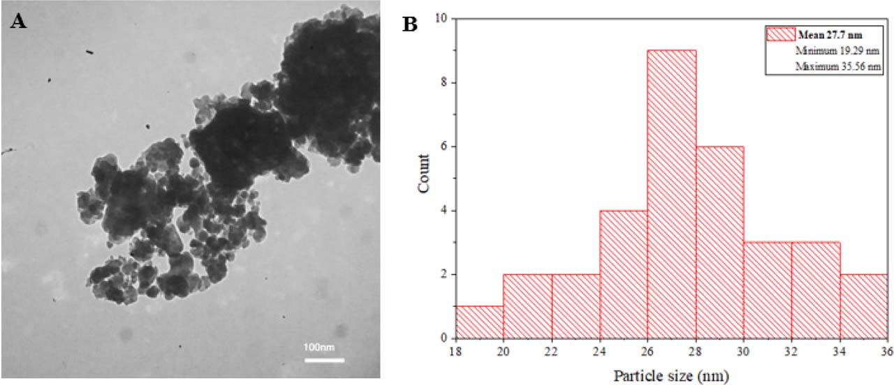
Fig. 12.
A: Representative TEM image showing aggregated but nearly spherical nanoparticles (scale bar = 100 nm). B: Particle size distribution histogram with Gaussian fitting, indicating a size range of 20–35 nm and an average diameter of ~27.7 ± 3.9 nm.
The observed particle sizes are consistent with previous reports on biosynthesized ZnO-NPs, such as curcumin-mediated ZnO-NPs with an average size of approximately 27.6 nm (Alallam et al. 2023). As expected, the hydrodynamic diameter measured by DLS exceeded the TEM-derived particle size, reflecting contributions from solvation layers and interparticle interactions in suspension (Sirelkhatim et al. 2015). Additionally, the slightly larger particle sizes obtained by TEM compared to XRD estimates were also expected, as the Scherrer equation reflects crystallite size rather than whole particle dimensions. Similar discrepancies between XRD and TEM size measurement have been reported for green synthesized ZnO-NPs using Solanum nigrum (Ramesh et al. 2015), Murraya koenigii (Lakshmikandhan 2020) and Aloe barbadensis (Sangeetha et al. 2011).
Overall, SEM, TEM, DLS, and zeta potential analyses collectively confirm that the ARVR-mediated ZnO-NPs are predominantly spherical, relatively uniform in size, and stable in aqueous suspension. These combined morphological, size, and surface charge characteristics demonstrate the effectiveness of plant-derived biomolecules in controlling nanoparticle formation and colloidal stability, supporting the potential utility of the synthesized ZnO-NPs in antimicrobial, antibiofilm, photocatalytic, and other applications, subject to further biocompatibility evaluation.
Antibacterial and antibiofilm activities of ZnO-NP
The biosynthesized ZnO-NPs were evaluated against all studied isolates and exhibited strong antibacterial activity. The MIC values were 156 μg/ml for S. epidermidis Charmo3 and K. aerogenes Charmo28, and 312 μg/ml for S. aureus Charmo4 and E. coli Charmo5 (Table III). In all cases, the MBC values were one dilution step higher than the corresponding MICs (312 and 625 μg/ml, respectively), confirming the bactericidal potential of the NPs.
Table III
Minimum inhibitory concentration (MIC) and minimum bactericidal concentration (MBC) values of ARVR-derived ZnO-NPs against clinical isolates. Values are expressed as mean ± SD (μg/ml, n = 3).
| Bacterial Isolates | MIC (µg/ml) | MBC (µg/ml) |
|---|---|---|
| Staphylococcus epidermidis Charmo3 | 156 ± 20 | 312 ± 10 |
| Klebsiella aerogenes Charmo28 | 156 ± 10 | 312 ± 20 |
| Staphylococcus aureus Charmo4 | 312 ± 10 | 625 ± 15 |
| Escheirchia coli Charmo5 | 312 ± 10 | 625 ± 10 |
These antibacterial concentrations are in good agreement with previous reports on biosynthesized ZnO-NPs. For instance, MIC values ranging from 150 to 450 μg/ml have been reported for S. aureus and E. coli using plant-mediated ZnO-NPs with an average particle size of approximately 22 nm (El-Habib et al. 2024). The slightly higher MIC value observed in this study could be attributed to the slightly larger nanoparticle size used here (27.7 nm), which is consistent with the well-established size-dependent antibacterial activity of ZnO-NPs (El-Habib et al. 2024). In addition, surface-associated biomolecules derived from the biological synthesis process may enhance nanoparticle dispersion and surface reactivity. This, in turn, facilitates closer interactions between the nanoparticles and bacterial cells. Comparable bactericidal concentrations have been reported for biologically synthesized ZnO-NPs against MDR strains, further supporting the robustness of the present findings (Rahman et al. 2023). In contrast, commercially available ZnO-NPs have been reported to require higher concentrations to achieve similar MIC effects, highlighting the enhanced antibacterial efficiency of the ARVR-mediated ZnO-NPs (Pangprasit et al. 2025). This improved performance highlights the advantages of plant-mediated approaches, where plant-derived compounds not only facilitate NPs formation but may also contribute to improved biological activity through surface functionalization.
Interestingly, the susceptibility pattern observed in this study did not strictly follow a Gram-positive versus Gram-negative classification. Although E. coli Charmo5 and K. aerogenes Charmo28 are both Gram-negative bacteria, K. aerogenes Charmo28 was more sensitive to ZnO-NPs. Similarly, among Gram-positive isolates, S. epidermidis Charmo3 exhibited lower MIC and MBC values than S. aureus Charmo4. These differences likely arise from species-specific variations in cell envelope composition, membrane permeability, and intrinsic stress-response mechanisms rather than Gram-type of the bacterium alone. For instance, S. aureus Charmo4 possesses well-developed antioxidant defense systems, including catalase and the carotenoid pigment staphyloxanthin, which can mitigate oxidative stress and may reduce susceptibility to ZnO-NPs-induced damage (Clauditz et al. 2006). In contrast, E. coli Charmo5 is known to limit NPs interaction through its outer membrane structure and active efflux systems (Martinez-Gutierrez et al. 2013), whereas K. aerogenes Charmo28 may possess membrane characteristics that facilitate greater interaction with ZnO-NPs and increased exposure to Zn2+ ions, as suggested in previous studies.
Comparative morphology studies have demonstrated that ZnO-NPs with spherical or complex morphologies tend to exhibit stronger antibacterial activity than rod-shaped or plate-like counterparts, an effect commonly attributed to increased surface area and enhanced contact with bacterial cell surfaces (Esparza González et al. 2021). In the present study, the predominance of spherical ZnO-NPs together with irregular flower-like structures may have enhanced ZnO-NP-bacteria interactions, contributing to the observed broad-spectrum antibacterial activity against both Gram-positive and Gram-negative bacteria. Although the MIC and MBC values of ZnO-NPs are higher than those of conventional antibiotics, these nanostructures are not intended to replace clinically used drugs but rather to complement them, particularly in the context of MDR and biofilm-associated infections. ZnO-NPs are generally regarded as less toxic and more biocompatible than other metallic nanoparticles, such as silver, supporting their potential suitability for biomedical applications (Siddiqi et al. 2018).
Following the pronounced antibacterial activity observed against planktonic cells, the antibiofilm potential of the biosynthesized ZnO-NPs was evaluated against the same clinical isolates.
All tested bacterial isolates exhibited strong biofilm-forming ability, as shown by optical density values exceeding four times the cut-off value (OD > 4×ODc), providing an ideal model for evaluating the antibiofilm activity of ZnO-NPs. As shown in Figure 13, ZnO-NPs exhibited pronounced antibiofilm activity against the studied isolates in a concentration-dependent manner. Near-complete biofilm inhibition (>98%) was achieved at 78 μg/ml for S. epidermidis Charmo3 and K. aerogenes Charmo28, whereas S. aureus Charmo4 and E. coli Charmo5 required a higher concentration of 156 μg/ml to reach comparable inhibition levels. This species-specific response closely mirrors trends observed in the MIC and MBC assays, indicating a consistent susceptibility pattern between planktonic and biofilm-associated cells.
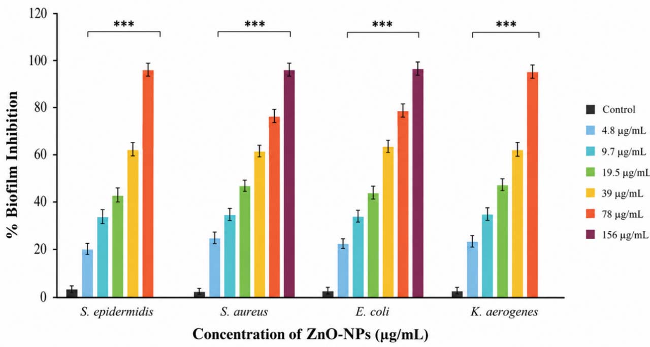
Fig. 13.
Dose-dependent antibiofilm activity of ARVR-derived ZnO-NPs against Staphylococcus epidermidis Charmo3, Staphylococcus aureus Charmo4, Escherichia coli Charmo5, and Klebsiella aerogenes Charmo28. Biofilm inhibition was quantified after 24 h of exposure to increasing ZnO-NPs concentrations (4.8–156 μg/ml). Data are expressed as mean ± SD from three independent biological replicates.
The observed differences in antibiofilm sensitivity may be related to variations in cell-envelope architecture and extracellular polymeric substance (EPS) composition, which have been reported to influence nanoparticle attachment, Zn2+ ion diffusion, and resistance to oxidative stress. Similar dose-dependent antibiofilm effects of ZnO-NPs have been reported against a wide range of pathogens, including Pseudomonas aeruginosa, Pseudomonas putida, and Candida tropicalis (Valadbeigi et al. 2023).
The antibacterial and antibiofilm effects of ZnO-NPs have been attributed in previous studies to multiple complementary mechanisms. Owing to their nanoscale size and high surface-to-volume ratio, previous studies have reported that ZnO-NPs can closely associate with bacterial cell surfaces, potentially compromising membrane integrity and facilitating antimicrobial interactions (Raghupathi et al. 2011). In aqueous environments, ZnO-NPs have also been reported to release Zn2+ ions, which may interfere with essential enzymatic processes and quorum-sensing pathways, thereby inhibiting bacterial metabolism and biofilm maturation (Sirelkhatim et al. 2015). Additionally, ZnO-NPs have been reported to generate reactive oxygen species (ROS), contributing to oxidative stress and weakening both bacterial cells and the EPS matrix that stabilizes biofilm structures (Brayner et al. 2006). Particle size and morphology further modulate these effects, with smaller, spherical, or complex surface structures often showing enhanced activity due to increased surface reactivity and more effective contact with bacterial cells (Sirelkhatim et al. 2015). However, these mechanisms were not directly investigated in the present study and are discussed based on previously published findings on ZnO-NPs.
Consistent with previous reports, higher ZnO-NPs concentrations were required to inhibit S. aureus and E. coli, organisms reported to possess strong oxidative-stress defenses and, in the case of E. coli, outer-membrane and efflux barriers that may limit NPs interaction (Martinez-Gutierrez et al. 2013). The antibiofilm assays were conducted at pH 8.0, a condition reported to favor ZnO-NPs dissolution and ROS-related activity, which may have contributed to the high levels of biofilm suppression observed at concentrations between 78 and 156 μg/ml (Espitia et al. 2012). Together with the MIC and MBC findings, these results suggest that antibiofilm efficacy may be influenced by an interplay between ZnO-NPs physicochemical properties (size, morphology, and surface chemistry) and species-specific bacterial traits.
Biofilm-associated infections are a major clinical challenge due to their tolerance to antibiotics and ability to evade immune responses. ARVR-mediated ZnO-NPs strongly inhibited biofilm formation, highlighting their potential as effective alternatives or adjuncts to conventional antimicrobial strategies. These findings support the promise of plant-based, green-synthesized ZnO-NPs for managing biofilm-associated and MDR infections.
Cytotoxic effect of ZnO-NPs on HEK-293 cells
The cytotoxic potential of the biosynthesized ZnO-NPs was evaluated using the HEK-293 cell line, a widely accepted in vitro model for preliminary nanotoxicological assessment. This cell line was selected due to its well-characterized growth behavior, reproducibility, and sensitivity to oxidative stress, making it suitable for evaluating nanoparticle-induced cellular damage. As kidney-derived cells, HEK-293 also provide relevant insight into potential nephrotoxic effects associated with nanoparticle exposure, and their responsiveness to metal oxide nanoparticle-induced oxidative stress has been well documented (Xia et al. 2008).
The MTT assay revealed a concentration-dependent cytotoxic effect of ZnO NPs on HEK 293 cells after 24 h exposure (Figure 14A). Control cells maintained approximately 100% viability, confirming normal metabolic activity. At 10 μg/ml only a slight and statistically non-significant reduction in viability of about 94% (p > 0.05) was observed, indicating minimal cytotoxicity. A sharp and statistically significant decline in cell viability was observed at 25 μg/ml, approximately 15.7% (p < 0.0001 vs. control), representing a substantial greater than 6-fold reduction. This decrease became more pronounced at 50 μg/ml, approximately 9.9%, with a statistically significant difference compared to 25 μg/ml (p < 0.001), indicating a continued dose dependent effect. At higher concentrations equal to or greater than 50 μg/ml, cell viability remained consistently low at approximately 6 to 10%, with no statistically significant differences observed among most concentrations from 50 to 1000 μg/ml (p > 0.05), indicating a plateau in cytotoxic response. However, a statistically significant difference was observed between 50 and 1000 μg/ml (p < 0.05); nevertheless, the magnitude of this difference was relatively small, suggesting limited biological relevance within the plateau range. This nonlinear response pattern suggests the presence of a critical concentration threshold beyond which cellular defense mechanisms are overwhelmed, resulting in an abrupt loss of viability, a behavior commonly associated with oxidative stress-mediated nanoparticle toxicity (Nel et al. 2006). Morphological observations (Fig. 14B) supported these findings, revealing progressive cellular damage with increasing nanoparticle concentration. Treated cells exhibited reduced cell density, cell shrinkage, and loss of normal morphology, along with decreased formazan crystal formation, all indicative of compromised mitochondrial activity and reduced cellular viability.
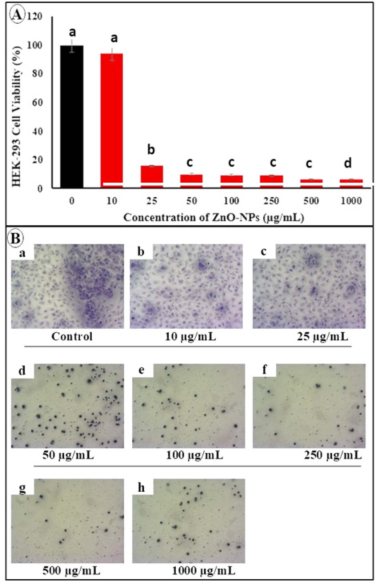
Fig. 14.
Cytotoxic and morphological effects of ZnO-NPs on HEK-293 cells. (A) Cell viability (%) after 24 h exposure to ZnO-NPs (0–1000 μg/ml), determined by MTT assay and expressed relative to control (100%). Data represent mean ± SD (n = 3).
Different letters indicate significant differences (p < 0.05; one-way ANOVA with Tukey’s post hoc test).
The IC50 value was 16.67 μg/ml. (B) Representative images of HEK-293 cells after 24 h treatment showing concentration-dependent morphological changes, including reduced cell density and cell shrinkage compared to control.
The calculated IC50 value (16.67 μg/ml) indicates a relatively high sensitivity of HEK-293 cells to ZnO-NPs. This value falls within the range reported for green-synthesized ZnO nanoparticles, supporting the reproducibility of the present findings (Khan et al. 2015). The observed cytotoxicity is consistent with the ROS-mediated mechanisms and Zn2+ ion release discussed earlier (Brayner et al. 2006), which contribute to mitochondrial dysfunction and subsequent cell death pathways.
A critical aspect of this study is the balance between antimicrobial efficacy and cytotoxicity. ARVR-mediated ZnO-NPs demonstrated antibacterial activity with MIC values of 156–312 μg/ml and MBC values of 312–625 μg/ml, while notable antibiofilm activity was observed at lower concentrations (78–156 μg/ml), indicating enhanced effectiveness against biofilm formation. However, the IC50 value obtained in HEK-293 cells (16.67 μg/ml) was substantially lower than the MIC range, indicating that cytotoxicity occurred at concentrations below those required to inhibit bacterial growth. This finding represents a limitation and suggests that the current ZnO-NPs formulation may not be suitable for systemic therapeutic applications without further optimization. Nevertheless, the pronounced antibacterial and antibiofilm activities observed against MDR isolates remain noteworthy. In particular, the strong inhibition of biofilm formation at concentrations lower than the bactericidal range highlights the potential utility of these ZnO-NPs in localized, coating-based, or surface-disinfection applications, where direct and prolonged exposure of mammalian tissues can be minimized. Such applications are especially relevant given the critical role of biofilms in persistent infections, antimicrobial resistance, and healthcare-associated contamination. Therefore, while the observed cytotoxicity limits the potential for systemic use, it does not preclude other practical antimicrobial applications. This interpretation is consistent with previous reports describing the dose-limiting cytotoxicity of ZnO-NPs in mammalian systems despite their potent antimicrobial activity (Xia et al. 2008).
Despite these promising findings, a number of limitations should be acknowledged. The cytotoxicity assessment was performed using a single cell line, and cellular responses may vary among different tissue types. Furthermore, in vitro conditions do not fully replicate the complexity of in vivo biological systems, where nanoparticle behavior is influenced by protein corona formation, biodistribution, metabolism, and clearance mechanisms (Monopoli et al. 2012). Therefore, further optimization, evaluation using additional human cell lines, and comprehensive in vivo studies are required to better define the safety profile, therapeutic window, and practical application potential of these nanoparticles. In addition, the antimicrobial evaluation was performed using only four MDR clinical isolates. Although these isolates represent clinically relevant Gram-positive and Gram-negative pathogens, the limited sample size does not permit broad generalization of the findings to all MDR bacteria. Future studies involving a larger and more diverse collection of clinical isolates are required to confirm the reproducibility and broader applicability of the observed antimicrobial and antibiofilm effects.
Conclusions
In this study, ZnO-NPs demonstrated significant antibacterial and antibiofilm activity against clinically relevant pathogens, accompanied by a clear dose-dependent cytotoxic effect on HEK-293 cells. The antimicrobial efficacy observed at relatively low MIC and MBC values highlights the strong bactericidal potential of the synthesized nanoparticles. However, the IC50 value was substantially lower than the MIC range, indicating that cytotoxic effects occurred at concentrations below those required to inhibit bacterial growth. This finding represents an important limitation and suggests that the current ZnO-NPs formulation may not be suitable for systemic therapeutic applications without further optimization. Despite this limitation, the strong antibacterial and antibiofilm activities observed against MDR pathogens support the potential utility of these nanoparticles in localized, coating-based, or surface-disinfection applications, where systemic exposure can be minimized. Overall, the results demonstrate that ARVR-mediated ZnO-NPs possess promising antimicrobial properties; however, their safety profile requires careful consideration. Further studies focusing on nanoparticle optimization, surface modification, targeted delivery systems, evaluation in additional mammalian cell models, and comprehensive in vivo investigations are necessary to improve biocompatibility, better define the therapeutic window, and establish the practical applicability of these nanoparticles.
Acknowledgment
The authors gratefully acknowledge Assist. Prof. Dr. Lanja Hewa Mustafa Khal and Dr. Rubar H. M. Salih, College of Agricultural Herbarium, University of Sulaimani, for their valuable assistance in the identification and authentication of the plant that was used in this study.
Notes
[1] Declaration of generative AI and AI-Assisted technologies in the manuscript preparation process
During the preparation of this work, the authors used ChatGPT (OpenAI) to assist with language refinement, grammatical editing, and improvement of clarity and readability of the manuscript. After using this tool, the authors carefully reviewed and edited the content as needed and take full responsibility for the content of the published article.