
Introduction
Entamoeba dispar is a commensal human intestinal protozoan that was described previously as non-pathogenic E. histolytica until recent recognition of E. dispar as a separate species (Diamond and Clark 1993). E. dispar is morphologically identical to E. histolytica, the causative agent of amebic colitis and amebic liver abscess. It has been estimated that approximately 10% of the world’s population is infected with E. histolytica annually, of which 90% were asymptomatic clinically (Petri and Singh 1999). The high prevalence of infection and the frequency of asymptomatic individuals are associated with inaccurate identification and diagnosis of the parasite based on traditional microscopic examination, which relies on morphological features of the cyst and/or trophozoites. These two species can be distinguished by isoenzyme analysis and molecular methods (Mahmood 2023), which were not used in the previous reports.
The prevalence of E. dispar is not well characterized; however, this species is thought to be responsible for most infections that were previously attributed to E. histolytica. Several epidemiological studies have indicated that both asymptomatic and symptomatic individuals are frequently colonized by E. dispar (Mojarad et al. 2010; Shahbazi and Rahimi-Esboei 2014; Yimer et al. 2017; Bahrami et al. 2019). The importance of accurate diagnosis of E. dispar stems from its widespread geographical distribution, high incidence, and association with unnecessary treatment. There is a need to characterize the prevalence and epidemiology of this non-pathogenic species of Entamoeba in nature, particularly in regions where E. histolytica is endemic. A few studies have reported the prevalence of E. dispar in other Iraqi cities, such as those in Diwanyha (south-central), Baghdad (central), and Al-Najaf (southwest of Iraq) (Yousif 2014; Hussein et al. 2015; Abid 2016; Al-Khalidi 2016); however, no data have been reported on the epidemiology of E. dispar in Sulaimaniyah (northern Iraq).
Another Entamoeba species is E. moshkovskii, which is indistinguishable from E. histolytica and E. dispar in both the trophozoite and cystic stages. It was first identified in 1941 in Moscow, Russia, as a free-living, non-pathogenic strain of amoeba in sewage. The first human isolate was named E. histolytica Laredo strain since it was detected in Laredo, Texas, in 1961. Subsequently, in 1991, biological and molecular studies revealed different profiles of polymorphism analysis of the SSU rRNA genes between E. histolytica and the Laredo strain, confirming that the E. histolytica Laredo strain is essentially a strain of E. moshkovskii, based on this DNA marker (Clark and Diamond 1991). Only a few studies have reported this species in south-central and north-central Iraq (Abid 2016; Mahmood and Baker 2020); however, no study has assessed the presence of this non-pathogenic species in Sulaimaniyah. Therefore, this study aimed to molecularly characterize microscopy-positive Entamoeba infections in symptomatic children and to evaluate the ability of PCR to differentiate morphologically indistinguishable species identified by conventional microscopy. The study estimates the prevalence of non-pathogenic Entamoeba species (E. dispar and E. moshkovskii) in the stool samples of patients with gastrointestinal discomforts.
Experimental
Materials And Methods
Study design and setting. In this cross-sectional study, 323 stool samples were collected from patients admitted to the Dr. Jamal Ahmad Rashid Pediatric Teaching Hospital, Sulaimaniyah, Iraq, between July 2024 and September 2025 due to gastrointestinal problems and/or dysentery, using a consecutive sampling technique. The study was conducted at the primary pediatric referral hospital in the city, which receives the majority of diarrheal cases in children, making it the most appropriate site for recruiting symptomatic patients.
Sample size calculation. The required sample size was determined using Cochran’s formula for cross-sectional studies, assuming a 95% confidence level, a 5% margin of error, and an estimated population proportion of 50% to ensure maximum variability (Ahmed 2024). This calculation yielded a minimum sample size of 323 participants.
Inclusion criteria. Patients aged < 1 to 14 yearswithgastrointestinal problems and/or dysentery.
Exclusion criteria. Patients with other microbiologically confirmed infections or chronic illnesses. All stool samples were cultured for common enteric bacterial pathogens, including Campylobacter, Shigella, Escherichia coli, and Salmonella. Samples that tested positive for these pathogens were excluded from the study.
Fecal sample collection. A stool sample (approximately 5 g) from each patient was collected aseptically and placed in a clean, sterile, well-sealed plastic container with a lid, labeled with the patient’s date and name, and transported directly to the laboratory without fixative in an ice box.
Microscopical examination. Using direct wet mount preparation, a drop of normal saline was placed in the centre of a clean microscope slide. Then, to a small portion of stool specimen was picked up with an applicator stick, mixed with normal saline, covered with a coverslip, and examined under a microscope using the objective lens at 10×, then 40× magnification power.Samples positive for Entamoeba species were stored at −80°C for further molecular analysis.
Extraction of DNA from stool specimens. Samples from patients whose microscopic examination revealed the presence of Entamoeba species were subjected to DNA extraction using QIAamp® Fast DNA Stool Mini Kit (Qiagen, Germany), which ensures purification of genomic DNA from both fresh and frozen stool samples according to the manufacturer’s protocol.
Nested Polymerase Chain Reaction (PCR) for detection of Entamoeba species. Pure genomic DNA was used as a source of nucleic acid for amplification, detection, and identification of E. dispar and E. moshkovskii. The PCR first targeted a conserved fragment of the Entamoeba genus by amplifying an 897-bp region of the 18S rRNA gene, subsequent PCR reactions targeted E. dispar, and another PCR primer targeted E. moshkovskii by amplifying 174-bp and 553-bp fragments, respectively (Table I) (Khairnar and Parija 2007). These primers targeted the 18S- ribosomal RNA gene was confirmed for specificity by the Basic Local Alignment Search Tool (BLAST) from NCBI, USA.
Table I
Primers used to detect Entamoeba species (Khairnar and Parija, 2007).
| Target name | Primer name | Product size | Primer sequence |
|---|---|---|---|
| Entamoeba | Forward | 898 bp | 5′ TAAGATGCACGAGAGCGAAA 3′ |
| Reverse | 5′ GTACAAAGGGCAGGGACGTA 3′ | ||
| E. dispar | Forward | 174 bp | 5′TCTAATTTCGATTAGAACTCT 3′ |
| Reversed | 5′ TCCCTACCTATTAGACATAGC 3′ | ||
| E. moshkovskii | Forward | 553 bp | 5′ GAAACC AAGAGTTTCACAAC 3′ |
| Reverse | 5′ CAATATAAGGC TTGGATGAT 3′ |
PCR amplification was performed using a thermal cycler (Techne Ltd., Cambridge, UK) with 25 µl reaction volumes that consisted of 13 µl Hot Start Master Mix (containing Taq DNA polymerase 1 unit/10 µl, 2× reaction buffer, 4 mM MgCl2, 0.5 mM each of dATP, dCTP, dGTP, dTTP, pH 9, and loading dye), 2 µl of the forward and reverse primers (10 ρmol for each), 3 µl extracted DNA, and 7 µl deionized water (GeNet Bio, South Korea). Cycling conditions were initial denaturation at 95°C for 30 s, annealing at 58°C for 30 s, and extension at 72°C for 50 s, followed by a final extension at 72°C for 5 min. Similar running conditions were followed in the second PCR round, except that the number of cycles was reduced to 30 and the annealing temperature was lowered to 52°C. Positive and negative controls were run in each PCR round. The PCR products were electrophoresed (70 V for 90 min) on 1% or 2% agarose gels in 1× Tris-borate-EDTA buffer and stained with 0.2 µg/ml GelRed (Sigma-Aldrich, Missouri, USA) to visualize a 100-bp DNA ladder (Promega Corp, Wisconsin, USA).
Sequencing of PCR products. To confirm the specificity of the PCR results, two random samples were selected and sequenced with primers of each species. The PCR product was sequenced in both forward and reverse directions using BigDye terminators and an ABI 3730XL sequencer (Macrogen® Corp., South Korea). After manual editing of the forward and reverse sequence reads, they were submitted to the nucleic acid sequence databases (GenBank).
Statistical analysis. Microscopic and molecular data were analyzed using Statistical Package for the Social Sciences (IBM, Chicago, USA, version 25). The analyzed data were expressed as frequencies and percentages. Correlations between variables were assessed using Fisher’s exact test and Chi-square tests. A p value < 0.05 was considered statistically significant.
Results
Microscopic examination. Of the 323 diarrheal stool samples, 56 (17.3%) were positive for Entamoeba. No co-infections with other intestinal protozoa, including Giardia species, were detected by microscopy. Among the examined samples, 28 revealed only cysts, 15 only trophozoites, while 13 showed both stages. Typically, cysts were spherical and measured 10–20 µm. Mature cysts had four nuclei, but immature cysts could contain one, two, or three nuclei. Trophozoites had indefinite boundaries, an asymmetrical shape, and measured about 12–60 µm. They also displayed directional motility during saline wet mount examination and possessed a single spherical nucleus. The nucleus had a single centrally located karyosome; however, this feature was not observed in all samples.
Molecular analysis. The results of nested PCR using the inner primer specific for E. dispar revealed 42 positive samples, equivalent to 75% of the positive samples by microscopy (n=56) (Table II and Fig. 1) and 13% of the total collected samples (n=323). While the remaining 14 samples were negative for E. dispar by PCR. However, the inner primers targeting E. moshkovskii amplified the positive control, but without any positive sample (Fig. 2). Analysis of E. dispar positivity showed significantly (p≤0.05) higher rates among females (n=25, 17.4%), urban residents (n=28, 17.3%), those using tap water or untreated water (n=30, 18.2%), and children aged 1 to 4 years (n=29, 17.3%) (Table III).
Table II
Frequency and percentage of Entamoeba species infection among symptomatic children.
| Microscopy Results for Entamoeba species | Frequency | Percentage |
|---|---|---|
| Negative | 267 | 82.7 |
| Positive | 56 | 17.3 |
| PCR Results for E. dispar | ||
| Negative | 281 | 87.0 |
| Positive | 42 | 13.0 |
| Total | 323 | 100 |
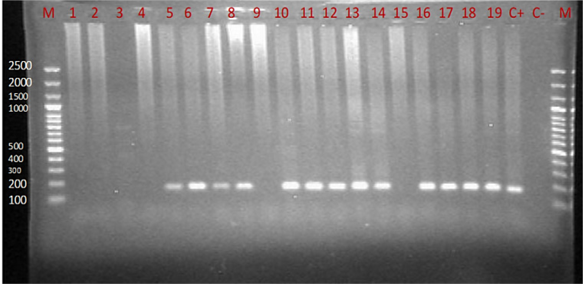
Fig. 1.
Gel electrophoresis under UV light using an agarose gel (2%) stained with 0.2 mg/ml of GelRed for analysis of the nested PCR product. Lanes (1–4, 9, and 15) are negative, lanes (5–8, 10–14, and 16–19) are 174 bp product representing positive samples using primers specific to amplify Entamoeba dispar. C+: Positive control, C-: Negative control, and M: 100 bp DNA marker.
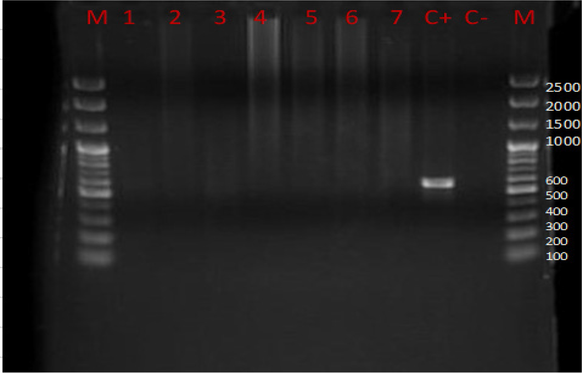
Fig. 2.
Gel electrophoresis under UV light using agarose gel (1%) stained with 0.2 mg/ml of GelRed for analysis of nested PCR product. Lanes (1–7) are negative, C+: Positive control revealed 550 bp product using primers specific to amplify Entamoeba moskovskii, C-: Negative control, and M: 100 bp DNA marker.
Table III
Sociodemographic factors of positive results by molecular examination for E. dispar.
| Parameters | Negative | Positive | p-value | |
|---|---|---|---|---|
| Frequency (percentages) | ||||
| Gender | Male | 162 (90.5) | 17 (9.5) | 0.037* |
| Female | 119 (82.6) | 25 (17.4) | ||
| Residence | Rural | 134 (82.7) | 28 (17.3) | 0.022* |
| Urban | 147 (91.3) | 14 (8.7) | ||
| Tape water or untreated water | Yes | 135 (81.8) | 30 (18.2) | 0.005* |
| No | 146 (92.4) | 12 (7.6) | ||
| Age group (Years) | <1 | 3 (100) | 0 (0.0) | 0.000* |
| 1–4 | 139 (82.7) | 29 (17.3) | ||
| 5–8 | 129 (96.3) | 5 (3.7) | ||
| ≥9 | 10 (55.6) | 8 (44.4) | ||
Sequencing of PCR products. The PCR products were sequenced using the Sanger method on an ABI 3730 × l DNA Analyzer (Macrogen, South Korea), and raw sequence files were assessed for quality using Finch TV (version 1.5.0, Geospiza Inc., USA). The obtained sequences of the positive PCR products were submitted to NCBI, which assigned accession numbers PX794898 and PX794899 to the GenBank database. The BLAST sequence analysis tool at NCBI showed that the sequenced E. dispar amplicons were 100% identical to the E. dispar strain MRY9.1 and the E. dispar voucher A7 small subunit ribosomal RNA gene (OP451884.1 and PV330538.1).
Discussion
The results showed that 17.3% of examined samples by microscopy were positive for Entamoeba species among symptomatic children; this high rate may be attributed to the fact that the Kurdistan region of Iraq is endemic for Entamoeba species, providing favorable environmental conditions for this protozoan (Chowdhury 2022). Similar results have been reported in different Iraqi regions, such as in Duhok (21.68%) (Hasan et al. 2023), Erbil (22.8%) (Abdullah 2017), Baqubah (46.01%) (Ibrahim et al. 2018), Thi-Qar (11.1%) (Flaih et al. 2021). In other countries, the infection rate was 18.6% in Malaysia (Anuar et al. 2012), 19.2% in the United Arab Emirates (Elbakri et al. 2013), and 19% in Northeast India (Nath et al. 2015). Lower prevalence rates were reported in regions of Iran that share borders with Iraq, for example, a study conducted in northwest of Iran reported 1.01% in 2016 (Bahrami et al. 2019) and 3.6% in 2014 (Mohammadzadeh et al. 2017); however, a higher rate (23.8%) was documented in Gorgan, northern Iran between 2010 and 2012 (Sharbatkhori et al. 2014). Variations in prevalence rates may be due to differences in sample size, environmental conditions, water sources, and personal hygiene practices.
Direct microscopy can reveal both pathogenic and non-pathogenic Entamoeba species present in the human gastrointestinal tract. However, only molecular analysis accurately differentiates between them. Routine laboratory diagnosis and stool examination in most developing countries is based on microscopy; therefore, all Entamoeba organisms are often identified as the pathogenic E. histolytica (Mahmood and Baker 2020; Mahmood 2023). Nevertheless, other Entamoeba species remain commensal and non-pathogenic, accounting for a large proportion of microscopically misdiagnosed stool examinations (Yimer et al. 2017; Bahrami et al. 2019). E. dispar is a distinct species lacking invasive potential; it shares morphological similarities with E. histolytica and also colonizes the human intestine. It is responsible for the majority of asymptomatic infections detected globally. The distinction between the two amoebae has had a significant impact on the epidemiology of amoebiasis (Singh et al. 2021). Metronidazole is considered the standard therapy for treating invasive amoebiasis, and misdiagnosed commensal E. dispar and E. moshkovskii are often treated unnecessarily. Adverse effects can occur even with conventional doses of metronidazole, such as headache, loss of appetite, metallic taste, nausea, and vomiting, while dizziness, convulsions, poor coordination, and numbness of the extremities are less common but more serious adverse effects that warrant discontinuation of metronidazole (Gonzales et al. 2019). In the present study, molecular analysis showed that 75% of microscopy-positive samples were E. dispar. Similar results were documented in Iraq (72.6%) (Abid 2016). Molecular studies in Turkey and Brazil revealed that all examined samples were E. dispar (Pinheiro et al. 2004; Dagci et al. 2007). A higher prevalence of E. dispar compared E. histolytica was reported in many studies in Bangladesh, Malaysia, Australia, Canada, India, Nigeria, Ghana, South Africa, Colombia, and Iran (Haque et al. 1997; Gonin and Trudel 2003; Verweij et al. 2003; Samie et al. 2006; Fotedar et al. 2007; Khairnar and Parija 2007; Mojarad et al. 2010; Anuar et al. 2012; López et al. 2015; Dawah et al. 2016; Bahrami et al. 2019). The occurrence of E. dispar was significantly greater in females than in males. Similar results have been reported in Erbil, Iraq, and in South Africa (Samie et al. 2006; Mahmood and Baker 2020). Given that E. dispar is a non-pathogenic commensal organism, its identification in symptomatic pediatric patients does not imply an etiological role in the development of gastrointestinal disease. The objective of this study was to determine the prevalence and molecular occurrence of E. dispar, rather than investigating the causes of the reported symptoms. Therefore, other infectious or non-infectious etiologies cannot be excluded and were beyond the scope of this investigation.
Of the 56 positive samples by microscopy, no E. moskoviskii was found at the molecular level, which could be related to the geographic distribution of the species and sample size of this study. A low rate of E. moshkovskii has been reported relative to E. dispar and E. histolytica in Erbil, Iraq (Mahmood and Baker 2020). Comparable results have been documented in western Iran, Northeast India, and Malaysia (Ngui et al. 2012; Nath et al. 2015; Bahrami et al. 2019).
According to the results, E. dispar was more prevalent in rural areas, since the most frequent sources of Entamoeba species and E. dispar are shared—including human feces, septic tanks, and raw sewage. The limited basic infrastructure in rural areas, including insufficient access to sanitation, inadequate chlorinated water supply, and a lack of health education and awareness, likely account for the high infection rates (Chowdhury 2022). Although E. dispar is a non-pathogenic gut commensal protozoan, its presence provides evidence for human fecal contamination of drinking water or poor hygienic practices (Chowdhury 2022). This interpretation is further supported by the significantly lower infection with E. dispar in people drinking bottled or chlorinated water in this study. The documented lower infection rates among individuals drinking bottled or filtered water compared with those drinking tap or untreated water may be due to insufficient chlorination of the municipal water supply or contamination of drinking water with sewage. Entamoeba cysts can survive in drinking water and have been documented in the water supply in Iraq (Chowdhury et al. 2022). Similar findings have been reported in Turkey (Kurt et al. 2008) and Pakistan (Karim et al. 2024), which documented that the prevalence of the Entamoeba species was lower among individuals drinking bottled water than among those drinking tap water.
Similarly, this study documented the highest infection rate in children aged one to four years. This trend likely reflects the level of childcare and supervision during early childhood. Also, older children, particularly those aged ≥ 9 years, tend to exhibit greater independence in their behavior, which may lead to increased exposure to contaminants through playing and eating outside the home. Age group categorization varies in several studies; for example, the 1–10-year age group in Pakistan and Iraq reported higher infection rates, followed by the 11–20 year and adult age groups (Hasan et al. 2023; Karim et al. 2024). A study in Thi-Qar, Iraq reported the highest proportion of infections in the 5–14-year age group (Flaih et al. 2021).
The limitations of this study include a moderate sample size, which may affect the generalizability of the study. Additionally, PCR was performed only on microscopy-positive samples, which prevents a proper evaluation of diagnostic performance and limits epidemiological conclusions.
Conclusions
The prevalence of E. dispar was directly correlated with source of drinking water, place of residence, and age. This study demonstrates that E. dispar is present among microscopy-positive samples from symptomatic children, highlighting the potential for misclassification of Entamoeba species when relying solely on microscopy.
Acknowledgments
The authors thank the College of Medicine, University of Sulaimani, for providing the workspace and laboratory equipment. Sincere gratitude is also extended to the Sulaimani Directorate of Health for providing support during sample collection at Dr. Jamal Ahmed Rashid Pediatric Teaching Hospital, Sulaimaniyah, Iraq.
Notes
[2] Ethical statement
This study was conducted in accordance with the Declaration of Helsinki – Ethical Principles for Medical Research, revised in 2008; and was approved by the Ethics Committee of the College of Medicine, University of Sulaimani, Sulaimaniyah, Iraq in meeting number (302/18), dated July 18, 2024. Written informed consent was obtained from the participants’ parents or guardians.
[3] Author contributions
SAFM: Conceptualization, methodology, and writing the original draft. SJA: Methodology, resources, and writing the original draft. SOB: Study registration, data collection, and writing the original draft. HMB: Formal analysis and supervision. FB: Review and editing of the final draft. SZSS: Writing the original draft. AZS: Writing the original draft. All authors approved the final version of the manuscript to be published.