
Table I
qPCR primer sequence.
| Primer and probe name | Primer sequence (5’-3’) | Modification |
|---|---|---|
| mip-F | GTTATCCCTGGATGGACAGAAG | / |
| mip-R | GCAAGACCTGAGGGAACATAA | / |
| mip-P | ATGCCAGCTGGATCAACTTGGGAA | 5’ 6-FAM/ZEN/3’ IBFQ |
Table II
Antibody information.
| Antibody Name | Lot number | Manufacturer |
|---|---|---|
| Rabbit Anti-STING Antibody | ab288157 | Abcam, UK |
| Rabbit Anti-RIPK3 Antibody | PA5-19956 | Invitrogen, USA |
| Rabbit Anti-MLKL Antibody | MA5-31971 | Invitrogen, USA |
| Rabbit Anti-phospho-MLKL Antibody | MA5-32752 | Invitrogen, USA |
| Rabbit Anti-β-actin Antibody | ab213262 | Abcam, UK |
| Goat Anti-Rabbit IgG (H&L) HRP-Conjugated Antibody | ab205718 | Abcam, UK |
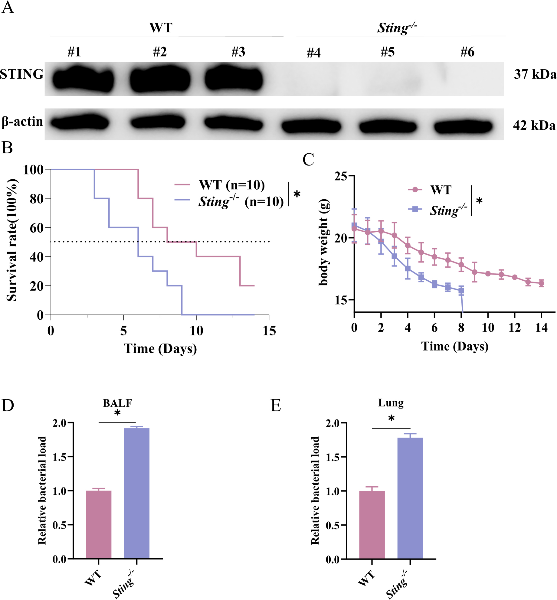
Fig. 1.
Sting deletion decreases survival rates following infection and exacerbates pulmonary bacterial load.
WT and Sting−/− mice were intranasally inoculated with a lethal dose of L. pneumophila to establish a pneumonia model; A: The knockout efficiency was evaluated via WB; B: Survival analysis of WT and Sting−/− mice over 14 days following L. pneumophila infection by log-rank; C: Body weight changes of WT and Sting−/− mice over 14 days following L. pneumophila infection; D: Relative bacterial loads in the BALF of WT and Sting−/− mice following infection was detected; E: Relative bacterial loads in the lung tissues of WT and Sting−/− mice following infection was detected; *, P < 0.05. Survival experiment (B and C) was independently repeated twice with 10 mice per group. All other experiments (A, D, E) were independently repeated three times with at least 3 mice per group. Data are shown as mean ± SD.
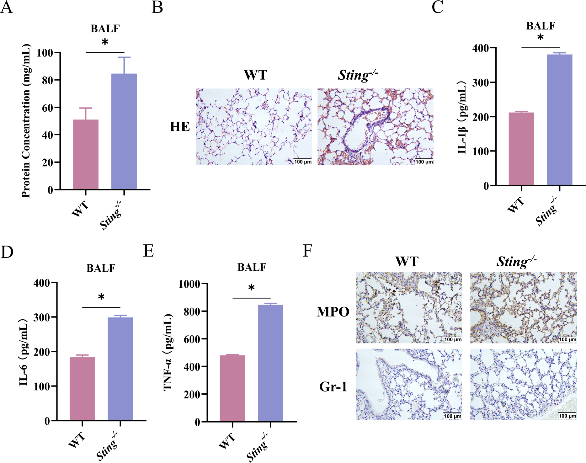
Fig. 2.
Sting deletion exacerbates infection-induced pulmonary inflammatory responses and tissue injury in mice.
WT and Sting−/− mice were intranasally inoculated with a sublethal dose of L. pneumophila to establish the pneumonia model; A: Total protein concentration in the BALF of mice was detected via ELISA; B: Analysis of lung tissue structure via H&E staining; C–E: Inflammatory cytokine concentrations IL-1β (C), IL-6 (D), and TNF-α (E) in the BALF of mice were detected via ELISA; F: MPO and Gr-1 expressions in lung tissues were detected via IHC; *, P < 0.05. All experiments were independently repeated three times with at least 3 mice per group. Data are shown as mean ± SD.
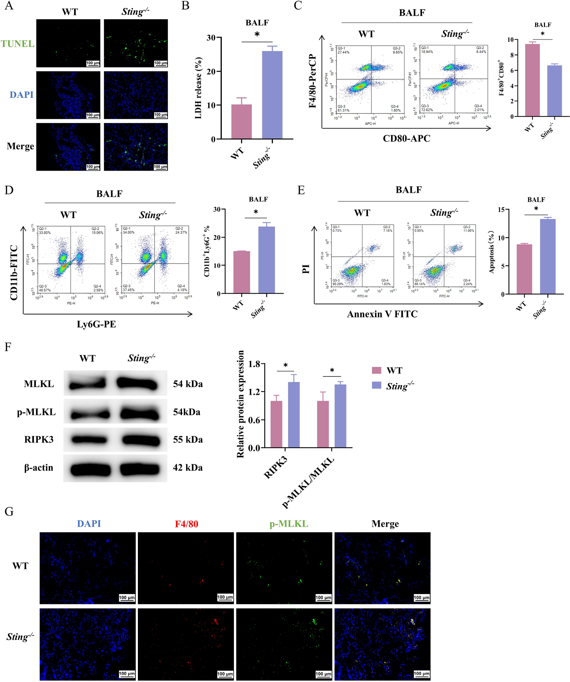
Fig. 3.
Sting deletion exacerbates infection-induced immune cell death.
WT and Sting−/− mice were intranasally inoculated with a sublethal dose of L. pneumophila to establish the pneumonia model;
A: Observation of cell apoptosis in mouse lung tissue via TUNEL staining; B: Assessment of cellular damage via an LDH assay kit; C; Proportion of macrophages in the BALF of mice was determined via flow cytometry; D: Proportion of neutrophils in the BALF of mice was determined via flow cytometry; E: Apoptosis rate of macrophages in the BALF of mice was determined via flow cytometry; F: Protein expression levels of necroptosis markers (RIPK3, p-MLKL, and MLKL) were detected via WB; G: IF co-localization of p-MLKL with the macrophage marker F4/80 in lung tissue sections. Nuclei were counterstained with DAPI; *, P < 0.05.
All experiments were independently repeated three times with at least 3 mice per group. Data are shown as mean ± SD.