
Introduction
Legionella pneumophila is a Gram-negative bacterium ubiquitously distributed in natural environments (Tomaskovic et al. 2022). When aerosols containing this bacterium are inhaled by susceptible individuals, the pathogen invades and colonizes alveolar macrophages, subsequently triggering Legionnaires’ disease (Graham et al. 2024). Serving as an atypical pneumonia, L. pneumophila-induced pneumonia is clinically characterized by severe disease progression and an elevated mortality rate, posing a persistent threat to public health (Mahgoub et al. 2025). During infection, L. pneumophila injects a repertoire of effector proteins into the cytoplasm of host cells via the type IV secretion system (T4SS), thereby enabling it to evade lysosomal degradation and to exploit endoplasmic reticulum resources to form specialized Legionella-containing vacuoles for intracellular replication and proliferation. The host detects this invasive behavior via pattern recognition receptors (PRRs) and initiates elaborate innate immune responses to combat the infection (García-Rodríguez et al. 2023; Yang et al. 2023). Nevertheless, the specific functional mechanisms of pivotal signaling hub molecules within the host innate immune signaling pathways in L. pneumophila pneumonia remain elusive.
During host defense against L. pneumophila infection, the immune system establishes a multi-layered defense architecture via precisely orchestrating cellular responses (Mascarenhas and Zamboni 2024). Serving as frontline sentinels, alveolar macrophages exert dual functions following pathogen recognition via PRRs. Beyond eliminating a subset of invading bacteria via direct phagocytosis, they also initiate pulmonary inflammatory responses and construct the initial immune microenvironment by secreting inflammatory cytokines (Liu and Shin 2019). Furthermore, these cytokines promote vascular endothelial cell activation and adhesion molecule expression, driving rapid recruitment of circulating neutrophils to the infection site, thereby forming the second wave of defense after macrophages (Effah et al. 2021). Upon arrival at the infection focus, neutrophils prominently enhance host bacterial elimination via diverse effector mechanisms, including the release of neutrophil extracellular traps, reactive oxygen species, and antimicrobial peptides (Koutantou et al. 2025).
Thus, a moderate inflammatory response is indispensable for effective infection control, whereas excessive or persistent inflammation disrupts the alveolar-capillary barrier, induces tissue edema, and impedes gas exchange function, thereby exacerbating lung injury (Moldoveanu et al. 2009; Stotts et al. 2023). Against this backdrop, an in-depth investigation of the pivotal molecular mechanisms modulating immune and inflammatory responses during L. pneumophila infection holds essential significance for elucidating host defense pathways and identifying potential therapeutic targets.
Notably, the stimulator of interferon genes (Sting), serving as a core signaling hub of innate immunity, plays critical roles in sensing intracellular pathogen invasion and orchestrating immune responses (Rousset 2023). The Sting pathway is broadly implicated in regulating host defense mechanisms during bacterial and protozoan infections (Louie et al. 2020; Wang et al. 2019; You et al. 2025). For instance, in Staphylococcus aureus-induced pneumonia, Sting deletion causes macrophage death, bacterial overproliferation, and exacerbated inflammation; conversely, pharmacological inhibition of necroptosis rescues the host defense defects in Sting-deficient mice(Liu et al. 2021). Nevertheless, while the protective effects of Sting against systemic bacterial infections have been well documented, its immunomodulatory functions during L. pneumophila-induced pneumonia, particularly in coordinating lung-specific immune responses, remains to be systematically elucidated.
The immunomodulatory effects of Sting on L. pneumophila-induced pneumonia have been demonstrated. Sting deletion prominently attenuates pulmonary bacterial elimination, potentiates inflammatory cytokine release and neutrophil infiltration, and promotes macrophage apoptosis alongside upregulation of necroptosis-related protein expression. Herein, the core status of Sting in host defense against L. pneumophila infection and multiple mechanisms, including orchestration of innate immune responses, regulation of inflammatory cascades, and maintenance of cellular homeostasis, are systematically elucidated, with a novel theoretical foundation for the development of targeted therapeutic strategies offered.
Experimental
Materials and Methods
Bacterial culture. L. pneumophila (strain Philadelphia-1, serogroup 1, ATCC 33152, American Type Culture Collection, USA) was cultivated on Buffered Charcoal Yeast Extract Agar (BCYE, HOPEBIO, China) at 37℃ for 3 days. They were subsequently subcultured into 10 ml of BCYE liquid medium (HOPEBIO, China) and incubated for 16 hours. Ultimately, the turbidity of the bacterial suspension was examined at a wavelength of 600 nm with a spectrophotometer (Thermo Fisher Scientific, USA), with the bacterial concentration determined via turbidity-to-concentration conversion.
Construction of murine infection models. For in vivo studies, 32 C57BL/6J wild-type (WT) and Sting−/− mice (6–8 weeks old, half male and half female) were derived from Shanghai Model Organisms Center, Inc. (China). All animal experiments were carried out according to the institutional guidelines and approved by the Animal Care and Use Committee (IACUC) in Shanghai Yishang Biotechnology Co., Ltd, approval number IACUC-2025-I-056. Mice were incubated under specific pathogen-free conditions. To evaluate post-infection survival, experimental mice were intra-nasally inoculated with a lethal dose of L. pneumophila suspension (30 μl, 1×10⁷ CFU per mouse), with 10 mice in each of the WT and Sting−/− groups. Survival rates of WT and Sting−/− mice were monitored continuously and recorded over 14 days, and body weights were recorded daily. Furthermore, a separate cohort of WT and Sting−/− mice was intranasally administered a sublethal dose of L. pneumophila (30 μl, 5×10⁶ CFU/per mouse), with 6 mice per group, to further investigate the correlation between survival rates and bacterial load. At 24 hours post-infection, mice were euthanized, with bronchoalveolar lavage fluid (BALF), lung tissue, and blood samples harvested for subsequent analyses.
Determination of bacterial load via PMA-qPCR. Samples were pretreated with ethidium monoazide bromide (PMA, absin, China) at a final concentration of 50 μM. Following incubation at 40℃ for 15 minutes in the dark, samples were exposed to a 500W halogen light source at a distance of 20 cm for 15 minutes to conduct the photo-crosslinking reaction. Free PMA was eliminated via centrifugation at 10,000×g for 10 minutes. DNA was extracted with a genomic DNA isolation kit (TIANGEN BIOTECH (BEIJING) CO., LTD, China) and applied as the template for qPCR. Specific primers and TaqMan probes targeting L. pneumophila were synthesized by Sangon Biotech (Shanghai) Co., Ltd, with primer sequences detailed in Table I. qPCR reactions were implemented in a 20 μl system with the following cycling conditions: initial denaturation at 95℃ for 15 minutes, followed by 40 cycles of denaturation at 94℃ for 10 seconds and annealing/extension at 60℃ for 1.5 minutes. The ABI 7500 Real-Time PCR system (Thermo Fisher Scientific, USA) was employed for detection. The obtained Ct values were converted to viable bacterial load (CFU/ml). All bacterial load determinations were performed on samples collected at 24 hours post-infection.
Table I
qPCR primer sequence.
| Primer and probe name | Primer sequence (5’-3’) | Modification |
|---|---|---|
| mip-F | GTTATCCCTGGATGGACAGAAG | / |
| mip-R | GCAAGACCTGAGGGAACATAA | / |
| mip-P | ATGCCAGCTGGATCAACTTGGGAA | 5’ 6-FAM/ZEN/3’ IBFQ |
Enzyme-Linked Immunosorbent Assay (ELISA). To evaluate lung tissue barrier function and the extent of the inflammatory response, BALF total protein and inflammatory cytokine concentrations were measured by ELISA. Following centrifuging the BALF samples to obtain the supernatant, the total protein concentration was determined with a Mouse Total Protein ELISA kit (COIBO Bio, China). This measurement served as an indicator for evaluating the permeability of the pulmonary capillary-alveolar barrier. Concurrently, to dissect local inflammation levels, the concentrations of specific inflammatory cytokines in the BALF supernatant were quantified with the corresponding Mouse IL-1β ELISA kit (Wuhan Fine Biotech Co., Ltd, China), Mouse IL-6 ELISA kit (Shanghai Enzyme-linked Biotechnology Co., Ltd., China), and Mouse TNF-α ELISA kit (Shanghai Enzyme-linked Biotechnology Co., Ltd., China). All detections were implemented with a microplate reader (Thermo Fisher Scientific, USA) to record absorbance at a wavelength of 450 nm. Concentrations of total protein and inflammatory cytokines were calculated according to standard curves. Cytokine and total protein levels were measured in BALF samples collected at 24 hours post-infection.
Hematoxylin and Eosin (H&E) staining. Mouse lung tissues were harvested and fixed in 4% paraformaldehyde (Beyotime Biotechnology, China) for 24 hours, then paraffin-embedded and sectioned. Sections were dewaxed in xylene and rehydrated via a graded ethanol series (absolute ethanol to 70% ethanol; Sinopharm Group Co., Ltd., China), then rinsed with tap water for 2–5 minutes. H&E staining was subsequently implemented: sections were first stained with hematoxylin (Beyotime Biotechnology, China) for 5–8 minutes, differentiated in 1% hydrochloric acid-ethanol for several seconds, rinsed under running tap water for bluing, and then counterstained with eosin (Beyotime Biotechnology, China) for 0.5–2 minutes. Post-staining, sections were dehydrated via a graded ethanol series (75%, 85%, 95%, and 100% ethanol; Sinopharm Group Co., Ltd., China), cleared in xylene (Beyotime Biotechnology, China), and ultimately mounted with neutral balsam (Beyotime Biotechnology, China). All sections were identified and imaged via a light microscope (Leica, Germany).
Immunohistochemical (IHC) analysis. To evaluate neutrophil infiltration in lung tissue, IHC analysis was employed to detect the neutrophil-specific markers myeloperoxidase (MPO) and granulocyte differentiation antigen-1 (Gr-1). In brief, lung tissue samples were fixed in 4% paraformaldehyde (Beyotime Biotechnology, China) for 24 hours, then paraffin-embedded and sectioned. Following dewaxing and rehydration, sections underwent antigen retrieval with sodium citrate buffer for 20 minutes. Endogenous peroxidase activity was then blocked, with sections blocked with 5% bovine serum albumin (BSA) (Gibco, USA) for 1 hour. Ultimately, sections were cultivated overnight at 4℃ with primary antibodies: rabbit anti-MPO (ab208670, Abcam, UK) and rabbit anti-Gr-1 (ab238132, Abcam, UK). Following rinsing, sections were incubated with a horseradish peroxidase (HRP)-conjugated goat anti-rabbit IgG H&L secondary antibody (ab205718, Abcam, UK) at 37℃ for 30 minutes. Chromogenic reaction was implemented with freshly prepared diaminobenzidine chromogen solution (Beyotime Biotechnology, China). Nuclei were counterstained with hematoxylin (Beyotime Biotechnology, China), and sections were then dehydrated, cleared, and mounted with neutral balsam (Beyotime Biotechnology, China). All sections were examined and imaged under a light microscope (Leica, Germany).
TUNEL staining. To evaluate the level of apoptosis in lung tissue, staining analysis was performed according to the instructions of the TUNEL assay kit (Beijing Solarbio Science & Technology Co., Ltd., China). Brief procedures were listed: Paraffin-embedded lung sections were dewaxed in xylene and rehydrated via a graded ethanol series. A 40 μg/mL Proteinase K solution diluted in phosphate-buffered saline (PBS) from a 2 mg/mL stock was applied to each section (100 μl per sample) and incubated at 37℃ for 30 minutes. The terminal deoxynucleotidyl transferase enzyme reaction mixture was subsequently added, followed by co-incubation at 37℃ for 60 minutes. Following thorough rinsing with PBS, sections were mounted with a DAPI anti-fade mounting medium (Beijing Solarbio Science & Technology Co., Ltd., China), which allowed simultaneous visualization of TUNEL green fluorescence signals and nuclear counterstaining. All sections were examined and imaged under a fluorescence microscope (Leica, Germany).
Lactate Dehydrogenase (LDH) determination in BALF. To assess the extent of alveolar cavity cellular damage following L. pneumophila infection at 24 hours post-infection, an LDH cytotoxicity assay kit (Beyotime Biotechnology, China) was employed. BALF was harvested from mice and centrifuged at 1000×g for 10 minutes at 4℃ to remove cellular debris, and the supernatant was collected. A 50 μl aliquot of the supernatant was pipetted into each well of a 96-well plate, followed by the addition of 50 μl of substrate mixture per well. The plate was incubated at room temperature in the dark for 30 minutes. Stop solution was subsequently added to each well to terminate the reaction. Absorbance at a wavelength of 490 nm was examined via a microplate reader (Thermo Fisher Scientific, USA), with the concentration of LDH released in BALF calculated according to standard curves.
Flow cytometric analysis. Flow cytometry was employed to quantify the populations of macrophages and neutrophils in the BALF of mice from both groups at 24 hours post-infection. For the identification of neutrophils, double staining was implemented with a rat anti-mouse CD11b-FITC antibody (FITC-65055, Proteintech, China) and a rat anti-mouse Ly6G-PE antibody (65140-1-Ig, Proteintech, China). Macrophages were identified and characterized with a rat anti-mouse F4/80-PerCP antibody (ab210252, Abcam, UK) and a rabbit anti-mouse CD80-APC antibody (APC-FcA98158, Proteintech, China).
To assess macrophage apoptosis, staining was implemented with an Annexin V-FITC/PI apoptosis detection kit (BioLegend, USA). All samples were analyzed via a flow cytometer (Agilent, USA) to determine the proportions of neutrophils and macrophages, as well as the rate of macrophage apoptosis.
Western Blot (WB) analysis. Lung tissue samples were homogenized in RIPA lysis buffer (Beyotime Biotechnology, China) containing protease inhibitors. Following centrifugation at 12,000×g for 10 minutes at 4℃, the supernatant was harvested. The protein concentration was determined with a bicinchoninic acid protein assay kit (Beyotime Biotechnology, China). Equal amounts of protein were separated by SDS-PAGE (Beyotime Biotechnology, China) and transferred onto PVDF membranes (Millipore, USA). The membranes were then blocked with 5% skim milk (Beyotime Biotechnology, China) and incubated with primary antibodies overnight at 4℃. Following rinsing three times with Tris-buffered saline containing Tween 20, the membranes were incubated with the corresponding HRP-conjugated secondary antibodies for 1 hour at room temperature. Protein bands were visualized via an enhanced chemiluminescence substrate (Thermo Fisher Scientific, USA). The antibodies employed are listed in Table II. Gray value quantification of WB bands was performed using ImageJ software (National Institutes of Health, USA). The gray value of each protein band was normalized to the corresponding internal reference protein, and the relative expression level was calculated and compared with the WT group. All data was obtained from no less than three independent experiments.
Table II
Antibody information.
| Antibody Name | Lot number | Manufacturer |
|---|---|---|
| Rabbit Anti-STING Antibody | ab288157 | Abcam, UK |
| Rabbit Anti-RIPK3 Antibody | PA5-19956 | Invitrogen, USA |
| Rabbit Anti-MLKL Antibody | MA5-31971 | Invitrogen, USA |
| Rabbit Anti-phospho-MLKL Antibody | MA5-32752 | Invitrogen, USA |
| Rabbit Anti-β-actin Antibody | ab213262 | Abcam, UK |
| Goat Anti-Rabbit IgG (H&L) HRP-Conjugated Antibody | ab205718 | Abcam, UK |
Immunofluorescence (IF). The IF colocalization assay was performed on lung tissue sections to identify cell types undergoing necroptosis. Paraffin-embedded lung sections were deparaffinized, rehydrated and subjected to antigen retrieval, followed by blocking with 5% BSA. The sections were incubated overnight at 4℃ with primary antibodies against p-MLKL (37333, Cell Signaling Technology, USA) and F4/80 (sc-377009, Santa Cruz Biotechnology, USA). After washing, the samples were incubated with Alexa Fluor 488-conjugated donkey anti-rabbit IgG (A-21206, Invitrogen, USA) and Alexa Fluor 555-conjugated donkey anti-rat IgG (A-21434, Invitrogen, USA) for 1 hour at room temperature away from light. Nuclei were counter-stained using DAPI-containing antifade mounting medium (Vector Laboratories, Burlingame, USA). All images were captured under a Leica fluorescence microscope, Germany.
Statistical analysis. Experimental data were dissected and plotted with GraphPad Prism 8.0 (Graph-Pad Software Inc., USA). Measurement data were presented as mean ± SD (n ≥ 3). Differences between the two groups were compared using the Student’s t-test, and comparisons among multiple groups were conducted using ANOVA. P < 0.05 indicated statistically significant.
Results
Sting deletion decreases survival rates following infection and exacerbates pulmonary bacterial load. A pneumonia model that was established by intranasal inoculation of a lethal dose of L. pneumophila in C57BL/6J WT mice and Sting knockout (Sting−/−) mice, validated by WB (Fig. 1A), was employed to investigate the impacts of Sting on mice with L. pneumophila pneumonia infection. The aim was to investigate the effects of Sting on host defense against L. pneumophila infection. Furthermore, qPCR results of mouse lung tissues revealed that mRNA levels of IFNB1 and ISG1 were significantly reduced in Sting−/− mice relative to wild-type mice (Fig. S1), further verifying successful establishment of Sting−/− mice. Monitoring of survival rates unveiled that Sting−/− mice exhibited a prominently lower survival rate compared to WT mice within 14 days post-infection (Fig. 1B), implying that Sting deletion attenuated host resistance to lethal L. pneumophila infection. Meanwhile, body weight monitoring showed that Sting−/− mice exhibited more dramatic weight loss than WT mice after infection (Fig. 1C). Furthermore, quantitative analysis of bacterial loads in BALF and lung tissue homogenates was implemented using PMA-qPCR. The relative bacterial loads in both BALF and lung tissues of Sting−/− mice were prominently higher than those in WT mice (Fig. 1D–E). Collectively, Sting exerted critical effects on the anti-L. pneumophila response in mice, with its deletion leading to impaired bacterial elimination and decreased host survival rates.
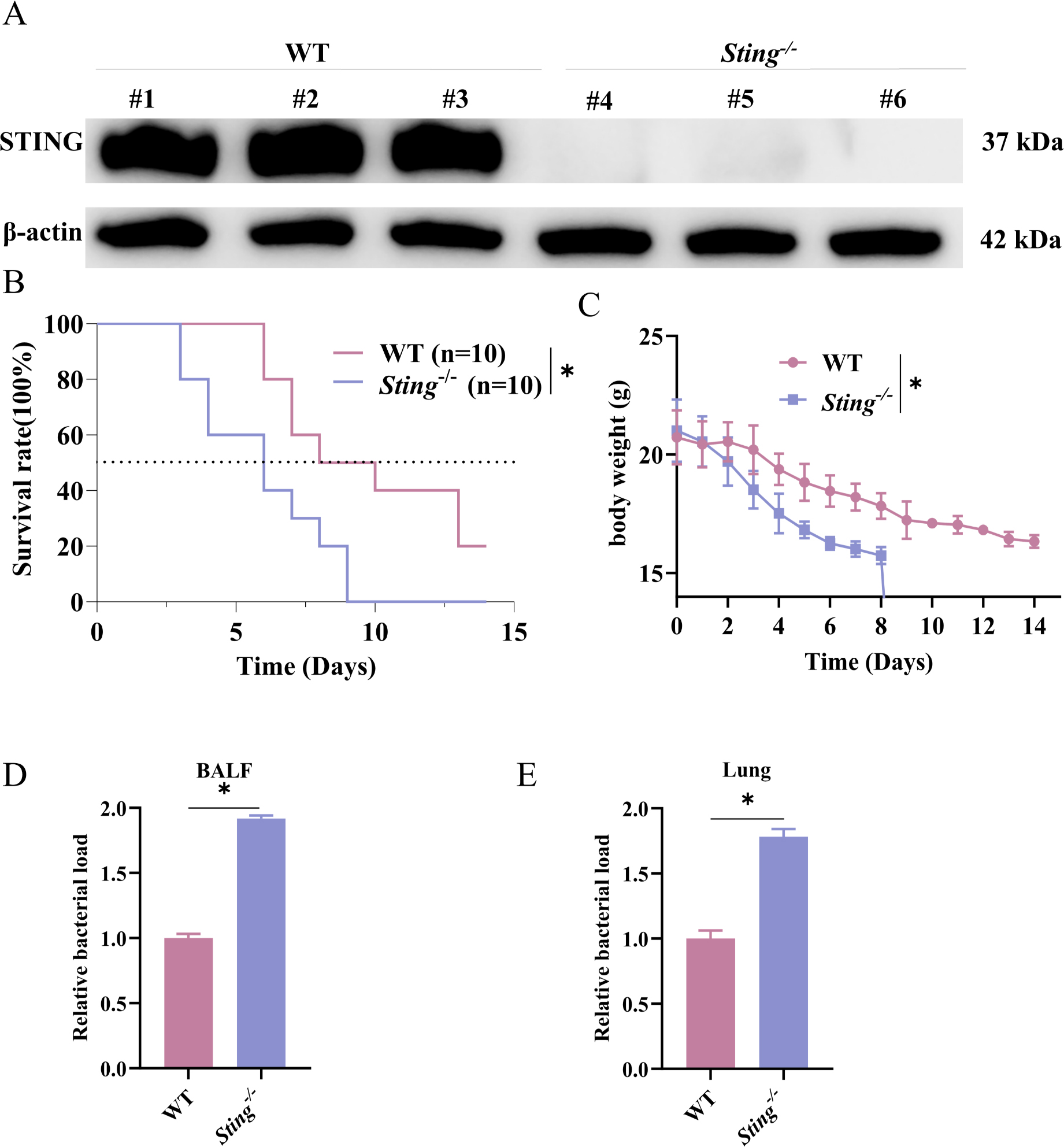
Fig. 1.
Sting deletion decreases survival rates following infection and exacerbates pulmonary bacterial load.
WT and Sting−/− mice were intranasally inoculated with a lethal dose of L. pneumophila to establish a pneumonia model; A: The knockout efficiency was evaluated via WB; B: Survival analysis of WT and Sting−/− mice over 14 days following L. pneumophila infection by log-rank; C: Body weight changes of WT and Sting−/− mice over 14 days following L. pneumophila infection; D: Relative bacterial loads in the BALF of WT and Sting−/− mice following infection was detected; E: Relative bacterial loads in the lung tissues of WT and Sting−/− mice following infection was detected; *, P < 0.05. Survival experiment (B and C) was independently repeated twice with 10 mice per group. All other experiments (A, D, E) were independently repeated three times with at least 3 mice per group. Data are shown as mean ± SD.
Sting deletion exacerbates infection-induced pulmonary inflammatory responses and tissue injury in mice. Given the critical effects of Sting on innate immunity, its regulatory mechanisms underlying pulmonary inflammatory responses during L. Pneumophila infection was further investigated. At the acute infection phase (24 hours), the pulmonary inflammatory levels and histopathological alterations in WT and Sting−/− mice were dissected. First, ELISA was employed to detect total protein concentration in BALF. The total protein content in BALF of Sting−/− mice exhibited a more prominent upregulation than that in WT mice (Fig. 2A), implying that Sting deletion impaired pulmonary vascular barrier function and elevated its permeability. Further histopathological examination of lung tissues via H&E staining unveiled that, compared with the WT group, Sting−/− mice exhibited more pronounced inflammatory cell infiltration in their lungs, more severe alveolar structural destruction, and more extensive interstitial thickening, indicating prominent pathological damage (Fig. 2B). In addition, to elucidate the effects of Sting on inflammatory cytokine secretion, the levels of key pro-inflammatory cytokines in BALF were measured. ELISA results demonstrated that the concentrations of IL-1β, IL-6, and TNF-α in BALF of Sting−/− mice were prominently higher than those in WT mice (Fig. 2C–E), indicating that Sting deletion promoted the release of pro-inflammatory cytokines. Since IL-1β and TNF-α were pivotal chemokines for neutrophil recruitment, the impacts of Sting deletion on pulmonary neutrophil infiltration were investigated. IHC analysis of neutrophil infiltration-related markers unveiled that the expression intensities of MPO and Gr-1 in lung tissues of Sting−/− mice were prominently higher than those in the WT group (Fig. 2F), indicating enhanced neutrophil recruitment in the lungs of Sting-deficient mice. Collectively, Sting deletion in the early stage of L. pneumophila infection led to impaired pulmonary barrier function, elevated pro-inflammatory cytokine release, and enhanced neutrophil infiltration, thereby exacerbating pulmonary tissue injury.
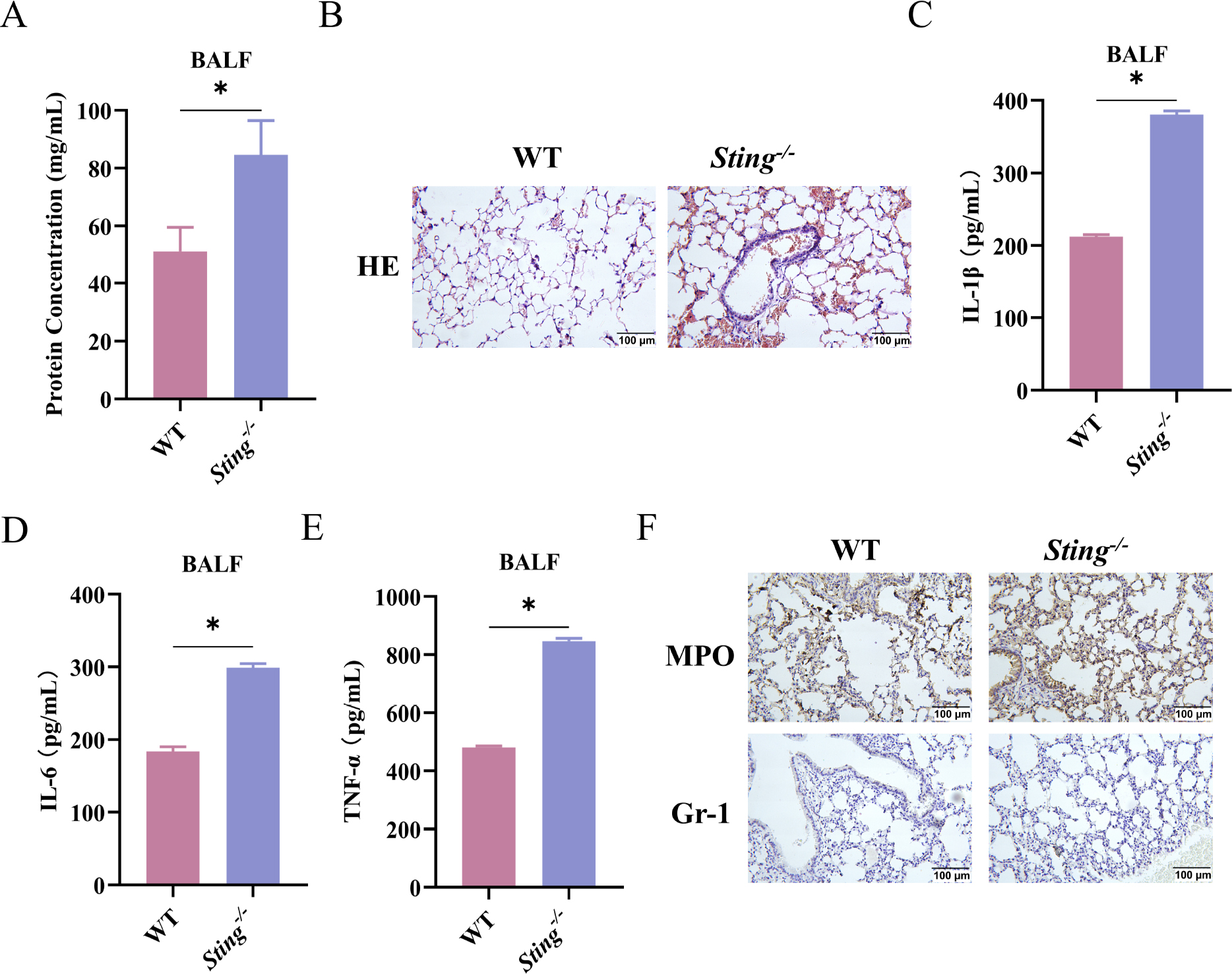
Fig. 2.
Sting deletion exacerbates infection-induced pulmonary inflammatory responses and tissue injury in mice.
WT and Sting−/− mice were intranasally inoculated with a sublethal dose of L. pneumophila to establish the pneumonia model; A: Total protein concentration in the BALF of mice was detected via ELISA; B: Analysis of lung tissue structure via H&E staining; C–E: Inflammatory cytokine concentrations IL-1β (C), IL-6 (D), and TNF-α (E) in the BALF of mice were detected via ELISA; F: MPO and Gr-1 expressions in lung tissues were detected via IHC; *, P < 0.05. All experiments were independently repeated three times with at least 3 mice per group. Data are shown as mean ± SD.
Sting deletion exacerbates infection-induced immune cell death. To investigate the impacts of Sting on immune cell death and its underlying mechanisms during L. pneumophila infection, TUNEL staining was employed to identify cell death in lung tissues. The count of TUNEL-positive cells in lung tissues of Sting−/− mice was prominently higher than that in WT mice (Fig. 3A). The LDH release in BALF was further detected to validate the extent of cell damage. Sting−/− mice exhibited prominently higher LDH activity in BALF compared to WT mice (Fig. 3B) Furthermore, quantitative analysis of immune cells (macrophages and neutrophils) in BALF was implemented with flow cytometry. The count of macrophages in Sting−/− mice was prominently lower than in WT mice (Fig. 3C), whereas the count of neutrophils was prominently elevated in Sting−/− mice (Fig. 3D). Notably, the apoptosis rate of macrophages was prominently higher in the Sting−/− group relative to the WT group (Fig. 3E).
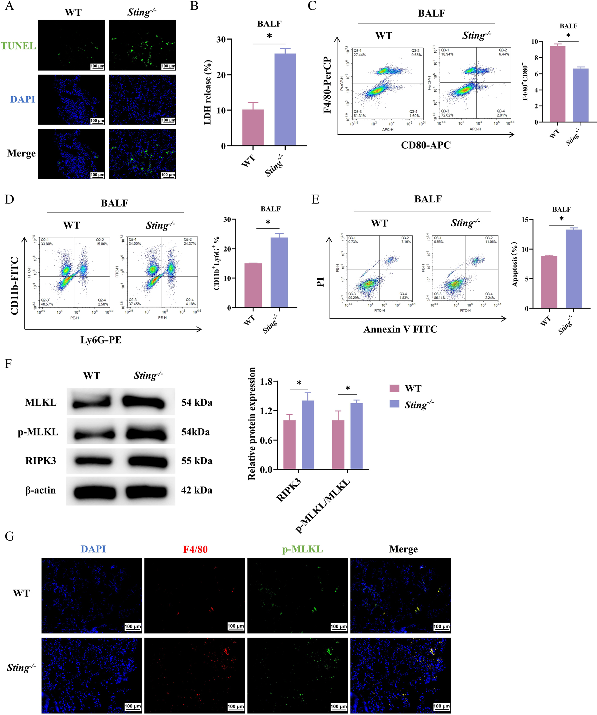
Fig. 3.
Sting deletion exacerbates infection-induced immune cell death.
WT and Sting−/− mice were intranasally inoculated with a sublethal dose of L. pneumophila to establish the pneumonia model;
A: Observation of cell apoptosis in mouse lung tissue via TUNEL staining; B: Assessment of cellular damage via an LDH assay kit; C; Proportion of macrophages in the BALF of mice was determined via flow cytometry; D: Proportion of neutrophils in the BALF of mice was determined via flow cytometry; E: Apoptosis rate of macrophages in the BALF of mice was determined via flow cytometry; F: Protein expression levels of necroptosis markers (RIPK3, p-MLKL, and MLKL) were detected via WB; G: IF co-localization of p-MLKL with the macrophage marker F4/80 in lung tissue sections. Nuclei were counterstained with DAPI; *, P < 0.05.
All experiments were independently repeated three times with at least 3 mice per group. Data are shown as mean ± SD.
Necroptosis served as a pivotal mediator of cell death in diverse pathogen infections (Liu et al. 2021; Zhang et al. 2022). Thus, the exacerbated cell death in Sting−/− mice might be linked to aberrant activation of the necroptosis pathway. The expression of pivotal necroptosis markers was detected via WB. The expression levels of RIPK3, p-MLKL, and total MLKL in lung tissues of Sting−/− mice were prominently upregulated (Fig. 3F). To clarify the specific cell type with necroptosis, we conducted IF colocalization analysis of p-MLKL and macrophage marker F4/80 on lung tissue sections (Fig. 3G). Obvious colocalization between p-MLKL and F4/80-positive macrophages was observed, suggesting that necroptosis mainly occurs in macrophages upon L. pneumophila infection. Sting deletion exacerbated macrophage apoptosis and immune cell exhaustion, thereby attenuating the capacity of the host’s anti-infectious immune defense.
Discussion
The core effects of the Sting signaling pathway on host defense against L. pneumophila-induced pneumonia were unveiled. Sting deletion impaired bacterial elimination, disrupted the inflammatory response, and promoted macrophage apoptosis, which in turn affected necroptosis. Collectively, the critical position of Sting in host defense via multi-dimensional immune regulatory mechanisms was elucidated, with novel insights into the therapeutic targeting of L. pneumophila-induced pneumonia offered.
Serving as a core component of innate immunity, Sting is a pivotal adaptor molecule in the sensing pathways for intracellular cyclic dinucleotides and DNA. It exerts critical effects on immune responses, primarily by inducing the production of type I inter-ferons and establishing an effective antiviral immune state (Dvorkin et al. 2024; Wang et al. 2025). The Sting pathway also plays a significant role in bacterial infections. For instance, CRISPR/Cas9 technology was employed to unveil that Sting knockout in a zebrafish model prominently elevated host susceptibility to bacterial infection (Sellaththurai et al. 2023). Furthermore, infection with carbapenem-resistant Acinetobacter baumannii induced mitochondrial damage via exosomes carrying the virulence factor OMP38, leading to the release of mitochondrial DNA into the cytosol. This DNA was subsequently recognized by cGAS, activating the Sting signaling pathway and ultimately triggering excessive inflammatory responses and lung tissue damage (Yang et al. 2024). In addition, Ruiz-Moreno et al. (2018) demonstrated that the cGAS-STING pathway plays a pivotal role in host defense against L. pneumophila in both mice and humans. STING-deficient mice harbored higher bacterial loads after infection, and the HAQ TMEM173/STING allele was markedly enriched in patients with legion-naires’ disease, which is consistent with our findings. However, the effects of Sting on pneumonia induced by L. pneumophila infection remained elusive. Collectively, Sting deletion decreases survival rates and elevates bacterial load in the lungs upon infection. The critical effects of the Sting pathway on host defense against bacterial infections are further validated.
Notably, infection and inflammation constitute a tightly interconnected pathological network during bacterial pneumonia progression, amplifying each other’s pathological effects via a positive feedback loop (Stotts et al. 2023). Specifically, when pathogens invaded the host lungs, their proliferation directly stimulated the innate immune system, triggering an inflammatory cascade reaction (Zhang et al. 2021). Conversely, a dysregulated inflammatory microenvironment exacerbated pathogen persistence and dissemination via disrupting tissue structural integrity and attenuating immune elimination capacity (Korkmaz and Traber 2023). Leveraging the pathological foundation, the Sting signaling pathway exerted core effects on modulating this critical link during L. pneumophila infection. Sting deletion leads to impaired bacterial elimination capacity linked to compromised macrophage function, elevated macrophage apoptosis, and the triggering of an overactive neutrophil response. Neutrophils served as the core effector cells of the acute inflammatory response. Their extensive activation and abnormal aggregation in lung tissue have emerged as a critical link in promoting the inflammatory cascade reaction (Herrero-Cervera et al. 2022; Xiao et al. 2025). Collectively, the critical effects of Sting on suppressing excessive neutrophil responses and mitigating inflammation-related tissue damage are elucidated.
Cellular death is an essential immune mechanism that enables the host to combat microbial invasion, exerting critical effects on controlling pathogen dissemination and maintaining tissue homeostasis (Newton et al. 2024). Necroptosis was a regulated form of cellular necrosis mediated by pivotal molecules such as RIPK1 and RIPK3, etc., which executed ultimate membrane rupture via the phosphorylation and oligomerization of MLKL, thereby holding essential significance in anti-infective immunity (Zhou et al. 2024). Notably, infection with the Gram-positive bacterium Streptococcus pneumoniae triggered necroptosis via its virulence factor pneumolysin, resulting in cardiomyocyte death and cardiac injury. Conversely, inhibitors of necroptosis prominently mitigated organ damage during invasive pneumococcal disease (Beno et al. 2020). Excessive activation of this pathway served as a pivotal mechanism underlying infection-related tissue damage; however, its effects on L. pneumophila pneumonia remained incompletely elucidated. Furthermore, Sting defends immune cells during L. pneumophila infection via suppressing the necroptosis pathway, with its deletion exacerbating immune cell depletion and necroptosis, thereby attenuating the host’s anti-infective immune defense capacity. Collectively, beyond exhibiting a novel mechanism by which the Sting pathway maintains immune cell homeostasis in L. pneumophila, this finding also offers a novel theoretical foundation for targeting the Sting-necroptosis axis in the treatment of intracellular pathogen infections.
Sting exerts a critical protective effect on host defense against L. pneumophila infection. Sting deletion triggers excessive inflammatory responses, along with apoptosis and necroptosis in immune cells, collectively leading to impaired alveolar barrier function and disrupted immune homeostasis, thereby compromising the host’s defensive capacity. However, certain limitations exist. Firstly, the specific downstream effector molecules by which Sting modulates immune cell apoptosis remain elusive. Secondly, while the research primarily focuses on neutrophils and macrophages, it remains unclear whether Sting affects the functions of other immune cell types during L. pneumophila infection warrants further investigation. Future research will prioritize the identification of critical downstream targets mediating Sting-regulated immune cell apoptosis. Furthermore, the precise functions of Sting within specific immune cell subsets were dissected by constructing myeloid cell-specific Sting knockout mice.
Notes
[1] Ethical statement
All animal experiments were carried out according to the institutional guidelines and approved by Animal Care and Use Committee (IACUC) in Shanghai Yishang Biotechnology Co., Ltd, approval number IACUC-2025-I-056.
[2] Contributed by Author contributions
Jiqing Lin: conceptualization, methodology, investigation, writing-original draft.
Gang Yan: validation, formal analysis, data curation.
Canhui Zhang: software, resources, visualization.
Jianhui Wu: formal analysis, investigation, project administration.
Qing Xue: formal analysis, investigation, visualization, funding acquisition.
Zhixin Liu: formal analysis, investigation, writing-review & editing.
Guiju Fang: conceptualization, supervision, writing-review & editing, project administration.
Funding
This work was supported by the Fujian Provincial Natural Science Foundation in 2023, Grant No. 2023J011089.
Supplementary materials are available on the journal’s website.