
Introduction
According to the latest estimates of the International Diabetes Federation, gestational diabetes mellitus (GDM), one of the most common metabolic disorders during pregnancy (Kautzky-Willer et al. 2023), affects approximately 10% of pregnant women worldwide (Sun et al. 2022). More than 90% of GDM cases occur in low-and middle-income countries (Sweeting et al. 2022). GDM can markedly increase adverse maternal perinatal and postpartum outcomes, including miscarriage, fetal malformations, preeclampsia, fetal growth restriction, fetal death, macrosomia, neonatal hypoglycemia, neonatal hyperbilirubinemia, and neonatal respiratory distress syndrome (Zhang et al. 2024, Wen et al. 2023, Regnault et al. 2024). Offspring of mothers with GDM are at increased risk of developing obesity, insulin resistance, and metabolic syndrome during childhood and adulthood (Kaza et al. 2025). GDM leaves identifiable characteristic “fingerprints” in the infant gut microbiota, in which the gut microbiota of female neonates exhibits a stronger potential for histamine and dopamine degradation (Wang et al. 2024). Maternal metabolic abnormalities may influence offspring neurodevelopment, immune regulation, and long-term metabolic health by altering the metabolic functions of the early neonatal microecology, thereby revealing a potential biological link among “maternal metabolism-fetal microecology-offspring health”. Researchers have begun to focus on gestational weight gain (GWG) as a key indicator reflecting metabolic load and changes in insulin sensitivity during pregnancy, which has been closely associated with GDM occurrence and progression, as well as perinatal outcomes (Zheng et al. 2022). However, the extent of weight gain during pregnancy regulates placental-fetal microbiota through the maternal metabolic environment and intrauterine microecology, which remains insufficiently investigated.
Microbial communities are crucial in energy metabolism, immune regulation, and inflammatory responses. Maternal gut, vaginal, and oral microecology undergo dynamic changes during pregnancy (Saadaoui et al. 2024). The occurrence of GDM can disrupt the balance of these microecological systems (Xia et al. 2025). Traditional views hold that the placenta is near-sterile (Perez-Munoz et al. 2017). Recent high-throughput sequencing studies have detected low-abundance bacterial DNA signals in placental samples, suggesting possible origins from maternal gut, oral, or genital tracts, or perinatal environmental exposure (Saadaoui et al. 2024, Aagaard et al. 2014). Nevertheless, as the placenta is a typical low-biomass sample, whether these bacterial DNA signals represent stable, active, tissue-resident microbial communities remains debated. Meanwhile, exogenous contamination during sampling, DNA extraction, PCR amplification, or sequencing may influence the results. Therefore, characterizing GDM-associated placental bacterial DNA communities with stringent negative controls and contaminant filtering may help explore potential links between maternal metabolic abnormalities and placental microenvironment changes (Stupak et al. 2023). We hypothesize that GDM-related placental microbial dysbiosis may alter neonatal meconium microbiota composition by affecting maternal-neonatal microbial interactions. However, studies on the placental microbial characteristics of women with GDM and their impact on the early neonatal gut microbiota remain very limited.
This study enrolled pregnant women and their newborns to evaluate GDM-associated alterations in placental bacterial DNA communities and neonatal meconium microbiota, assess the placental source contribution to neonatal meconium based on community similarity, examine GWG’s modulation of GDM-related maternal-neonatal microbial changes, and analyze links between key bacterial taxa and short-term neonatal hospitalization. The findings may provide new evidence for understanding GDM-related maternal-neonatal microbial alterations and their perinatal clinical significance.
Experimental
Materials and Methods
Study population
As an observational prospective cohort study, it included 104 pregnant women who delivered at our hospital between March 2025 and August 2025. The study flow is shown in Fig. 1. Inclusion criteria were age 18–40 years, delivery at 37–42 weeks of gestation, singleton pregnancy with natural conception, establishment of prenatal medical records and regular antenatal examinations, completion of oral glucose tolerance test screening during pregnancy, and provision of written informed consent and voluntary participation. Exclusion criteria were multiple pregnancies or conception via assisted reproductive technologies such as in vitro fertilization, preexisting hyperglycemia or glucose metabolism abnormalities identified before 20 weeks of gestation, severe pregnancy complications during the current pregnancy (such as placenta previa or placenta accreta), chromosomal abnormalities in either parent or the fetus, antibiotic use within 3 months prior to delivery, premature rupture of membranes, presence of any of the following conditions: autoimmune diseases, congenital heart disease, hematological diseases, chronic hypertension, chronic kidney disease, insulin resistance, thyroid dys-function, or other severe medical or surgical disorders, or fewer than 500 valid 5-region 16S rRNA sequence reads. This study was approved by the Ethics Committee of Hangzhou Linping District Maternal & Child Health Care Hospital and was conducted in accordance with the Declaration of Helsinki. All participants provided written informed consent.
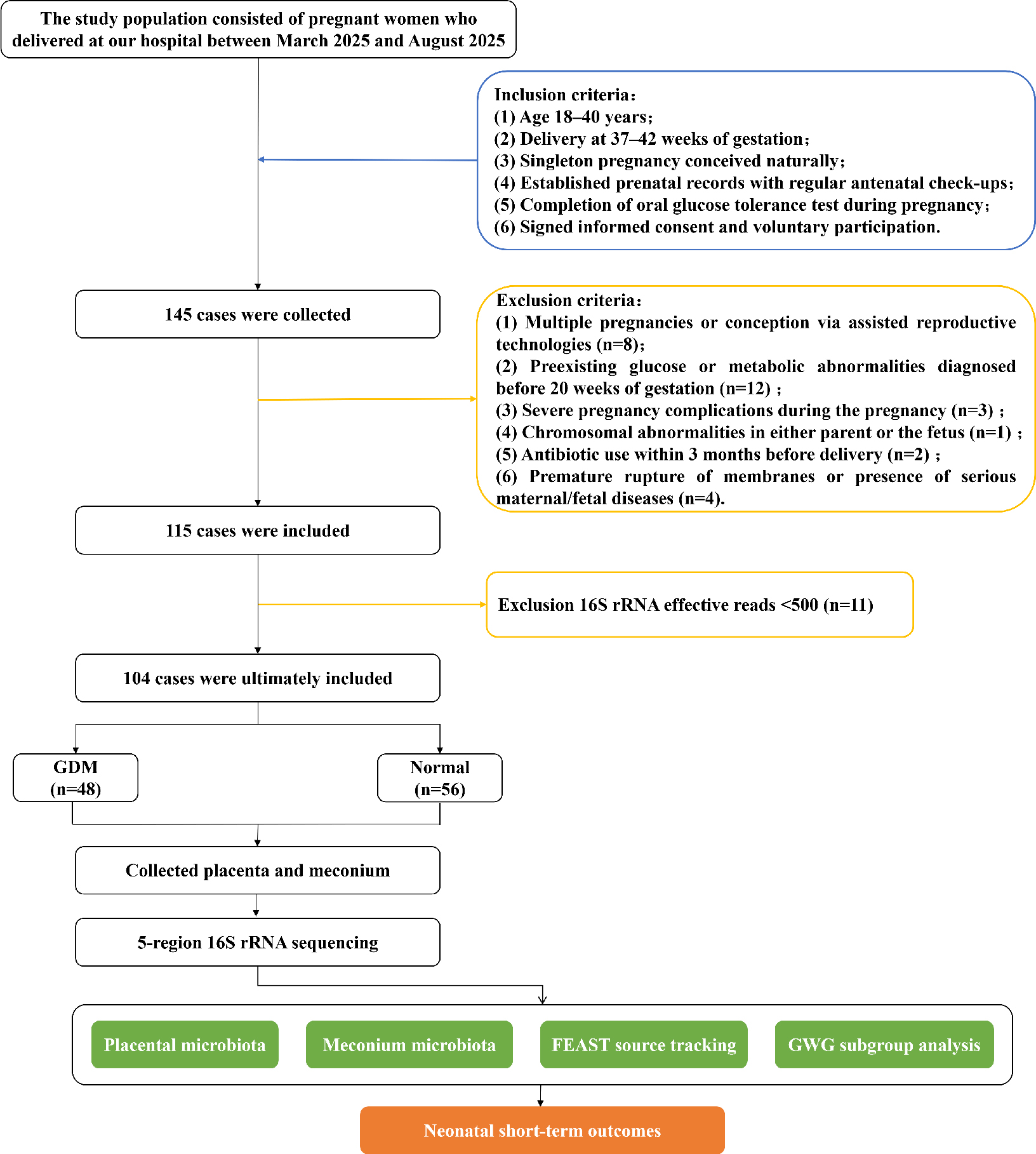
Fig. 1.
Study design and participant enrollment flowchart.
Sample size calculation
According to the literature, the operational taxonomic unit counts of 16S rRNA placental microbiota were 159.5 ± 34.57 in the GDM group and 176.25 ± 27.86 in the control group (Zheng et al. 2017). Using the two-sided t-test formula for comparing the means of two independent samples: n=2×(Z1-α/2+Z1-β)2σ2/δ2, with a significance level of α = 0.05, power 1 – β = 0.80, pooled standard deviation σ ≈ 31.40, and mean difference between groups δ = 16.75, the calculated sample size per group was approximately 56 cases, yielding a total sample size of 112. Considering an anticipated dropout or noncompliance rate of approximately 20%, the final sample size was set at 70 per group, for a total of 140 participants. The study initially planned to enroll 140 participants, but due to the limited number of subjects who met all inclusion and exclusion criteria and completed maternal-infant paired sample collection during the study period, 115 were preliminarily enrolled. After sequencing quality control (QC), 104 cases were finally analyzed.
Data collection
All clinical and laboratory data were collected from the medical records of all participants and reviewed by experienced obstetricians. Maternal demographic and anthropometric information included age, pre-pregnancy weight, pre-delivery weight, GWG, and body mass index (BMI). Other data collected included dietary habits, education level, and mode of delivery.
Early pregnancy biochemical markers included fasting blood glucose (FBG), postprandial blood glucose (PBG), glycated hemoglobin (HbA1c), total cholesterol (TC), triglycerides (TG), total bilirubin, and total bile acids. All pregnant women underwent a 75-g oral glucose tolerance test between 24 and 28 weeks of gestation. After at least 8 hours of overnight fasting, 5 mL of fasting venous blood was collected in the morning. Blood samples were centrifuged at 3,000 rpm for 15 minutes. Biochemical parameters were measured using a fully automated biochemical analyzer (AU5800, Beckman Coulter, USA) according to standard procedures. PBG levels were measured following the same protocol after ingestion of standardized glucose to ensure comparability of glycemic control.
Short-term neonatal outcomes included birth weight, neonatal Apgar scores, and neonatal hospitalization.
Disease and group definitions
The diagnosis of GDM was based on the 75-g oral glucose tolerance test performed at 24–28 weeks of gestation, with any of the following criteria confirming GDM: FPG ≥5.1 mmol/l, 1-hour postprandial glucose ≥10.0 mmol/l, or 2-hour postprandial glucose ≥8.5 mmol/l (Hod, Kapur 2015). GWG was calculated as the difference between maternal weight at delivery and pre-pregnancy weight to reflect weight changes during pregnancy (Jia, Bo 2025). GWG was categorized according to the 2009 Institute of Medicine guidelines for GWG, as shown in Supplementary Table I. Education level was classified into four categories based on the 2011 International Standard Classification of Education: lower secondary or below, upper secondary, post-secondary non-tertiary (college), and university degree.
Table I
General clinical characteristics of the GDM and Normal groups.
| Characteristics | GDM group (n = 48) | Normal group(n = 56) | P-value |
|---|---|---|---|
| General information | |||
| Age (years) | 30.21 (3.38) | 29.46 (4.27) | 0.386 |
| Pre-pregnancy BMI (kg/m2) | 23.76 (3.07) | 21.40 (2.81) | <0.001 |
| Gestational weight gain (kg) | 12.93 (5.80) | 14.52 (4.38) | 0.027 |
| Educational level | 0.525 | ||
| Lower secondary or below | 13 (27.08%) | 10 (17.86%) | |
| Upper secondary | 18 (38.51%) | 19 (33.92%) | |
| Post-secondary non-tertiary (college) | 4 (8.33%) | 10 (17.86%) | |
| University degree | 13 (27.08%) | 17 (30.36%) | |
| Dietary preference | 0.368 | ||
| Light | 14 (29.17%) | 23 (41.07%) | |
| Moderate | 18 (37.50%) | 20 (35.71%) | |
| Heavy | 16 (33.33%) | 13 (23.21%) | |
| Pharmacological intervention | 0.001 | ||
| Yes | 8 (16.67%) | 0 | |
| No | 40 (83.33%) | 56 (100.00%) | |
| Mode of delivery | 0.018 | ||
| Vaginal delivery | 6 (12.50%) | 18 (32.14%) | |
| Cesarean section | 42 (87.50%) | 38 (67.86%) | |
| Gestational hematological parameters | |||
| Fasting blood glucose (mmol/l) | 4.99 (0.65) | 4.48 (0.28) | <0.001 |
| Postprandial glucose (mmol/l) | 9.37 (1.43) | 7.49 (0.71) | <0.001 |
| Glycated hemoglobin (%) | 5.84 (0.40) | 5.24 (0.24) | <0.001 |
| Total cholesterol (mmol/l) | 6.48 (1.16) | 6.55 (1.30) | 0.776 |
| Triglycerides (mmol/l) | 4.52 (2.39) | 3.43 (1.23) | 0.024 |
| Total bilirubin (μmol/l) | 9.56 (2.83) | 10.76 (4.59) | 0.082 |
| Total bile acids (μmol/l) | 2.51 (3.10) | 2.36 (1.36) | 0.214 |
| Neonatal outcomes | |||
| Birth weight (g) | 3,413.13 (481.50) | 3,299.11 (326.38) | 0.216 |
| Apgar score | 10 (0) | 10 (0) | / |
| Neonatal hospitalization | 0.003 | ||
| Yes | 19 (39.58%) | 8 (14.29%) | |
| No | 29 (60.42%) | 48 (85.71%) | |
Based on GDM diagnosis, study participants were divided into GDM (n = 48) and normal control (n = 56) groups. To better understand how GWG influences pregnant women with GDM, we stratified the GDM group into inadequate (n = 17), appropriate (n = 16), and excessive (n = 15) GWG subgroups.
Sample collection
Placental samples were collected within 30 minutes after delivery. A region approximately 3 cm from the placental edge was selected. From the maternal side, a rectangular tissue piece measuring approximately 0.5 cm in width, 3 cm in length, and 1-1.5 cm in depth was excised, avoiding penetration through the fetal membrane surface. The superficial layer of approximately 0.25–0.5 cm from the maternal side was then removed. Nine cubic tissue blocks, each with an edge length of approximately 0.5 cm, were collected as study samples. Neonatal first-pass meconium samples were collected within a few hours after birth, approximately 200 mg per sample, ensuring that sterilized diapers were used by the infant to capture the initial gut microbial colonization as accurately as possible. All samples were placed in sterile, labeled cryotubes, rapidly frozen in liquid nitrogen, and stored at -80°C for DNA extraction and microbiota analysis.
5-Region 16S rRNA sequencing
The study employed 5-region 16S rRNA sequencing, targeting multiple variable regions (V2, V3, V5, V6, and V8) of the bacterial 16S rRNA gene to characterize bacterial communities in placental and neonatal meconium samples. Compared with single-region 16S sequencing, the multi-region strategy enhances taxonomic coverage and resolution, making it particularly suitable for exploratory detection of bacterial DNA signals in low-biomass samples such as the placenta (Fuks et al. 2018, Jiang et al. 2025). All negative controls were processed following the identical workflow. To account for potential contamination from the hospital and laboratory environment, as well as during different stages of sample processing, various types of controls were included, including sampling controls, DNA extraction controls, and no-template PCR amplification controls.
DNA was extracted from frozen samples using the CTAB method with the DP302-02 kit (TianGen, China). DNA concentration was measured using a Qubit 3.0 fluorometer (Invitrogen, USA) (Li et al. 2025). PCR amplification of the 16S rRNA gene was performed targeting the V2, V3, V5, V6, and V8 variable regions (Wang et al. 2025). The PCR reaction conditions were as follows: initial denaturation at 95 °C for 3 min; 35 cycles of 95 °C denaturation for 30 seconds, 55°C annealing for 30 seconds, and 72°C extension for 30 seconds; a final extension at 72°C for 5 minutes; and storage at 4°C.
The PCR products were quantified using a Qubit 3.0 fluorometer and pooled at equimolar concentrations. The pooled amplicons were purified with AMPure XP magnetic beads (Beckman Coulter, USA) to remove residual primers and impurities. Library concentrations were measured using the KAPA Library Quantification Kit (KAPA Biosystems) on a Roche LightCycler 480 real-time PCR system (Roche, Switzerland). The final libraries were sequenced on the NovaSeq 6000 platform (LC Biotechnology Co., Ltd., Hangzhou, China) using paired-end sequencing (2 × 150 bp).
Microbiome data analysis
Raw sequence data were processed in QIIME2. Adapter and primer sequences were first removed using the “cutadapt” plugin. The Short Multiple Regions Framework was applied to integrate reads from the five amplified regions into a unified microbial community profile, to enhance the taxonomic resolution of multi-region 16S rRNA sequencing data. QC and amplicon sequence variant identification were performed using the “DADA2” plugin.
Bacterial taxonomic annotation was performed based on the Greengenes 2 database (2024) constructed by Nejman et al. to identify the most likely 16S sequences and calculate relative abundances. To reduce the impact of low-abundance noise on subsequent analyses, sample read counts were normalized, samples with fewer than 500 total valid reads (including negative controls) were excluded, and bacterial features with relative abundance <10−4 were filtered out. Using a stringent contaminant filtering workflow (Li et al. 2025), with a 30% prevalence threshold, potential experimental and environmental contaminants were identified and removed. Remaining signals were considered to originate from genuine tissue-resident microbial communities. We set a threshold of <500 reads for QC to exclude samples with insufficient sequencing depth, where community composition estimates are unstable and prone to random noise.
Given that placental tissue is a low-biomass sample prone to bacterial DNA contamination from reagents, environments, and experimental procedures, we included sequencing reads from the negative control samples in the QC and contaminant-identification pipeline, along with the study samples. Controls were not used for biological comparisons between the GDM and Normal groups, but to assess background bacterial DNA signals and identify contaminants. Following contaminant filtering strategies from previous low-biomass microbiome research (Li et al. 2025), we adopted a prevalence-based identification method using negative controls: a bacterial feature detected in ≥30% of negative control samples was designated as a potential contaminant and removed prior to downstream analysis. After low-abundance filtering and contaminant removal, the remaining bacterial features were used to characterize bacterial DNA communities in placental and meconium samples. Of note, while this pipeline helps reduce the impact of potential contaminant signals, it cannot completely rule out residual contamination in low-biomass placental samples.
To assess the composition and distribution of dominant bacteria across taxonomic levels and study groups, we calculated relative abundances of each taxon and visualized them using stacked bar plots. α-diversity, evaluated using Shannon and Simpson indices, reflected within-sample richness and evenness of microbial community. β-diversity was used to assess differences in microbial community composition between samples, calculated using the Bray-Curtis distance, which reflects dissimilarity in both composition and relative abundance. Differences in β-diversity between groups were tested using permutational multivariate analysis of variance (PERMANOVA), which is typically used to determine whether statistically significant differences existed in overall microbial community structure between groups, with results visualized through principal coordinates analysis (PCoA). To identify differentially abundant microbial taxa between groups, linear discriminant analysis effect size (LEfSe) was applied, which assesses both the statistical significance of differential microbiota and their contribution to intergroup differences, with thresholds set at linear discriminant analysis score > 2 and P < 0.05.
Assessment of microbial sharing between placenta and meconium
To evaluate the degree of microbial sharing between the placenta and meconium, the microbial source tracking algorithm FEAST (v0.1.0) was used (Shenhav et al. 2019). FEAST is a source tracking model that estimates the relative contribution of candidate source samples to a target sample based on similarities in microbial community composition (Gao et al. 2023). Of note, FEAST primarily estimates potential source contributions based on compositional similarity between samples; the results reflect relative source contribution patterns and are not equivalent to actual biological transmission strength. To ensure reliability, only samples with a sequencing depth ≥5,000 reads were included. FEAST was run with 1,000 expectation-maximization iterations. The microbial source contributions were quantified per sample, averaged, and normalized so that they sum to 100%. A shared taxon was defined as a bacterial species detected in both a mother’s placental sample and her infant’s meconium sample. The proportion of shared taxa was calculated as the percentage of participants with at least one shared species among the total number of participants evaluated at that time point.
Visualization and statistical analysis
All downstream data processing and analyses were conducted in R (v4.3.1). Data are presented as mean ± standard deviation or median (interquartile range) for continuous variables, compared using t-tests or nonparametric tests, and as n (%) for categorical variables, compared using χ2 or Fisher’s exact test. Statistical tests were conducted using the “rstatix” package (v0.7.2) (Kassambara 2023). Visualization analyses were performed using the “ggplot2” (v3.4.4) and “ComplexHeatmap” (v2.15.4) packages (Gu 2022). All figures were generated from normalized data to ensure comparability and reproducibility.
Given that delivery mode may affect neonatal meconium microbiota composition and source tracking results, we performed subgroup analyses by delivery mode to assess the robustness of GDM-associated microbial alterations and placental source similarity-based contribution estimates. Due to the limited number of vaginal deliveries, particularly in the GDM group, these stratified analyses were considered exploratory.
Results
General patient information
The study initially enrolled 115 patients, of whom 11 were excluded for having fewer than 500 valid sequencing reads in either placental or meconium samples, resulting in a final analysis of 104 patients. Baseline characteristics are shown in Table I. No statistically significant differences were observed between the GDM and Normal groups in terms of age, education level, or dietary habits (P > 0.05). In the GDM group, pre-pregnancy BMI was significantly higher compared to the Normal group (23.76 ± 3.07 vs. 21.40 ± 2.81, P < 0.001), while their GWG was lower (12.93 ± 5.80 vs. 14.52 ± 4.38, P = 0.027). Among hematological and biochemical indicators during pregnancy, the GDM group showed significantly higher FBG, mean postprandial glucose, HbA1c, and TG levels compared to the Normal group (all P < 0.05), whereas TC, total bilirubin, and total bile acids did not differ significantly between groups (P > 0.05).
Neonatal birth weights did not differ significantly between the two groups and were all within the normal range. All neonates had Apgar scores of 10, with no cases of asphyxia or low scores observed. The proportion of neonatal hospitalizations was significantly higher in the GDM group than in the Normal group (39.58% vs. 14.29%, P = 0.003).
Alterations in placental microbiota composition in GDM
Negative controls were included alongside study samples in the QC and contaminant identification pipeline. Potential contaminant features (detection rate ≥30% in negative controls) were identified and removed (Supplementary Table II). The remaining bacterial features were used for subsequent analysis of placental and meconium samples. Analysis of the top 30 genera in placental microbiota revealed significant differences in community structure between the GDM and Normal groups (Fig. S1A). Among these, Brevundimonas was the most markedly decreased in the GDM group, followed by Bifidobacterium and Stutzerimonas. In contrast, genera such as Lactobacillus, Sphingomonas, Pseudomonas, and Methylobacterium showed a pronounced increase in the GDM group.
Diversity analysis of the placental microbiota revealed that GDM significantly altered the placental microecological structure. For α-diversity, both the Shannon index (P = 0.004, Fig. 2A) and the Simpson index (P < 0.001, Figure 2B) were significantly higher in the GDM group than in the Normal group, indicating greater community diversity. β-diversity analysis confirmed a significant separation in microbial composition between the two groups (P = 0.001, Fig. 2C). To identify key microbial taxa associated with GDM, LEfSe analysis identified 31 genera with significant differences: 14 were significantly enriched in the Normal group and 17 in the GDM group (P < 0.05; Fig. 2D). A volcano plot illustrated that genera such as Brevundimonas and Asprobacter had higher relative abundance in the Normal group, whereas Micrococcus, Moraxella, and Rhodococcus were significantly enriched in the GDM group (Fig. S2A).
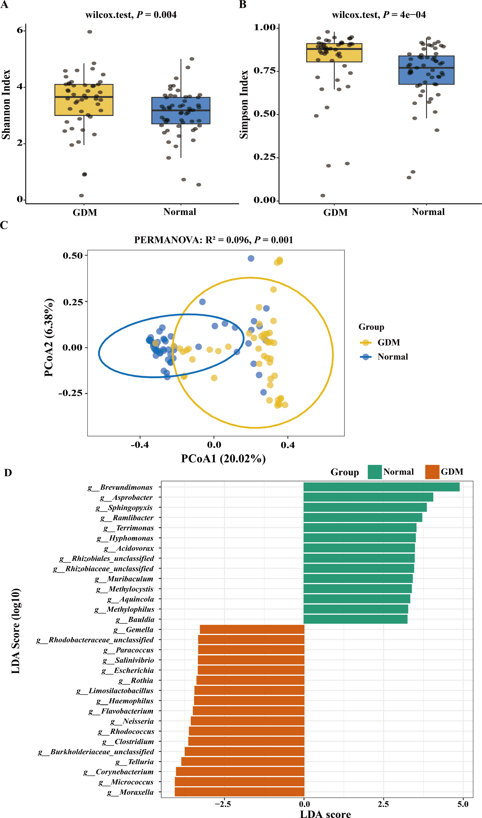
Fig. 2.
GDM-associated changes in placental microbiota. A-B) Placental α-diversity; C) Placental β-diversity; D) Differential bacterial genera in the placenta identified by LEfSe
Characteristics of neonatal meconium microbiota
Analysis of the relative abundance at the genus level in the neonatal meconium microbiome revealed significant differences in the top 30 community structures between the GDM and Normal groups (Fig. S1B). The GDM group was significantly enriched in several potentially opportunistic pathogenic or inflammation-associated genera, including Staphylococcus, Streptococcus, and Clostridium.
Diversity analysis revealed a significant impact of GDM on neonatal gut microecology. For α-diversity, the gut microbiota of neonates in the GDM group exhibited significantly higher diversity than that of the Normal group (Shannon index, P < 0.001; Simpson index, P = 0.003, Fig. 3A–B). β-diversity analysis showed a clear spatial separation in gut microbial composition between the two groups (P = 0.004, Fig. 3C), indicating systematic differences in overall community structure. LEfSe analysis identified a total of 17 genera with significant differences, of which 13 were significantly enriched in the Normal group, and four were significantly enriched in the GDM group (P < 0.05, Fig. 3D). The volcano plot visualized the impact of GDM on early neonatal gut colonization using differential abundance (Fig. S2B). Specifically, Phocaeicola and Azospirillum were significantly enriched in the gut of neonates in the GDM group. Bauldia showed a significantly decreasing trend in the GDM group.
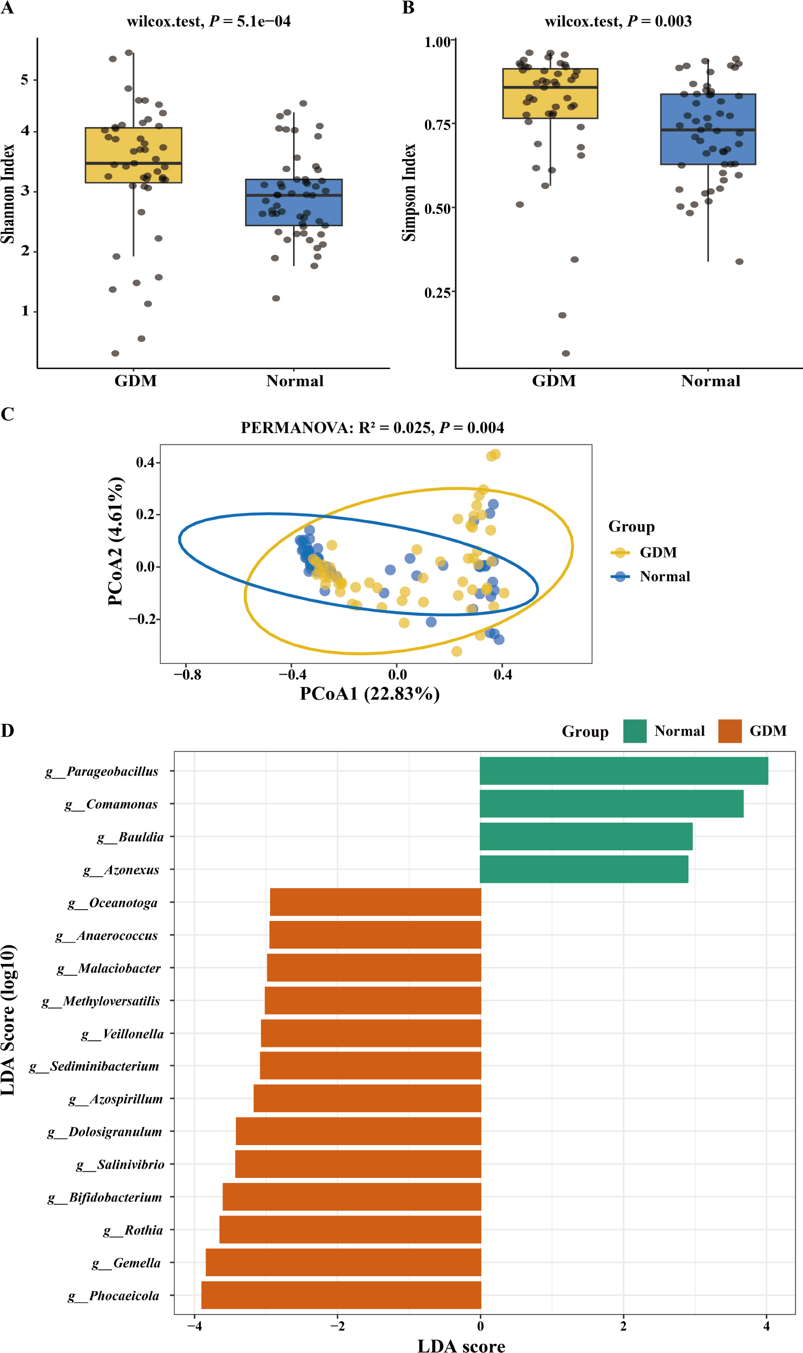
Fig. 3.
GDM-associated changes in neonatal meconium microbiota. A–B) α-diversity of neonatal meconium; C) β-diversity of neonatal meconium, D) Differentiated bacterial genera in neonatal meconium identified by LEfSe
Exploratory analysis of delivery mode effects on placental and meconium microbiota
Given that delivery mode differed between GDM and normal groups and may influence early neonatal gut microbiota, we further assessed its association with placental and meconium microbial structures. In placental samples, α-diversity showed no significant differences between delivery modes (Fig. S3A-B), whereas PCoA revealed a modest but significant community separation (PERMANOVA: R2 = 0.019, P = 0.012; Fig. S3C). In neonatal meconium samples, neither α-nor β-diversity showed consistent significant changes by delivery mode (Fig. S3D-F). These findings suggest that delivery mode may have a limited effect on placental bacterial DNA community structure, with low explanatory power. Given the small number of vaginal deliveries, particularly in the GDM group, this analysis should be considered exploratory.
Exploratory analysis of GWG effects on placental and neonatal meconium microbiota in patients with GDM
To explore the effects of the extent of GWG on the maternal-neonatal microbiota in GDM patients, we performed subgroup analyses within the GDM group. α-diversity analysis showed that, for both placental communities (Fig. S4A-B) and neonatal gut communities (Fig. S4C-D), no statistically significant differences were observed in Shannon or Simpson indices among the three GWG subgroups. This suggests that GWG had a limited overall effect on microbial community diversity. β-diversity analysis similarly indicated that placental (Fig. S4E) and neonatal gut communities (Fig. S4F) largely overlapped across different GWG subgroups, with no significant differences in overall community structure. Despite a relatively stable overall community structure, LEfSe analysis identified GWG subgroup-specific differentially enriched genera (Fig. S4G-H), suggesting that GWG may exert fine-scale regulatory effects on specific microbial taxa. However, given the small sample sizes across subgroups, these results should be considered exploratory.
Potential contribution of placental microbiota to early neonatal gut microbiota
Using FEAST-based source-tracking analysis, we evaluated the community-similarity-based contribution of the placental microbiota to neonatal meconium, aiming to determine whether GDM status alters the association between placental and meconial microbial composition. Results showed that the placenta-attributed similarity contribution to neonatal meconium was significantly lower in the GDM group than in the Normal group (86.78% [61.76%, 93.22%] vs. 93.81% [79.70%, 98.31%], P = 0.003, Fig. 4). Given the potential impact of delivery mode on neonatal gut microbiota, we performed stratified analysis. The GDM-associated reduction in placental similarity contribution persisted in the cesarean subgroup (P = 0.0053, Figure S5A) but not in the vaginal subgroup (P = 0.09, Fig. S5B), likely due to limited sample size.
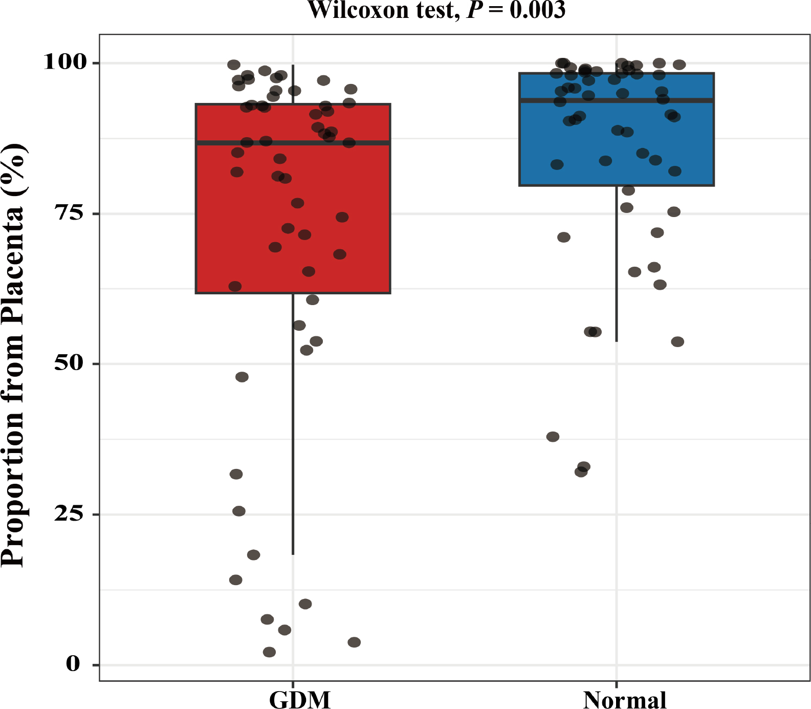
Fig. 4.
Analysis of the potential source contribution of placental microbiota to neonatal meconium microbiota based on community similarity.
In contrast, no significant difference in placental contribution to neonatal meconium was observed across GWG subgroups (74.16% [54.42%, 86.29%] vs. 87.70% [59.12%, 95.82%] vs. 86.84% [69.46%, 92.90%], P = 0.370, Fig. S6).
Associations between placental and neonatal gut microbiota and neonatal hospitalization
Spearman’s correlation analysis was used to quantify associations between placental and neonatal meconium microbiota abundance and neonatal hospitalization (Fig. 5). With in the placental microbiota, the relative abundances of Escherichia (ρ = 0.176, P = 0.016) and Curvibacter (ρ = 0.170, P = 0.020) were significantly and positively correlated with the occurrence of neonatal hospitalization. In contrast, Pelomonas (ρ = -0.168, P = 0.022) and Asprobacter (ρ = -0.160, P = 0.029) showed negative correlations. In the neonatal meconium microbiota, the relative abundance of Prevotella was significantly and positively correlated with neonatal hospitalization (ρ = 0.180, P = 0.011), whereas Sphingopyxis (ρ = -0.261, P = 0.002) and Paracoccus (ρ = -0.160, P = 0.023) were significantly and negatively correlated with neonatal hospitalization.
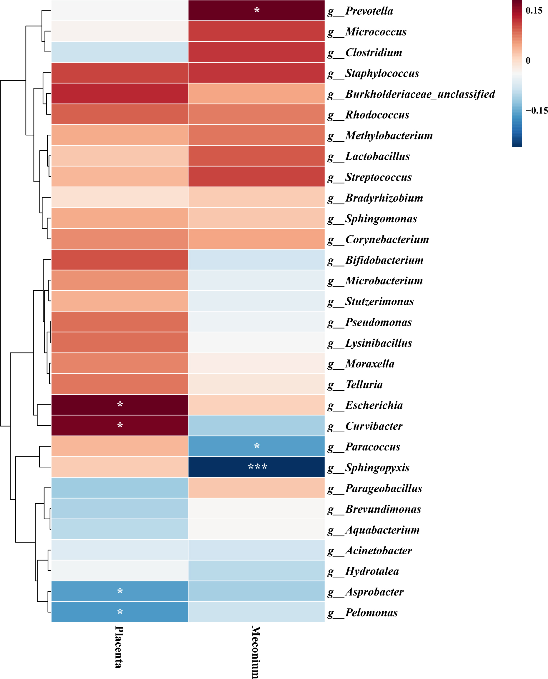
Fig. 5.
Association of differential microbial genera in placenta and neonatal meconium with the risk of neonatal hospitalization.
Discussion
The study focused on the placental-neonatal gut microbial axis to systematically evaluate the associations of GDM with maternal-neonatal microbiome structure, microbial continuity, and early neonatal clinical outcomes. Results showed that GDM was associated with altered placental bacterial DNA community composition, characterized by increased α-diversity and shifts in community structure. Meanwhile, the reduced placenta-attributed similarity contribution suggests weakened microbial continuity between the placenta and neonatal meconium microbiota, which was further associated with early neonatal hospitalization. These findings indicate that GDM-related metabolic abnormalities may be linked to alterations in early maternal-neonatal microbial characteristics and may have potential implications for early neonatal health. Of note, whether the placenta harbors stable, active, tissue-resident microbial communities remains debated. As placental tissue is a low-biomass sample, bacterial DNA signals derived from it are susceptible to exogenous contamination during sampling, reagent processing, and sequencing. Therefore, the placental findings in this study should be interpreted as bacterial DNA community profiles detected under negative control monitoring and contaminant filtering, rather than direct evidence of viable or stably colonizing microbial communities in the placenta.
In the low-biomass placenta, bacterial DNA signals are subject to multiple influences, including maternal metabolism, immune status, and perinatal environmental exposures; however, shifts in their composition typically mirror changes in maternal metabolism and immunity (Saadaoui et al. 2024, Aagaard et al. 2014). We found a significant reduction in Brevundimonas and Bifidobacterium in the placentas of GDM patients, along with enrichment of genera such as Lactobacillus, Pseudomonas, and Sphingomonas, which are associated with environmental or opportunistic pathogens. Bifidobacterium, a well-recognized probiotic, contributes to host health through antioxidant activity, antimicrobial effects, cholesterol metabolism regulation, and maintenance of immune homeostasis (Ferris et al. 2025, Insel et al. 2025, Chen et al. 2025). Its reduction in GDM placentas suggests decreased DNA signals of potentially protective bacterial genera under GDM conditions. Meanwhile, increased α-diversity and significant β-diversity separation in GDM placentas indicate potential disruption of placental microbial stability. Hyperglycemia can greatly alter the placental microenvironment through oxidative stress, local inflammation, and vascular dysfunction, potentially affecting the composition of detectable bacterial DNA signals in placental samples (Basak et al. 2024). Unlike in the adult gut, where higher diversity is generally associated with health, in studies of the placenta and early neonatal microbiota, increased diversity often indicates reduced niche control and impaired barrier function rather than a protective change (Zakis et al. 2022).
The neonatal gut microbiota in the GDM group was enriched in potentially opportunistic, pathogenic, or inflammation-associated genera, including Staphylococcus, Streptococcus, and Clostridium. Abnormal proliferation of Clostridium in the infant gut is often associated with inflammatory states and adverse health outcomes, representing an unfavorable microecological feature (Neri et al. 2021, Alshammari et al. 2020). Additionally, Farooqi et al. (2022) reported that a high proportion of Staphylococcus aureus positivity could be detected in both term and preterm placental samples, indicating that these genus-associated signals are reproducibly detectable in placental and perinatal specimens. The enrichment of similar taxa in the neonatal gut in this study supports the notion that GDM-asso-ciated placental microecological alterations may contribute to aberrant early gut colonization.
Vertical transmission of microbiota between mother and infant is widely recognized. Delivery and the perinatal period are considered critical windows for establishing the neonatal gut microbiota (Mirpuri and Neu 2021, Tian et al. 2023), and even regarded as a “microbial-level epigenetic mechanism”. (Gilbert 2014). Using a source-tracking model, we further revealed a significant reduction in the community-similarity-based contribution attributed to the placental source in neonatal meconium microbiota of the GDM group (93.81% to 86.78%, P = 0.003), providing exploratory evidence that GDM may affect the compositional link between placental and neonatal meconium microbiota. We hypothesize that the weakened maternal-infant microbial association may result from remodeling of placental microecology or alterations in placental barrier and immune regulatory functions, thereby affecting microbial transmission to the fetal environment.
The gut microbiota in early life is a key regulator shaping host metabolic and immune homeostasis. Through multi-organ axes such as the gut-liver-muscle-brain axis, it establishes a highly dynamic bidirectional communication network with the host. Microbial metabolites, including short-chain fatty acids, are fundamental in maintaining host homeostasis, acting via specific receptors and signaling pathways to modulate inflammation, energy metabolism, and the establishment of immune tolerance (Milani et al. 2017, Matara et al. 2022, Matara et al. 2022). This regulatory system is not fully developed and is highly sensitive to external disturbances in neonates. Aberrant microbial exposures can readily induce dysbiosis and disrupt immune system development (Sokou et al. 2024). Neonates born to mothers with GDM exhibit abnormalities in immunoglobulin levels and T lymphocyte subset composition (Xie 2024), suggesting a compromised innate immune defense. The study further linked changes in the abundance of multiple microbial genera in the placenta and neonatal gut to neonatal hospitalization, suggesting that GDM-related maternal-neonatal microbial remodeling may compromise neonatal immune homeostasis and increase inflammatory susceptibility, potentially leading to a higher risk of hospitalization. This underscores the potential value of community-similarity-based maternal-neonatal microbial linkage in preventing GDM-related adverse neonatal outcomes.
Several limitations should be acknowledged. First, our results should be considered exploratory, given the limited sample size of this single-center prospective cohort, particularly the insufficient statistical power for subgroup analyses by GWG extents and delivery mode. Delivery mode imbalance between GDM and control groups may confound the composition of neonatal meconium microbiota and source-tracking results. Although our exploratory stratified analyses of the patients with GDM confirmed persistently lower placental contribution in the cesarean subgroup, residual confounding cannot be fully excluded due to the limited number of vaginal deliveries in the GDM population. Second, our 5-region 16S rRNA sequencing of bacterial communities cannot resolve microbial functional potential and does not cover virome, mycobiome, or other non-bacterial microbial components. Even with negative controls and contaminant filtering, residual contamination cannot be completely ruled out due to the placenta’s low biomass. Thus, placental findings represent bacterial DNA profiles following QC and contaminant removal, rather than evidence of viable or stably colonizing microbial communities. The <500 reads threshold, on the one hand, ensures the reliability of microbial composition estimates; on the other hand, it may introduce exclusion bias by omitting low-biomass samples. Furthermore, FEAST infers source contributions from community similarity and may overestimate placental contribution, given limited candidate sources, and therefore does not reflect true biological transmission intensity. Finally, the lack of long-term follow-up regarding maternal-neonatal outcomes precludes assessment of associations between early microbial alterations and subsequent growth or metabolic risks, warranting future longitudinal studies. Further investigations integrating comprehensive maternal and environmental source samples, metagenomics, metabolomics, host immune markers, and longitudinal follow-up are needed to validate the biological and clinical significance of these findings.
Conclusion
The study suggests that, from the perspective of maternal-neonatal microbial continuity, GDM is associated with altered placental bacterial DNA communities, neonatal meconium microbiota composition, and their community-similarity-based linkage, which may correlate with short-term neonatal hospitalization. These findings offer a microbial perspective on GDM-related perinatal risks and suggest that future gestational metabolic management should consider its impact on early maternal-neonatal microbiota and neonatal outcomes.
Abbreviations
- GDM
Gestational Diabetes Mellitus
- GWG
Gestational Weight Gain
- BMI
Body Mass Index
- FBG
Fasting Blood Glucose
- PBG
Postprandial Blood Glucose
- HbA1c
Glycated Hemoglobin
- TC
Total Cholesterol
- PCoA
Principal coordinates analysis
- PERMANOVA
Permutational Multivariate Analysis of Variance
- LEfSe
Linear Discriminant Analysis Effect Size
- FEAST
Fast Expectation-maximization for Microbial Source Tracking
Notes
[2] Contributed by Authors’ contributions
Conceptualization: Yujia Qian, Dong Xu
Data curation: Xiaowen You
Formal analysis: Yujia Qian, Dong Xu
Investigation: Fangqin Tu
Methodology: Yujia Qian, Dong Xu
Project administration: Ye Tao
Resources: Ye Tao
Software: Minjuan Zhong
Supervision: Minjuan Zhong
Validation: Xiaowen You
Visualization: Xiaowen You
Writing – original draft: Yujia Qian, Xiaowen You, Fangqin Tu
Writing – review & editing: Ye Tao, Minjuan Zhong, Dong Xu
[3] Ethics approval
This study was approved by the Ethics Committee of Hangzhou Linping District Maternal & Child Health Care Hospital (LLSC-KYKT-2025-0006-A) and was conducted in accordance with the Declaration of Helsinki. All participants provided written informed consent.
[4] Contributed by Conflict of interest
The authors do not report any financial or personal connections with other persons or organizations, which might negatively affect the contents of this publication and/or claim authorship rights to this publication.