
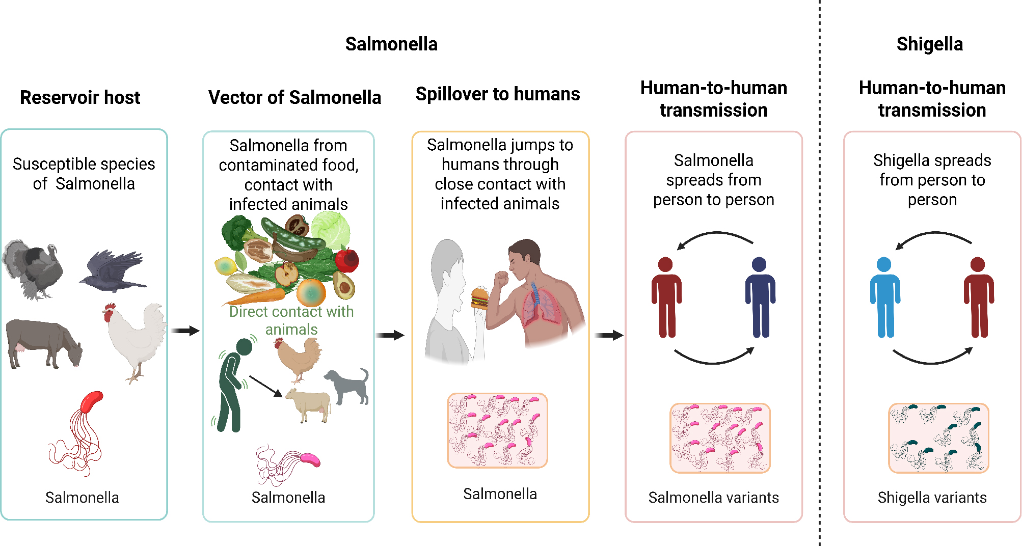
Fig. 1.
Reservoirs and transmission pathways of multidrug-resistant Salmonella and Shigella leading to human infection. Salmonella (left) originates from food-producing animals (poultry, cattle, pigs) contaminating meat, eggs, and produce. Shigella (right) is human-adapted, transmitted via fecal-oral route, contaminated water, or food. Created with BioRender.com.
Table I
Comparative epidemiology of Salmonella and Shigella.
| Feature | Salmonella Typhi/Typhoidal | Salmonella (non-typhoidal) | Shigella | References |
|---|---|---|---|---|
| Primary reservoirs | Human-adapted: human intestine (chronic carriers) | Zoonotic: food-producing animals (poultry, cattle, pigs) | Human | Percival et a. 2014; Crump et al. 2015 |
| Infective dose | Very low: 103 organisms | High: 103–106 organisms | Very low: 10–100 organisms | Levine et al. 1973; Blaser et al. 1980 |
| Transmission route | Fecal-oral (water, food, person-to-person) | Foodborne (meat, eggs, dairy, contaminated produce) | Fecal-oral (person-to-person, contaminated water/food) | Le boa et al. 2023; CDC 2024 |
| Outbreak settings | Waterborne/endemic; travelers, children | Foodborne outbreaks from animal products; all ages | Childcare centers, schools, refugee camps, prisons | Crump et al. 2015; CDC 2024 |
| Typical clinical presentation | Typhoid fever: sustained fever, relative bradycardia, abdominal pain | Watery diarrhea, nausea, vomiting, fever (gastroenteritis) | Dysentery: frequent bloody/mucoid stools, tenesmus, high fever | Dougan et al. 2014; Wilson et al. 2021; CDC 2024 |
| MDR prevalence trends | High fluoroquinolone resistance globally; XDR strains emerging | High in poultry/food isolates; fluoroquinolone resistance increasing | High multidrug resistance; often >80% to first-line agents in children | Wang et al. 2025; Tilahun et al. 2025 |
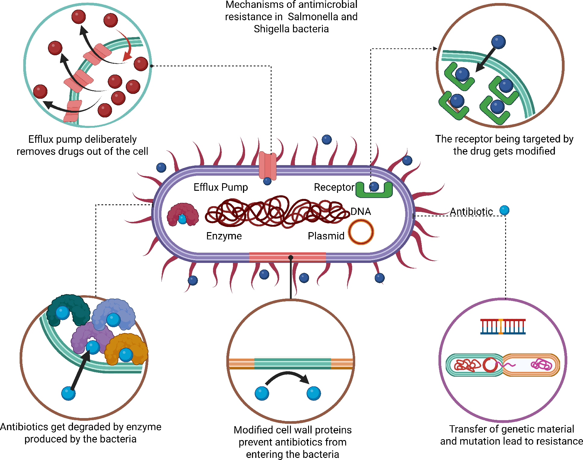
Fig. 2.
Major molecular mechanisms of antimicrobial resistance in Salmonella and Shigella. Six key strategies: (1) Enzyme-mediated antibiotic degradation (β-lactamases, carbapenemases); (2) Target site modifications (gyrA/parC mutations); (3) Reduced membrane permeability (porin loss); (4) Enhanced efflux pump activity (AcrAB-TolC system); (5) Biofilm formation; (6) Lipopolysaccharide modifications (reducing polymyxin susceptibility). MDR, multidrug-resistant. Created with BioRender.com.
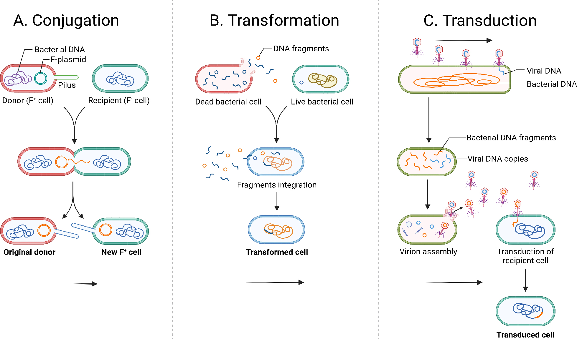
Fig. 3.
Horizontal gene transfer mechanisms facilitating dissemination of antibiotic resistance genes. Three routes: (A) Conjugation – plasmid transfer via pili; (B) Transformation – uptake of free DNA; (C) Transduction – bacteriophage-mediated transfer. Integrons and transposons enable rapid dissemination of multidrug resistance. Created with BioRender.com.
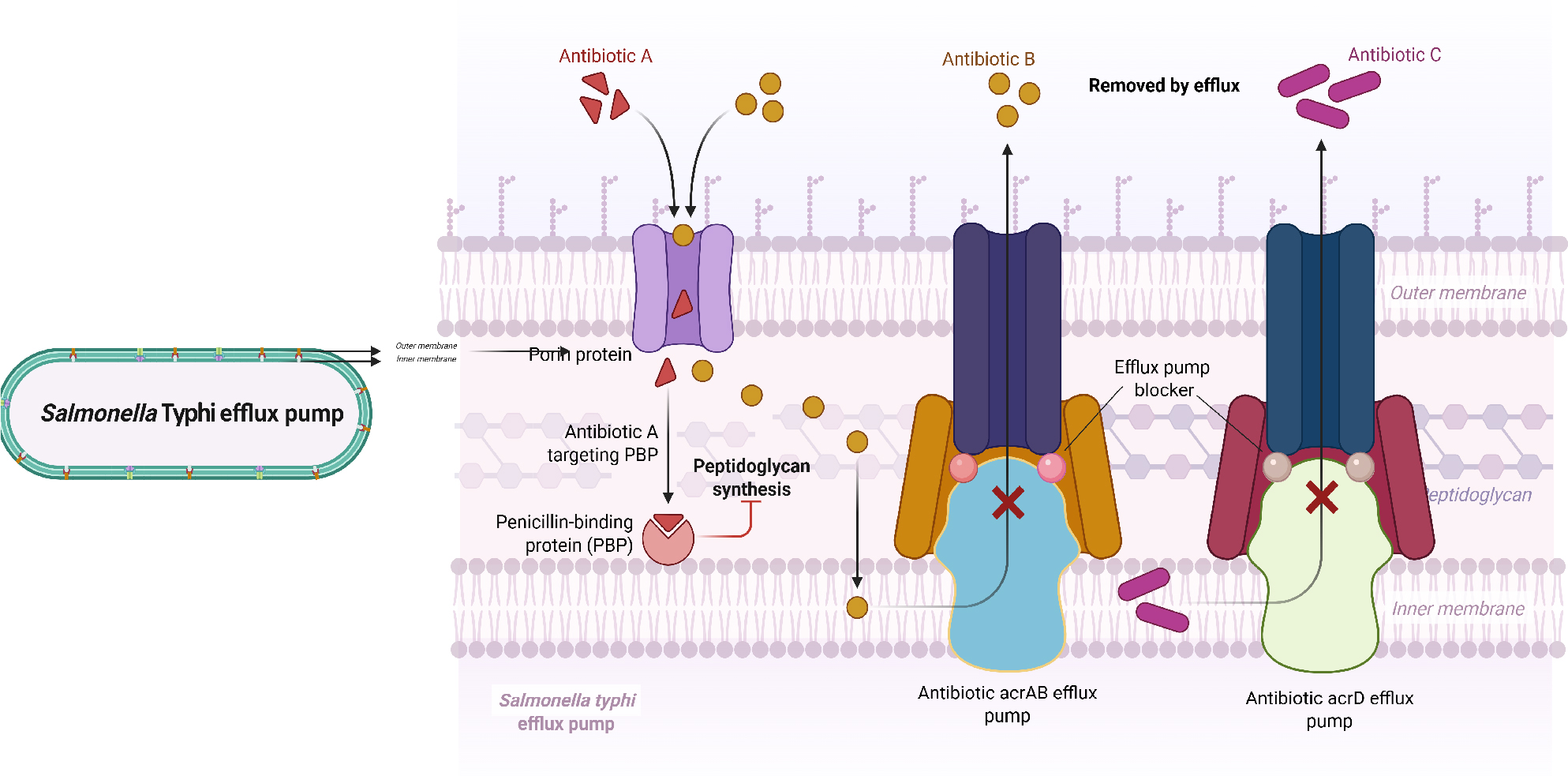
Fig. 4.
AcrAB-TolC multidrug efflux pump system in Salmonella and Shigella. The tripartite RND-family pump (AcrB inner membrane transporter, AcrA membrane fusion protein, TolC outer membrane channel) expels diverse antibiotics (β-lactams, quinolones, tetracyclines) from the cell. Overexpression confers multidrug resistance. IM – inner membrane; OM – outer membrane; PP – periplasm. Created with BioRender.com.
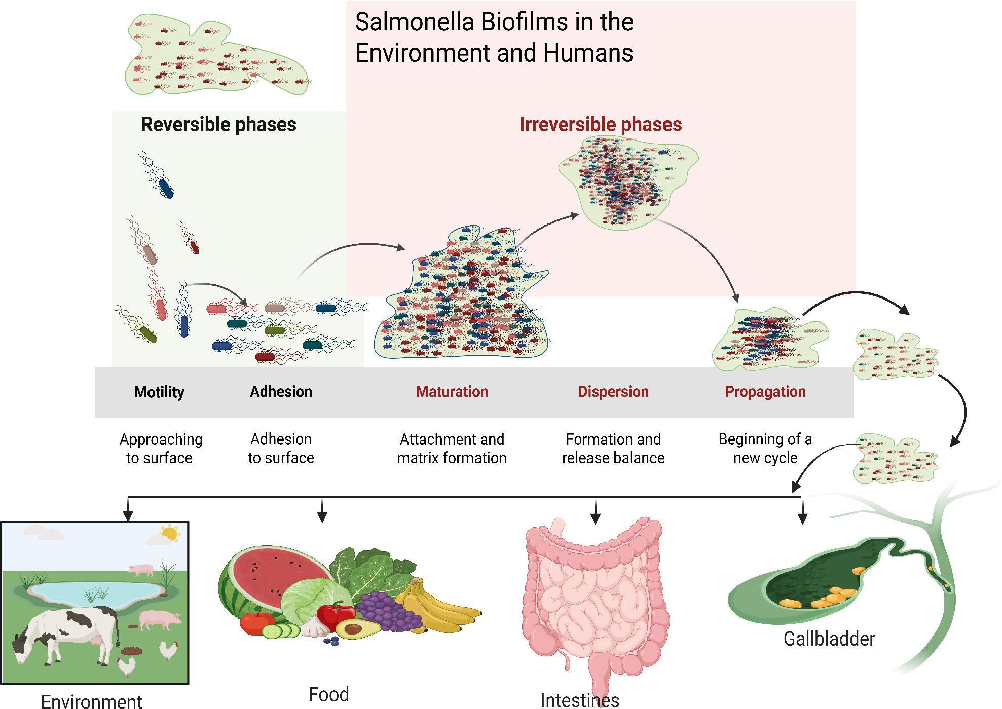
Fig. 5.
Biofilm formation cycle in Salmonella and its role in antimicrobial resistance. Five stages: (1) Reversible attachment; (2) Irreversible adhesion and EPS production (cellulose, curli, eDNA); (3) Microcolony formation; (4) Maturation into 3D communities (up to 1000-fold increased antibiotic tolerance); (5) Dispersion. Biofilms enhance survival in food/environmental settings and facilitate persistent infections. Created with BioRender.com.
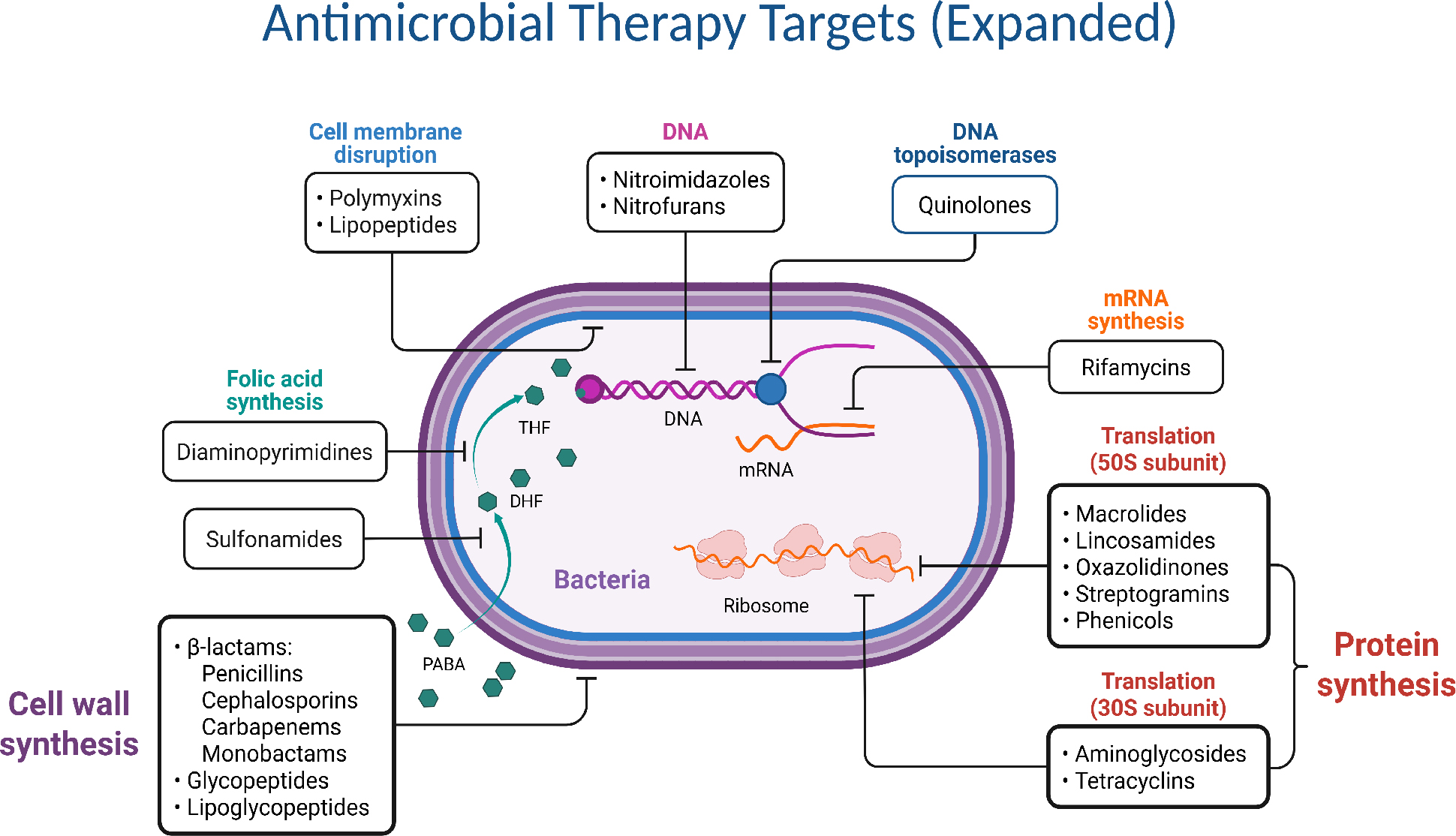
Fig. 6.
Cellular targets of major antibacterial drug classes. Six primary sites: (1) Cell wall synthesis (β-lactams, glycopeptides); (2) Protein synthesis – 30S subunit (aminoglycosides, tetracyclines); (3) Protein synthesis – 50S subunit (macrolides, chloramphenicol); (4) Nucleic acid synthesis (quinolones, rifamycins); (5) Folate metabolism (sulfonamides, trimethoprim); (6) Cell membrane (polymyxins).
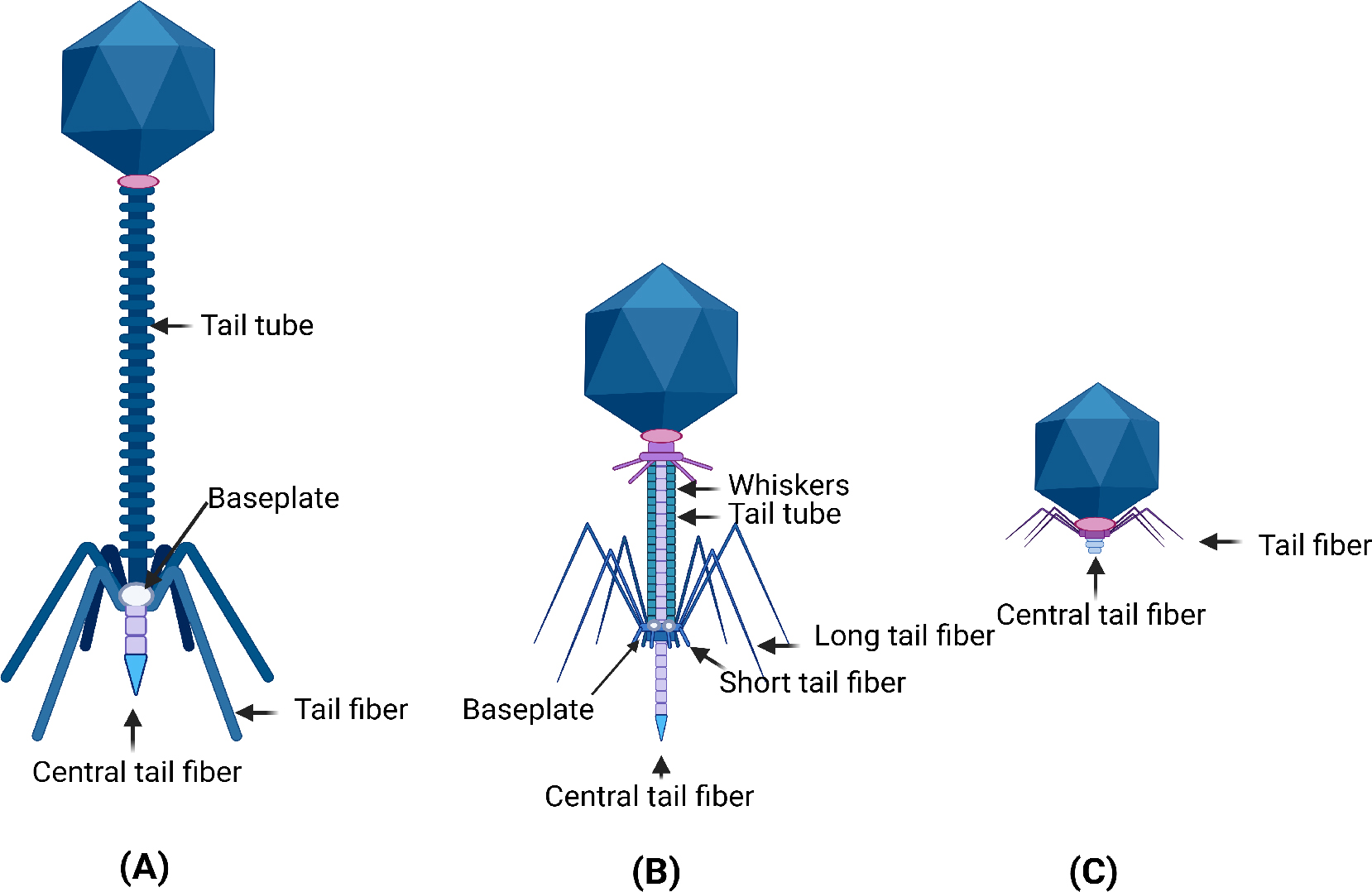
Fig. 7
Morphological diversity of bacteriophages from order Caudovirales used in phage therapy. Three main families: (A) Siphoviridae – long flexible non-contractile tails; (B) Myoviridae – long contractile tails with sheath; (C) Podoviridae – short non-contractile tails. Phage cocktails combining multiple types broaden host range and minimize resistance. Created with BioRender.com.
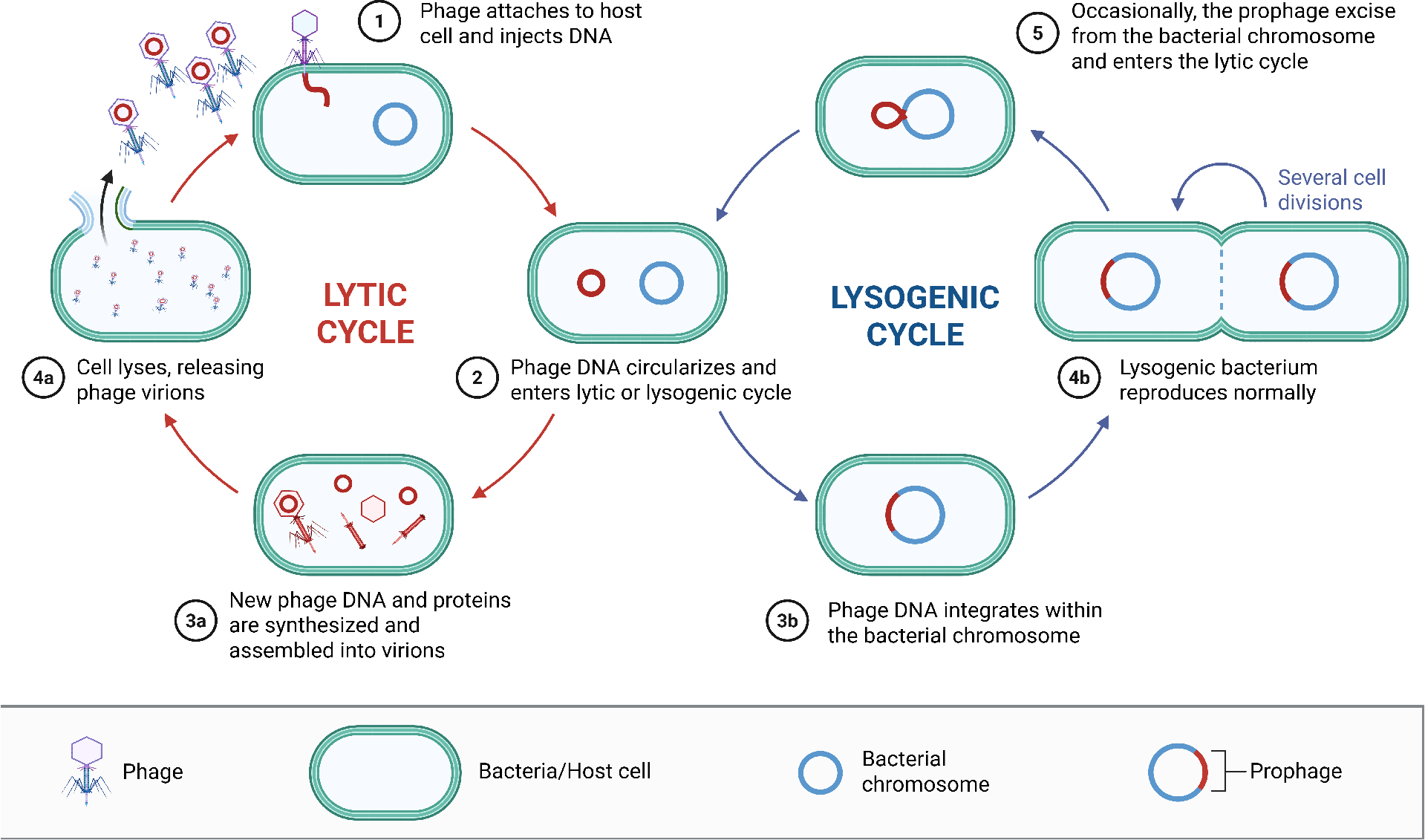
Fig. 8.
Bacteriophage life cycles: lytic versus lysogenic replication. Lytic cycle (left): Adsorption → genome injection → replication → assembly → lysis. Lysogenic cycle (right): Genome integration as prophage → passive replication with host → possible induction to lytic cycle. Strictly lytic phages are preferred for therapy to avoid transferring resistance genes. Created with BioRender.com.
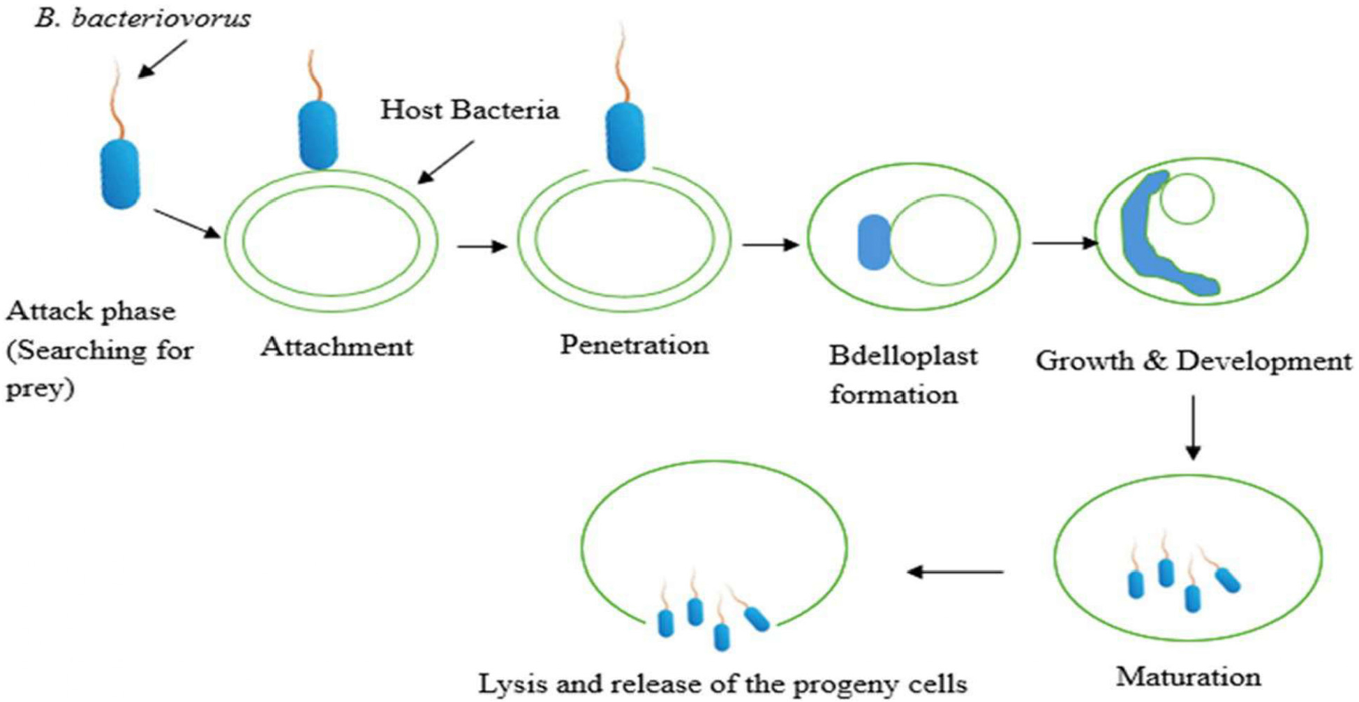
Fig. 9.
Predatory life cycle of Bdellovibrio bacteriovorus as a living antibiotic against Gram-negative pathogens. Six stages: (1) Attack phase – predator attaches to prey (e.g., Shigella, Salmonella); (2) Entry into periplasm; (3) Establishment – bdelloplast formation; (4) Growth – filamentous elongation; (5) Septation into progeny; (6) Release of motile predators. Exhibits broad-spectrum activity against MDR Enterobacterales, penetrates biofilms, and is non-pathogenic to humans. Adapted from Al-maaqar et al. (2025).