
Introduction
Traditionally, the human body hosts an estimated 10–100 trillion microorganisms, contributing to its internal ecosystem. Microbes and their collective microbial genome reflect an individual’s health status. Notably, microbial dysbiosis can lead to a variety of diseases, including inflammatory bowel disease and diabetes (Elinav et al. 2019), and increasing evidence supports the contribution of the microbiota in cancer development, with 15–20% of cancers driven by specific microbes (Sears and Garrett 2014). The microbiota influences cancer risk through multiple mechanisms, including regulation of cellular proliferation, immune function, and host metabolism (Sears and Garrett 2014). Among these, chronic inflammation is recognized as a critical factor in tumor invasion, promotion, and dissemination, which may further affect treatment efficacy (Arthur et al. 2012; Kostic et al. 2013).
Substantial research has focused on the associations between microbiota and specific cancer types (Rizzo et al. 2022; Sobstyl et al. 2022; Yang, et al. 2023), with oral and gut microbiota being the two most extensively studied subsets in the context of head and neck tumors and gastrointestinal tumors, respectively. This study specifically focuses on intra-tumor microbiota (encompassing microbial communities present within tumor tissues, which may originate from both oral and gut microbiota depending on the tumor’s anatomical location) rather than limiting the scope to oral or gut microbiota alone. The goal is to identify shared and distinct intra-tumor microbial signatures between head and neck tumors and gastrointestinal tumors, and to explore their value.
In head and neck tumors, the immunosuppressed microenvironment induced by oral microbiota may disrupt the healthy microbial balance. A comprehensive systematic review by Metsäniitty et al. (2021) proposed that several oral microbes, such as Fusobac-terium nucleatum and Porphyromonas gingivalis, are associated with malignant lesions. Furthermore, based on oral microbiota data from 745 healthy controls and 309 patients with cancer, Nouri et al. (2023) found that reduced levels of Prevotella, Abiotrophia, Leuconostoc, and Streptococcus, along with increased levels of Neisseria and Haemophilus, were associated with cancer development. In contrast, gastrointestinal tumors, including esophageal cancer (ESCA), gastric cancer (STAD), colon cancer (COAD), and rectal cancer (READ), have been closely associated with gut microbiota dysbiosis. Despite differences in their anatomical locations, local pH levels, and specific microbial compositions, these tumors share common microbial disruptions, thereby contributing to various diseases (Hold 2016; Gill et al. 2022). Asili et al. (2023) emphasized that oral microbiome dysbiosis not only affects oral health but also exhibits potential associations with gastrointestinal cancers, highlighting the crosstalk between oral and gut microbial communities in cancer pathogenesis. Elghannam et al. (2025) further reviewed the interactions between the gut microbiota and gastric cancer, noting that specific gut microbes (e.g., Helicobacter pylori) can drive gastric carcinogenesis through mechanisms such as inducing chronic inflammation and DNA damage. For colorectal cancer (CRC), Liu et al. (2023) conducted an independent evaluation of microbiome 16S rRNA sequence analysis methods in a CRC patient cohort, providing robust evidence for the role of gut microbiota (e.g., increased abundance of Fusobacterium, Streptococcus, Peptost-reptococcus, and Haemophilus and reduced abundance of Roseburia, Faecalibacterium, and Butyricicoccus) in CRC development. Additionally, Gopalakrishnan et al. (2018) noted that the gut microbiome can modulate anti-tumor immunity and influence the efficacy of cancer immunotherapy in gastrointestinal tumors, further underscoring the critical role of gut microbiota in gastrointestinal cancer progression and treatment. Flemer et al. (2018) even found that the oral microbiota (such as Streptococcus, Prevotella spp., and Lachnospiraceae) can serve as a predictive marker for CRC, suggesting potential microbial transmission or shared dysbiotic patterns between the oral cavity and gastrointestinal tract.
ESCA, STAD, COAD, and READ develop in distinct microenvironments. For instance, the stomach maintains a strongly acidic pH (1–3), favoring acid-tolerant microbes like H. pylori, whereas the colon and rectum maintain a neutral pH and harbor a more diverse microbial community dominated by Bacteroi-detes and Firmicutes (Sheh and Fox 2013; Hunt et al. 2015). The esophagus, as a transitional organ between the oral cavity and the stomach, harbors a microbial community influenced by both oral flora and gastric reflux (Li et al. 2023). Despite these differences, in this study, we grouped these four cancer types into a single “gastrointestinal tumor” category for three key reasons related to our research goals. Anatomically, the esophagus, stomach, colon, and rectum constitute the continuous digestive tract, and their microbial communities exhibit “vertical migration” (Hollister et al. 2014; Yang et al. 2025). Oral microbiota can enter the gastrointestinal tract via swallowing, while gut microbiota can influence the esophageal microenvironment through reflux, forming an interconnected microbial ecosystem that differs fundamentally from the oral cavity-dominated microenvironment of head and neck tumors. From a microecological mechanism perspective, all four gastrointestinal cancer types share a key feature of “gut microbiota dysregulation” as a driver of carcinogenesis, which distinguishes them from head and neck tumors (primarily driven by oral microbiota dysbiosis) (Zhou et al. 2021; Xie et al. 2024). For example, all four types show abnormal increases in pro-inflammatory microbes (e.g., Fusobacterium) and disruptions in anti-inflammatory microbial communities (e.g., Lachnospiraceae), with carcinogenesis mediated through shared pathways such as “microbial metabolite-immune regulation-chronic inflammation” (He et al. 2023; Zhao et al. 2023; Cao et al. 2024). Our primary aim was to identify shared intra-tumor microbial signatures that distinguish head and neck tumors from gastrointestinal tumors, rather than exploring microbial differences among gastrointestinal tumor subtypes. Given the limited sample size of individual gastrointestinal subtypes (e.g., ESCA and READ each have only 45 cases in our dataset), grouping them into a single cohort (a total of 309 cases) ensures sufficient statistical power to match the 154 cases of head and neck tumors, which is essential for constructing stable machine learning-based classification models and conducting reliable prognostic analyses. This grouping strategy does not negate the unique microbial characteristics of each gastrointestinal tumor subtype but prioritizes addressing the core research question of inter-group differences between head and neck and gastrointestinal tumors.
While existing studies have explored microbiota diversity across different cancer types. For example, Nejman et al. (2020) identified distinct microbial compositions in 1526 tumors across seven cancer types. Battaglia et al. (2024) performed a pan-cancer analysis of the microbiome in metastatic cancer, and Galeano Niño et al. (2022) investigated the effect of the intratu-moral microbiota on spatial and cellular heterogeneity in cancer. However, there remains a lack of targeted research comparing intra-tumor microbiota signatures between head and neck tumors and gastrointestinal tumors. Most previous studies have either focused on a single cancer type or employed broad pan-cancer analyses, without specifically addressing the shared and unique microbial features between these two clinically relevant tumor groups. This gap limits our understanding of whether intra-tumor microbiota can serve as a discriminative marker or as a shared prognostic factor for these tumor types.
Rather than limiting the scope to oral or gut microbiota alone, this study specifically focused on intra-tu-mor microbiota (encompassing microbial communities present within tumor tissues that may originate from both oral and gut microbiota, depending on the tumor’s anatomical location). Therefore, in the present study, we used data from the Cancer Microbiome Atlas (TCMA) and The Cancer Genome Atlas (TCGA) to screen for intra-tumor microbiota and clinical information for five tumor types: head and neck squamous cell carcinoma (HNSC), ESCA, STAD, COAD, and READ. Based on their anatomical location, these tumors were divided into head and neck tumors (HNSC) and gastrointestinal tumors (ESCA, STAD, COAD, READ). We aimed to: (1) screen for characteristic intra-tumor microbial communities that distinguish between the two tumor groups; (2) construct a microbiota-based classification model to differentiate head and neck tumors from gastrointestinal tumors; and (3) analyze the correlation between the distribution of these characteristic microbial communities and clinical prognostic factors. Our preliminary findings indicate that eight intra-tumor microbial communities (Actinobacteria, Bacteroidales, Capnocytophaga, Clostridia, Fusobacteria, Lachnospiraceae, Prevotella, and Prevotellaceae) are valuable for distinguishing head and neck tumors from gastrointestinal tumors, and that high levels of Capno-cytophaga, Lachnospiraceae, and Bacteroidales may be associated with longer overall survival.
Materials and Methods
Source of microbial data
On April 22, 2023, microbial detection data for HNSC, ESCA, STAD, COAD, and READ were downloaded from the TCMA database (Dohlman et al. 2021), encompassing microbial communities at the phylum, class, order, family, and genus levels. Clinical data of the five kinds of cancers were also downloaded from the TCGA database (Tomczak et al. 2015) and effective samples of the microbiota were subsequently selected based on their corresponding sample numbers.
Given the limited sample sizes of individual gastrointestinal subtypes (e.g., ESCA and READ each have only 45 cases in our dataset), grouping them into a single cohort (totaling 309 cases) ensured sufficient statistical power to match the 154 cases of head and neck tumors. This is essential for constructing stable machine learning-based classification models and conducting reliable prognostic analyses. This grouping strategy does not negate the unique microbial characteristics of each gastrointestinal tumor subtype but prioritizes addressing the core research question of inter-group differences between head and neck and gastrointestinal tumors.
Differential distribution of microbial communities in each cancer type
For each tumor type, samples were categorized into tumor and control groups based on their source, yielding a total of five comparison groups (HNSC vs. control, ESCA vs. control, STAD vs. control, COAD vs. control, and READ vs. control). The Wilcoxon test in R (version 3.6.1) was used to analyze the differential distributions of microbial communities between tumor and control samples within each comparison group, with a threshold of P < 0.05. Thereafter, the microbial communities with significantly different distribution levels in each tumor were compared to identify shared and unique microbial communities in various tumors using the UpSetR package in R3.6.1 (Conway et al. 2017).
Differential distribution of microbial communities between head and neck and gastrointestinal tumors
Based on location, the five tumor types were divided into two groups: head and neck tumors (HNSC) and gastrointestinal tumors (ESCA, STAD, COAD, READ), with 154 and 309 tumor samples, respectively. Microbial communities with significantly different distributions between head and neck tumors and gastrointestinal tumors were identified using the Wilcoxon test in R 3.6.1.
Construction of a classification model based on the optimal microbial communities
In this analysis, the 463 tumor samples were randomly divided into training and validation datasets in a 7:3 ratio, maintaining the same proportion across both tumor groups. The training dataset was designed to train and build the microbial-based classification model, while the validation dataset was used to assess the model’s performance.
In the training dataset, univariate logistic regression was performed using the rms package (version 6.3-0, https://cran.r-project.org/web/packages/rms/in-dex.html) in R3.6.1, based on the aforementioned microbial communities that showed significantly different distributions between head and neck tumors and gastrointestinal tumors. Microbial communities with P <0.05 were retained.
The retained microbial communities were submitted for selection of the optimal microbial communities using the least absolute shrinkage and selection operator (LASSO) algorithm in the lars package (version 1.2, https://cran.r-project.org/web/packages/lars/in-dex.html) (Goeman 2010), and the recursive feature elimination (RFE) algorithm using the caret package (version 6.0-76, https://cran.r-project.org/web/pack-ages/caret) in R3.6.1 (Deist et al. 2018). Afterward, the combined findings from the LASSO and RFE microbial communities were identified as optimal and used for subsequent analyses.
Finally, a classification model was constructed using the Support Vector Machine (SVM) method in the e1071 package (https://cran.r-project.org/web/pack-ages/e1071) in R3.6.1, based on the optimal microbial communities (Core: Sigmoid Kernel; Cross: 100-fold cross-validation) (Wang and Liu 2015). The performance of the SVM-constructed classification model in both the training and validation datasets was evaluated using receiver operating characteristic (ROC) curves generated by the pROC package (version 1.12.1) in R3.6.1 (Robin et al. 2011).
To further evaluate the predictive ability of each microbial community, a nomogram model was built based on the optimal microbial community distribution data using the rms package (version 6.3-0) in R3.6.1 (Pan et al. 2021). Afterward, the corrected polyline of the proposed nomogram model was drawn, and the efficiency of the proposed nomogram model was assessed using the C-index parameter (Chaudhary et al. 2018).
Clinical correlation analysis
First, the clinical information for the samples in both tumor groups was statistically analyzed, and the chi-square test in R3.6.1 was used to compare the distributions of clinical information between the two groups (head and neck tumors vs. gastrointestinal tumors). Then, combined with the clinical survival prognosis information of the samples, the correlation between the distribution of important characteristic taxonomic microbial communities and survival prognosis was analyzed using the Kaplan-Meier curve in the survival package (version 2.41-1, http://bioconductor.org/packages/surviv-alr/) of R3.6.1 (Wang et al. 2016).
Results
Differential distribution of microbial communities in each cancer type
After matching valid samples of microbial communities, the dataset included HNSC: 161 samples (154 tumor samples and 7 controls); ESCA: 50 samples (45 tumor samples and 5 controls); COAD: 138 samples (117 tumor samples and 21 controls); READ: 50 samples (45 tumor samples and 5 controls); and STAD: 111 samples (102 tumor samples and 9 controls). The distributional statistics for each microbial community in each cancer, including the 25th, 50th, and 75th percentiles, mean, and standard deviation, are shown in Table SI.
Compared with their corresponding control samples, a total of 13, 26, 20, 9, and 14 differentially distributed microbial communities were identified in HNSC, ESCA, COAD, READ, and STAD tumors, respectively (Table SII). These differentially distributed microbial communities were compared, and no microbial communities were significantly differently distributed across all five comparison groups (Fig. 1A). As shown in Fig. 1A, only 8, 12, 16, 7, and 8 distinct microbiota were differentially distributed in the HNSC, ESCA, COAD, STAD, and READ tumors, respectively. In addition, we observed that Proteobacteria showed significant differences in distribution between STAD, COAD, or ESCA and their corresponding controls (Fig. 1A). Compared with their corresponding controls, the levels of Proteobacteria in tumor samples were significantly decreased in ESCA and STAD (P < 0.05), but evidently increased in COAD (P < 0.05, Fig. 1B). The distributional statistics of each significantly differentially distributed microbial community in each cancer were displayed in Table SIII.
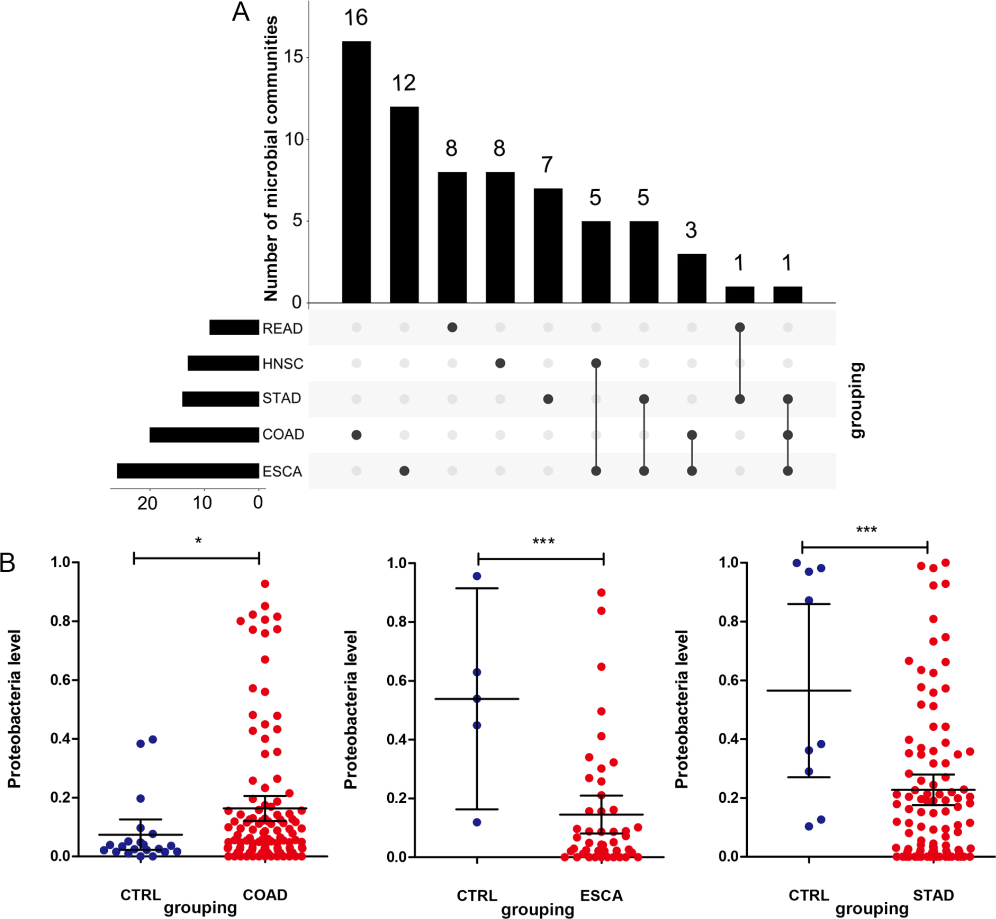
Fig. 1.
Differential distribution of microbial communities in each kind of cancer. (A) The distribution of microbial communities among head and neck cancer (HNSC), esophageal cancer (ESCA), stomach cancer (STAD), colon cancer (COAD), and rectal cancer (READ) using the UpSetR package in R3.6.1, which compares the elements within each set. (B) Comparison of Proteobacteria distribution in COAD, ESCA, and STAD. CTRL: control. * P < 0.05, *** P < 0.005, vs. control.
Differential distribution of microbial communities between head and neck tumors and gastrointestinal tumors
Based on the tumor location, tumors were grouped into head and neck tumors (tumor type 1) and gastrointestinal tumors (tumor type 2). After comparison using the Wilcoxon test, 23 microbial community types showed significantly different distributions between head and neck tumors and gastrointestinal tumors (Fig. 2). The relative abundance of Bacteroidales, Bacteroidia, Lachnospiraceae, Epsilonproteobacteria, Campylobacterales, Clostridiales, Clostridia, and Lactobacillus was significantly higher in the gastrointestinal tumors (tumor type 2) than in the head and neck tumors (tumor type 1) (P < 0.05). Conversely, the relative abundance of the other 15 microbial communities (such as Fusobacterium, Prevotella, Actinobacteria, and Veillonellales) was evidently reduced in the gastrointestinal tumors (tumor type 2) compared to the head and neck tumors (tumor type 1) (P < 0.05, Fig. 2).
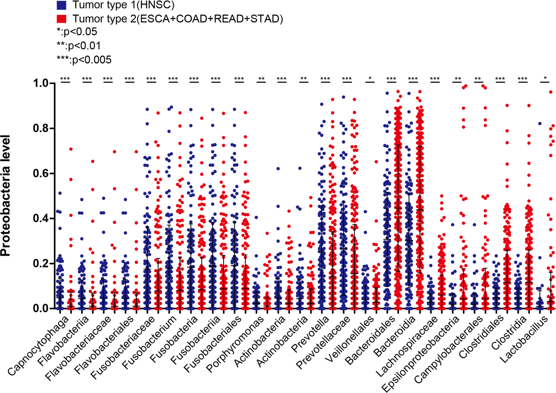
Fig. 2.
Comparison of differential distribution of bacterial communities between head and neck tumors (tumor type 1) and gastrointestinal tumors (tumor type 2). * P < 0.05, ** P < 0.01, *** P < 0.005, vs. tumor type 1.
Construction and evaluation of a classification model based on the optimal microbial communities
The training and validation datasets, respectively, contained 324 and 139 tumor samples, and the sample grouping information is shown in Table SIV. The 23 microbial communities with significantly different distributions between head and neck tumors and gastrointestinal tumors were subsequently submitted for further analysis. The univariate logistic regression analysis identified 18 distinct microbial communities associated with survival performance (P < 0.05, Fig. 3A). Thereafter, the RFE and LASSO algorithms identified 15 (Fig. 3B, Table SV) and 9 (Fig. 3C, Table SV) distinct microbial communities that were differentially distributed. After comparing the results of RFE and LASSO, eight overlapping microbial communities were selected as optimal, including Actinobacteria, Bacteroi-dales, Capnocytophaga, Clostridia, Fusobacteria, Lach-nospiraceae, Prevotella, and Prevotellaceae.
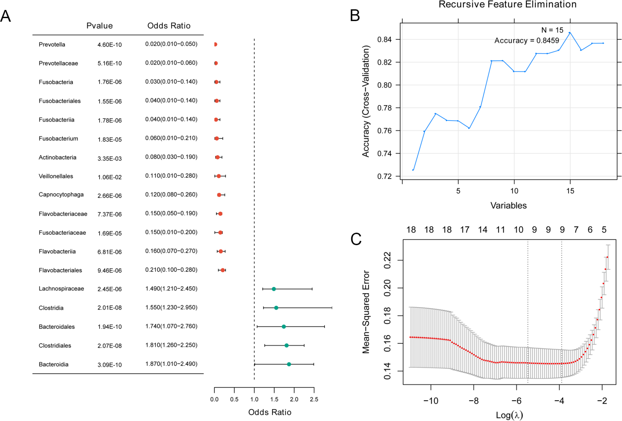
Fig. 3.
Screening of the optimal bacterial communities. (A) A total of 18 kinds of differentially distributed microbial communities associated with survival performance using univariate logistic regression analysis. (B) The screening parameter diagram of the recursive feature elimination (RFE) algorithm. (C) The screening parameter diagram of the LASSO algorithm.
A classification model based on the eight optimal microbial communities was then constructed, and ROC curves were generated for the training and validation datasets. In the training dataset, the area under the ROC curve (AUC) values of Actinobacteria, Bac-teroidales, Capnocytophaga, Clostridia, Fusobacteria, Lachnospiraceae, Prevotella,% and Prevotellaceae were 0.808, 0.722, 0.728, 0.734, 0.721, 0.814, 0.763, and 0.780, respectively. The AUC value of the combination of these eight optimal microbial communities (0.937) was higher than that of each individual microbial community (Fig. 4). Furthermore, the performance of the eight individual microbial communities and the combination was verified in the validation dataset, and the AUC of the combination was 0.856 (Fig. 4), indicating that the classification model constructed using the combination of the eight optimal microbial communities demonstrated superior diagnostic performance. This was effectively verified in the validation dataset, where the results were consistent with those from the training dataset.
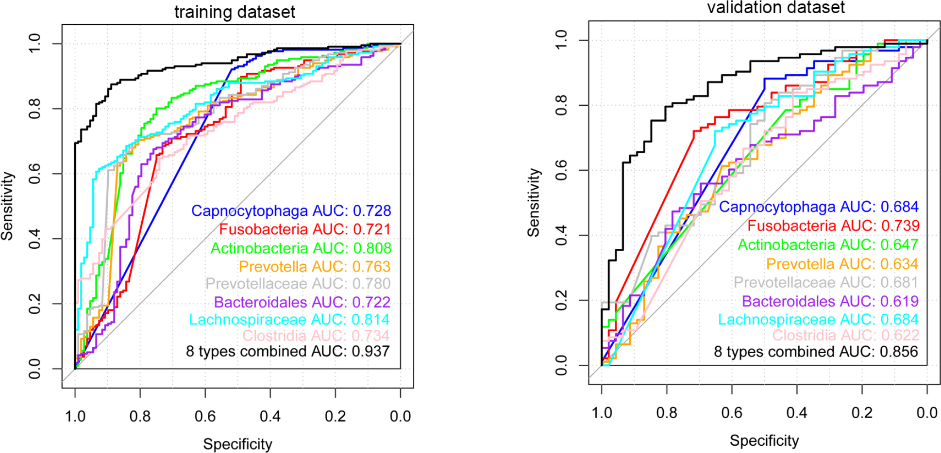
Fig. 4.
Receiver operating characteristic (ROC) curves based on the optimal eight bacterial communities in head and neck tumors and gastrointestinal tumors in the training and validation datasets.
A nomogram model was also developed for each microbial community, combining data from both the training and validation datasets. It was observed that the predictive performance of the nomogram derived from microbial communities in the training dataset (C-index = 0.8944, Fig. 5A) and in the validation dataset (C-index = 0.8023, Fig. 5B) was highly consistent with the clinical outcomes. Notably, Fusobacteria, Prevotella, Prevotellaceae, Lachnospiraceae, and Bacte-roidales had the highest scores in the nomogram model, resulting in high predicted probabilities (Fig. 5).
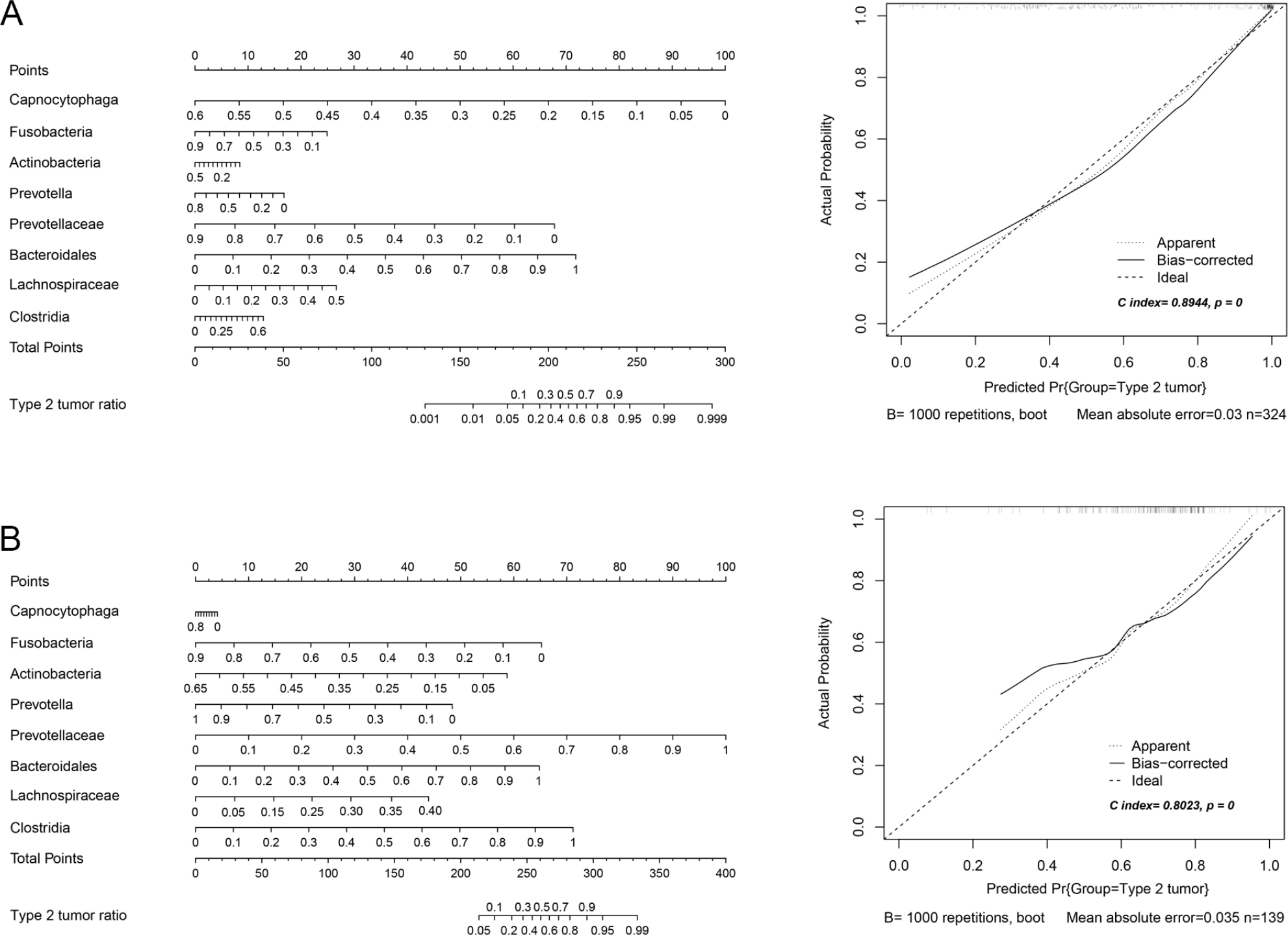
Fig. 5.
The nomogram model proposed based on the eight optimal microbial communities. (A) A nomogram model and the corrected polyline of the proposed nomogram model in the training dataset. (B) A nomogram model and the corrected polyline of the proposed nomogram model in the validation dataset.
Correlation between the microbial communities and clinical background
Clinical characteristics of the two tumor groups were statistically analyzed (Table SVI). After comparison using the chi-square test, significant differences were found in age, gender, pathologic M, N, and T stage, and vital status between head and neck tumors and gastrointestinal tumors (P < 0.05, Table I).
Table I
Comparison of clinical factors in different types of tumor groups.
| Characteristics total cases | Cancer type | P value | |
|---|---|---|---|
| Type 1 (HNSC) | Type 2 (COAD+ESCA+READ+STAD) | ||
| Age (years) | |||
| ≤ 60 | 82 | 97 | 6.15E-05 |
| > 60 | 71 | 193 | |
| Gender | |||
| Male | 112 | 170 | 1.66E-04 |
| Female | 41 | 120 | |
| Pathologic M | |||
| M0 | 151 | 239 | 1.66E-04 |
| M1 | 1 | 30 | |
| Pathologic N | |||
| N0 | 71 | 150 | 9.20E-06 |
| N1 | 26 | 78 | |
| N2 | 50 | 38 | |
| N3 | 3 | 14 | |
| Pathologic T | |||
| T1 | 7 | 12 | 8.48E-10 |
| T2 | 54 | 76 | |
| T3 | 40 | 159 | |
| T4 | 51 | 34 | |
| Pathologic stage | |||
| Stage I | 4 | 54 | 2.20E-16 |
| Stage II | 33 | 109 | |
| Stage III | 31 | 75 | |
| Stage IV | 84 | 35 | |
| Vital status | |||
| Dead | 62 | 80 | 1.64E-02 |
| Alive | 91 | 199 | |
Combined with the clinical survival prognosis, the samples were thereafter stratified into low (< the median) and high (≥ the median) distribution groups based on the median value for each microbial community. Using Kaplan-Meier curve analysis, the correlation between the eight optimal microbial communities and survival prognosis, and three microbial communities (Capnocytophaga, Lachnospiraceae, and Bacteroidales) with significant correlation with survival prognosis were identified (Fig. 6). The patients with high abundance of Capnocytophaga (Fig. 6A), Lachnospiraceae (Fig. 6B), and Bacteroidales (Fig. 6C) had significantly longer overall survival time than those with low abundance (P < 0.05).
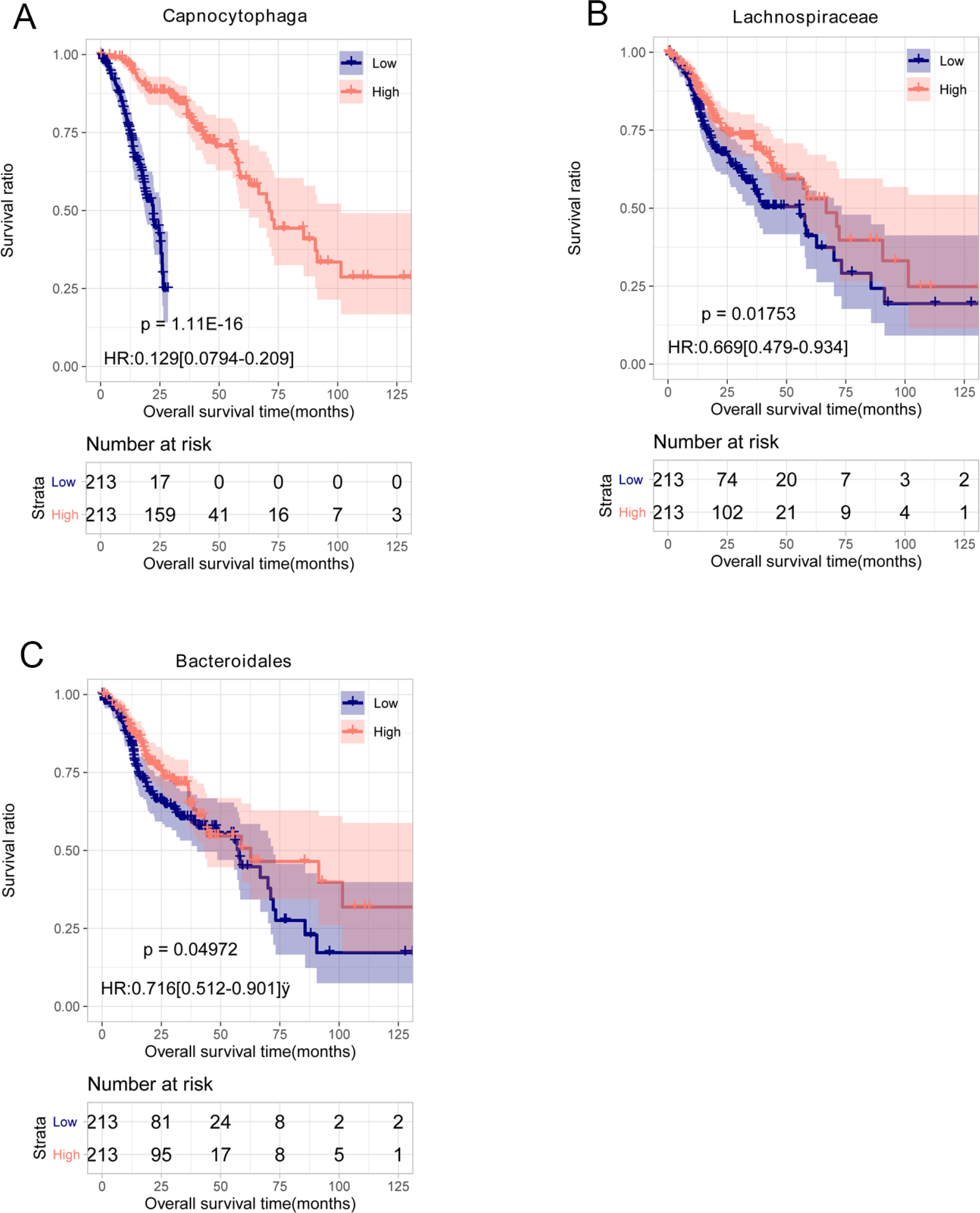
Fig. 6.
Kaplan-Meier (KM) curves of the microbial communities with different levels in tumors. (A) The KM curves of high- and low-Capnocytophaga levels. (B) The KM curves of high- and low-Lachnospiraceae levels. (C) The KM curves of high- and low-Bacteroidales levels.
Discussion
The relationship between microorganisms and tumor formation has received increasing attention. The present study identified eight optimal intra-tumor microbial communities, including Actinobacteria, Bacteroidales, Capnocytophaga, Clostridia, Fusobacteria, Lachnospiraceae, Prevotella, and Prevotellaceae, that effectively distinguish head and neck tumors (HNSC) from gastrointestinal tumors (ESCA/STAD/COAD/READ). The SVM classification model based on these microbes achieved AUCs of 0.937 in the training dataset and 0.856 in the validation dataset, while the nomogram model demonstrated high predictive accuracy (C-index = 0.8944 in training, 0.8023 in validation). These results align with and extend previous pan-cancer microbiome studies (Nejman et al. 2020; Battaglia et al. 2024). However, our study advances this field by focusing on the clinically relevant contrast between head and neck and gastrointestinal tumors—two groups with distinct anatomical and microbial regulatory origins—and identifying a shared signature that transcends gastrointestinal tumor subtype differences.
The biological basis for this discriminative power lies in the distinct microbial origins of the two tumor groups. Head and neck tumors are primarily shaped by the oral microbiota (Stasiewicz and Karpiński 2022), and our results show that HNSC samples exhibit higher abundances of oral-associated microbes, such as Fusobacterium and Prevotella. This finding is consistent with Nouri et al. (2023), who reported reduced Prevotella levels in patients with head and neck cancer but noted its persistent role as an oral-gut bridging microbe. In contrast, gastrointestinal tumors are dominated by gut microbiota-derived communities. Bacteroidales and Lachnospiraceae, two key members of the signature, are well-documented core gut taxa (Vacca et al. 2020; Jiang et al. 2025) whose dysregulation is linked to gastrointestinal carcinogenesis (Zafar and Saier 2021). For example, Lachnospiraceae produces butyrate, a short-chain fatty acid with anti-inflammatory and anti-tumor effects (Hexun et al. 2023). Its higher abundance in gastrointestinal tumors (vs. HNSC) may reflect a compensatory response to chronic gut inflammation—an observation supported by Elghannam et al. (2025), who linked dysregulation of gut microbial metabolites to gastric cancer progression.
Notably, Capnocytophaga, a genus traditionally associated with oral microbiota (Zhu et al. 2024), was also identified as a key discriminative taxon. This finding aligns with Flemer et al. (2018), who reported oral microbiota (including Capnocytophaga-related taxa) as predictive markers for CRC, suggesting microbial “vertical migration” across the digestive tract. Such migration may explain why a single microbial signature can distinguish the two tumor groups: oral microbes colonize head and neck tissues directly, while a subset (e.g., Capnocytophaga) and gut-native taxa (e.g., Bacteroidales) collectively shape the intra-tumor microbiome of gastrointestinal cancers.
It is crucial to emphasize that the primary objective and achievement of our SVM model was to classify a given tumor sample as either originating from the head and neck region or the gastrointestinal tract, based on its intratumoral microbiota profile. This is distinct from diagnosing the presence of cancer itself from normal tissue. Dohlman et al. (2021) constructed a bacterial signature unique to CRC. Wang and his colleagues reported a 10-microbe signature that showed value in distinguishing esophageal squamous cell carcinoma from esophageal adenocarcinoma (Wang et al. 2021). Another study by Shao et al. (2019) showed esophageal squamous cell carcinoma contained more Fusobacterium and less Streptococcus, and gastric cardia adenocarcinoma had more Helicobacter compared with non-tumor tissues, based on data from Chinese patients. These data supported the roles of host-microbe in tumor diagnosis. Similarly, our classification model with eight optimal microbial communities, which were valuable for distinguishing head and neck tumors from gastrointestinal tumors, provides a promising direction for constructing non-invasive classification tools to differentiate between head and neck tumors and gastrointestinal tumors.
Further, Kaplan-Meier analysis revealed that high intra-tumor abundance of Capnocytophaga, Lachno-spiraceae, and Bacteroidales correlated with longer overall survival in both tumor groups, which adds prognostic depth to existing microbiome-cancer associations. For Lachnospiraceae, its higher abundance correlates with elevated immunoscores in advanced CRC, likely via butyrate-mediated immune modulation (Hexun et al. 2023). Bacteroidales, a dominant gut order, has dual roles in cancer. While some species promote tumorigenesis via pro-inflammatory metabolites (Zitomersky et al. 2011), our results suggest that their intra-tumor enrichment may reflect a “protective” subcommunity, possibly through competition with pathogenic microbes like H. pylori (Shang et al. 2016) or regulation of gut barrier function (Wang et al. 2021).
The prognostic role of Capnocytophaga is particularly noteworthy. A previous study identified C. gingivalis as a potential promoter in oral cancer (Zhu et al. 2024), but our study found that its high intra-tumor abundance was associated with better survival. This discrepancy may reflect tissue-specific microbial functionality. In oral tissues, Capnocytophaga drives inflammation via immune escape (Shin et al. 2007), but in gastrointestinal tumors, it may interact with gut microbiota to suppress carcinogenesis (Lo et al. 2018). Together, these results highlight that intra-tumor microbial prognostic value is not universal but depends on tumor anatomical context and microbial community interactions. Collectively, it can be inferred that Cap-nocytophaga, Lachnospiraceae, and Bacteroidales may be closely linked to the prognosis of head and neck tumors and gastrointestinal tumors, and diet-mediated microbial modulation may represent a potential treatment strategy for improving the prognosis of cancers (John Kenneth et al. 2023).
A critical question raised regarding the present findings is whether the protective bacteria (Capnocytophaga, Lachnospiraceae, Bacteroidales), whose high abundance correlates with longer overall survival, represent a long-term attempt by the microbiome to mitigate adverse host responses during cancer progression. This hypothesis aligns with the core tenets of microbial-host symbiosis, in which gut and local tumor microbiota evolve dynamic adaptive responses to preserve host microecological homeostasis against pathological disturbances such as chronic inflammation, oxidative stress, and malignant transformation, all key adverse processes in cancer development. For Lachnospiraceae, a core butyrate-producing taxon, its high intra-tumor abundance is most likely a direct microbial compensatory response to the chronic inflammatory microenvironment of tumors. Butyrate, the major metabolite of Lachnospiraceae, exerts potent anti-inflammatory effects by inhibiting NF-κB activation and reducing pro-inflammatory cytokines (e.g., TNF-α, IL-6) secretion, while also alleviating oxidative stress by upregulating host antioxidant enzymes (e.g., SOD, GSH-Px) (Hodgkinson et al. 2023; Recharla et al. 2023). In the context of cancer progression, the host tumor microenvironment is characterized by persistent inflammatory and oxidative stress, and the enrichment of Lachnospiraceae may represent a microbial adaptive response to counteract these adverse host reactions, thereby reducing tumor cell proliferation and invasion. For the dominant gut microbial order Bacteroidales, its intra-tumoral enrichment as a protective factor may reflect a community-level compensatory response to dysregulation of the tumor microenvironment. Its species compete with pathogenic microbes (e.g., Helicobacter pylori, Fusobacterium nucleatum) for nutrients and ecological niches in tumor tissues to inhibit their pro-tumorigenic colonization, and some secrete short-chain fatty acids and polysaccharide metabolites that modulate host immune function (e.g., promoting anti-tumor CD8+ T cell and M1 macrophage activation) (Sun et al. 2019; Inamura 2021), representing a microbial attempt to restore the host’s impaired anti-tumor immune response during cancer progression. For Capnocytophaga, a taxon traditionally linked to the oral microbiota, its protective role across both head and neck and gastrointestinal tumors suggests a tissue-independent microbial compensatory response to host malignant transformation. While C. gingivalis has been reported to drive oral inflammation in some studies (La Rosa et al. 2020, Lawal and Baer 2021), our findings of its prognostic protective value imply a context-dependent functional shift: in established tumors, Capnocytophaga may alter its metabolic and signaling profiles to mitigate adverse host responses, such as inhibiting tumor cell epithelial-mesenchymal transition (EMT) or reducing tumor angiogenesis, a unique microbial adaptive response to the pathological tumor microenvironment. Collectively, these observations suggest the identified protective microbes are likely not only passive prognostic biomarkers but also active participants in the microbial compensatory response to host adverse reactions during cancer progression. This interpretation places our findings in a broader micro-ecological context, highlighting the dynamic symbiotic relationship between the intra-tumoral microbiota and the host tumor microenvironment. However, the causal relationship between microbial compensatory responses and host cancer progression remains to be confirmed by functional experiments (e.g., in vivo animal models with microbial depletion/colonization and longitudinal monitoring of tumor progression and host immune/inflammatory status).
Despite these promising findings, this study has several limitations that should be acknowledged. First, all data were derived from TCMA and TCGA, which lack detailed clinical metadata (e.g., patient diet, antibiotic use, smoking history, cancer severity, disease duration, and longitudinal progression status) known to influence microbiota composition. These confounding factors may have affected the abundance of key microbes (e.g., Lachnospiraceae, which is sensitive to diet). More importantly, the intra-tumoral bacteriome is a dynamic ecosystem that undergoes substantial alterations during cancer initiation and progression, and the absence of progression-related clinical data prevents us from exploring temporal changes in the identified microbial signatures during disease development and their potential association with cancer stage migration. Second, the relatively small sample size of the study cohort (463 tumor samples in total, with only 45 cases for ESCA and READ each) limits the power of subgroup analysis based on cancer progression and severity; larger cohorts with stratified clinical data are needed to validate the stability of the microbial signature across different disease stages. Third, obvious heterogeneity was observed in patient demographics and clinical characteristics, such as age, gender, pathological stage, and vital status. Fourth, and importantly, our study identifies a signature to differentiate between established tumor types, not to distinguish tumors from normal tissues. Consequently, our findings cannot inform on early diagnosis or prevention, which requires prospective studies beyond our retrospective design. Fifth, this study is purely bioinformatic and focused exclusively on the bacterial component of the intra-tumoral microbiome. The TCMA database primarily provides bacterial 16S rRNA sequence data, excluding fungi (mycobiome) and viruses (virome), two key microbial kingdoms increasingly recognized as contributors to cancer risk and progression. Fungi (e.g., Candida albicans, Malassezia) and viruses (e.g., HPV, EBV, bacteriophages) can directly drive tumorigenesis via chronic inflammation, DNA damage, or immune modulation, or indirectly shape bacterial community composition and function (e.g., via phage-mediated bacterial lysis, fungal-bacterial niche competition). These unmeasured mycobiome and viriome components may represent confounding factors, as their interactions with bacteria could have altered the abundance of the eight identified bacterial markers or modified their prognostic associations. Thus, our findings should be interpreted as specific to the bacterial microbiome, with the understanding that multi-kingdom microbial interactions may have influenced the overall results. Moreover, functional experiments (e.g., in vitro co-culture of tumor cells with key microbes and in vivo animal models) are required to confirm the causal roles of the identified microbial communities in tumor progression and survival. Future studies should collect clinical samples with complete longitudinal follow-up data on cancer progression, severity, and microbial dynamics, integrate multi-kingdom microbiome data (bacteria, fungi, viruses), and combine in vitro and in vivo experiments to clarify the temporal and spatial changes of intra-tumoral microbiota during disease development and the synergistic or antagonistic interactions between different microbial kingdoms.
In conclusion, our work demonstrates that eight intra-tumor microbial communities (Actinobacteria, Bacteroidales, Capnocytophaga, Clostridia, Fusobacteria, Lachnospiraceae, Prevotella, and Prevotellaceae) constitute a robust signature for distinguishing head and neck tumors from gastrointestinal tumors, with high diagnostic accuracy validated across training and validation datasets. Additionally, high intra-tumor abundance of Capnocytophaga, Lachnospiraceae, and Bacteroidales may be associated with improved overall survival, highlighting their potential as prognostic biomarkers and potentially as key participants in the microbial compensatory response to host adverse reactions during cancer progression. These findings have important clinical implications: the identified microbial signature could be translated into non-invasive classification tools (e.g., plasma or tissue biopsy-based microbial panels) for distinguishing head and neck tumors from gastrointestinal tumors in ambiguous clinical cases, while the prognostic association of Capno-cytophaga, Lachnospiraceae, and Bacteroidales suggest that these microbes may potentially serve as biomarkers for patient stratification. It is important to note that our findings are limited to the bacterial component of the microbiome; the mycobiome and virome may have influenced the observed associations through interactions with bacteria or by directly contributing to cancer progression. Future functional studies should not only investigate targeted modulation of these bacterial communities (e.g., via probiotics or dietary interventions) but also integrate multi-kingdom microbiome data to clarify the synergistic roles of bacteria, fungi, and viruses in tumor development and survival.
Abbreviations
- HNSC
head and neck cancer
- ESCA
esophageal cancer
- STAD
stomach cancer
- COAD
colon cancer
- READ
rectal cancer
- SVM
support vector machine
- TCMA
the Cancer Microbiome Atlas
- TCGA
the Cancer Genome Atlas
- RFE
recursive feature elimination
- ROC
receiver operating characteristics
Notes
[2] Availability of data and material
The datasets used and/or analyzed during the current study are available from the corresponding author upon reasonable request.
[3] Contributed by Authors’ contributions
Conception and design of the research: HG; Acquisition of data: HG; Data analysis and interpretation: JHW; Analysis and interpretation of data: YQN; Statistical analysis: FQL; Drafting the manuscript: HG; Revision of manuscript for important intellectual content: JHW. All authors approved and reviewed the final manuscript.
[4] Contributed by Conflict of interest
The authors do not report any financial or personal connections with other persons or organizations, which might negatively affect the contents of this publication and/or claim authorship rights to this publication.