
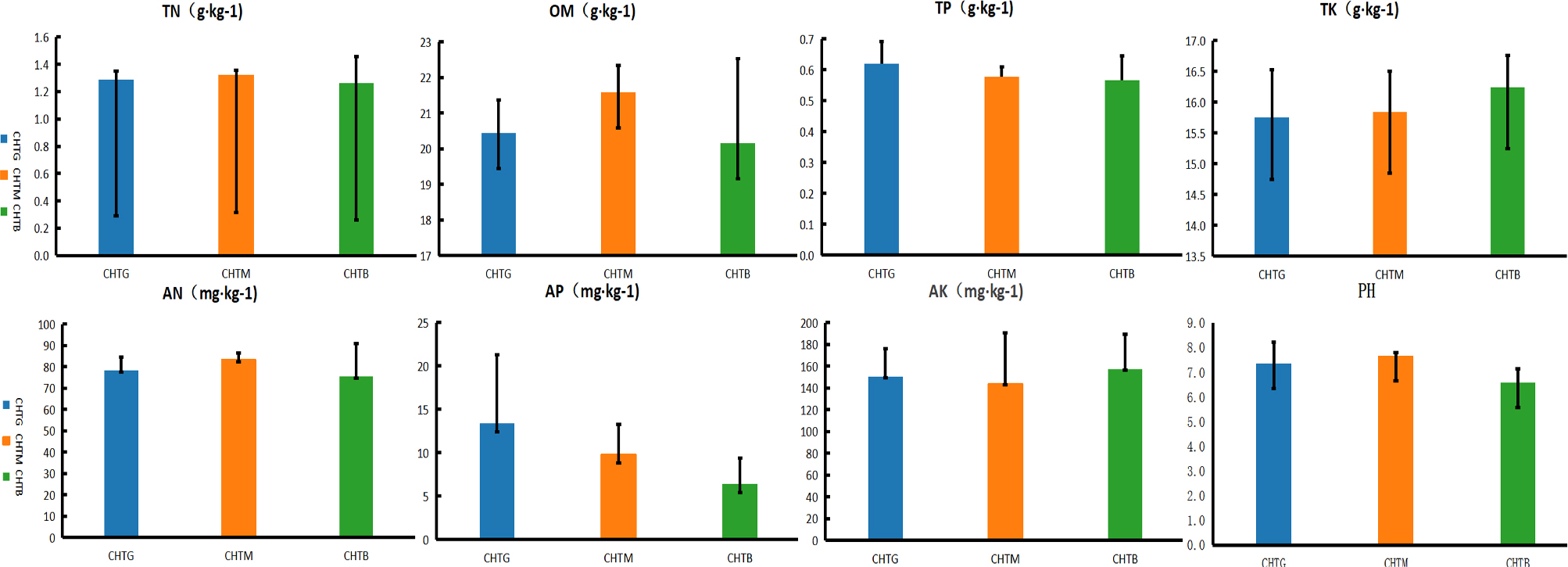
Fig. 1.
Soil nutrient content of three soil samples.
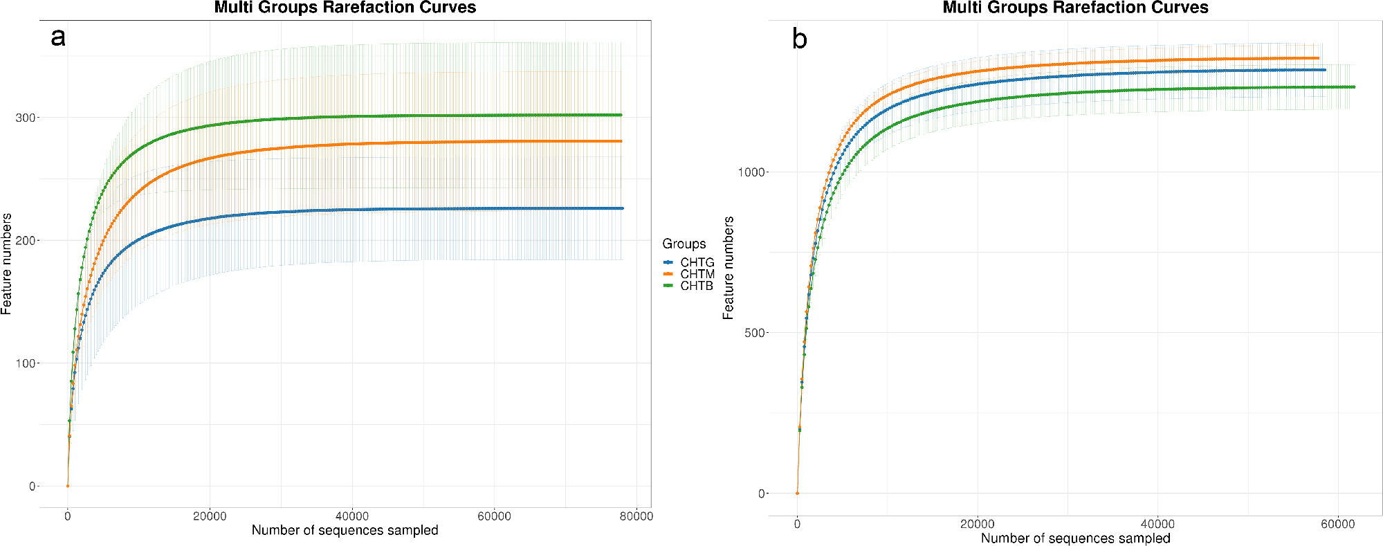
Fig. 2.
Samples rarefaction Curve.
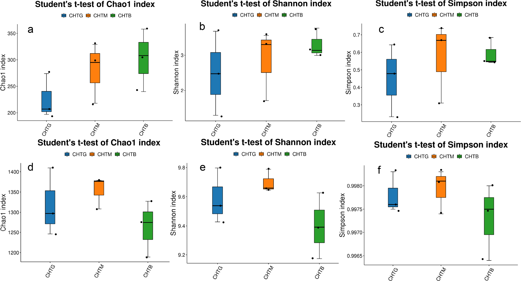
Fig. 3.
Soil microbial diversity index of fungi (a, b, c) and bacteria (d, e, f).
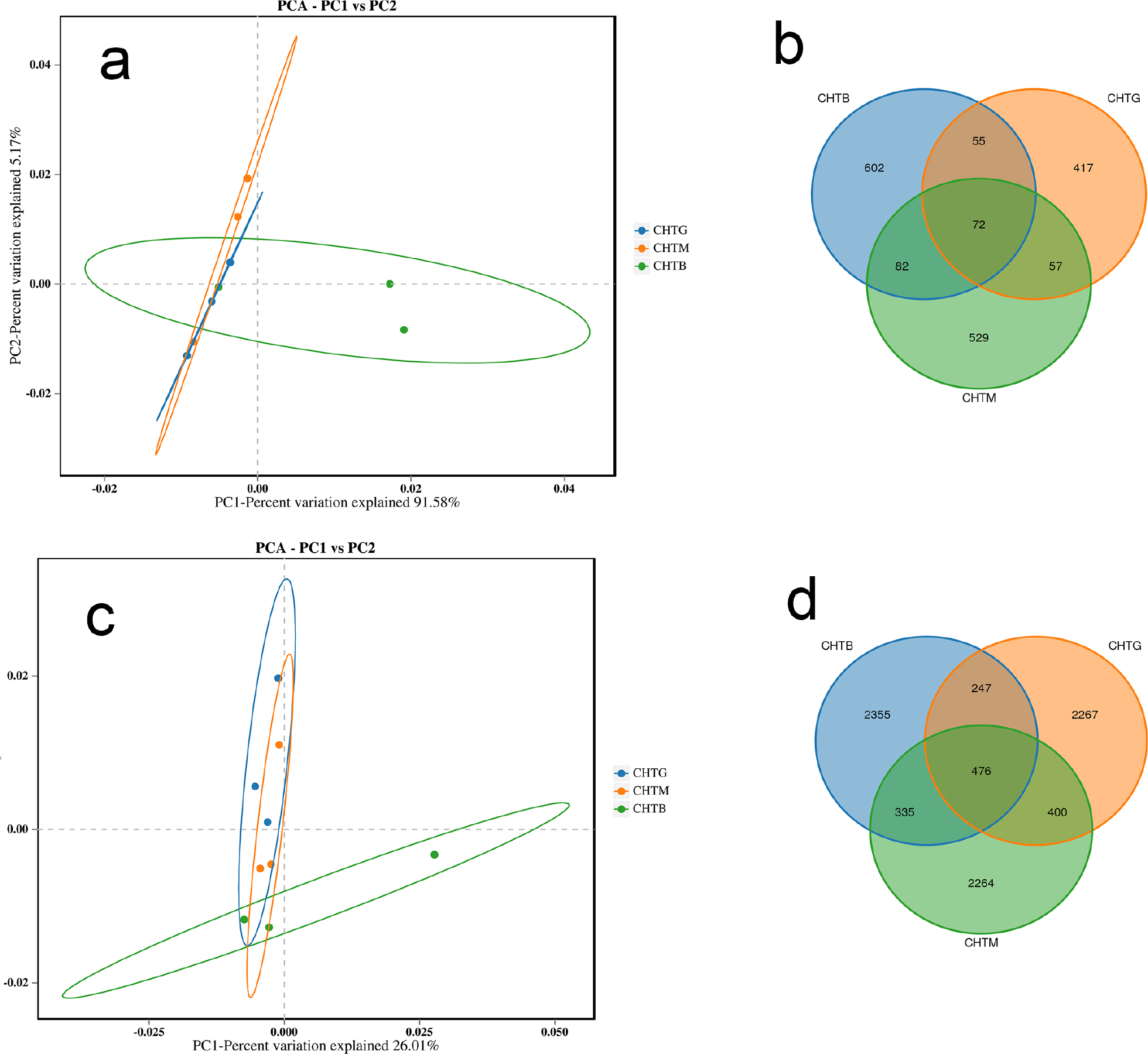
Fig. 4.
Comparative analysis of fungal and bacterial communities through Principal Component Analysis (PCA) and OTU-level Venn diagrams: fungal communities (a, b); bacterial communities (c, d).
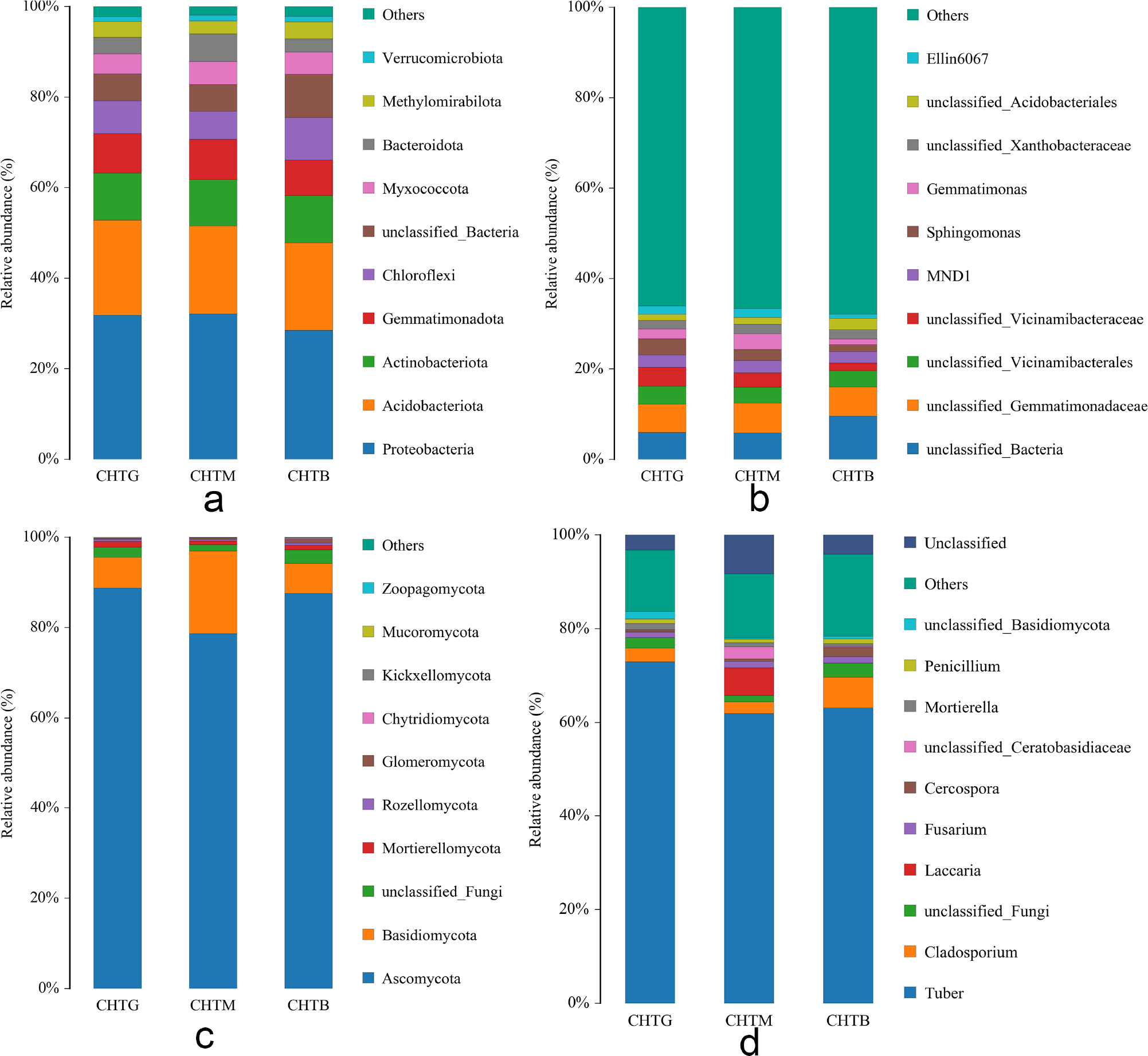
Fig. 5.
Taxonomic profiling of rhizosphere microbial communities: Bacterial composition at phylum and family levels (a, b); Fungal composition at phylum and genus levels (c, d).
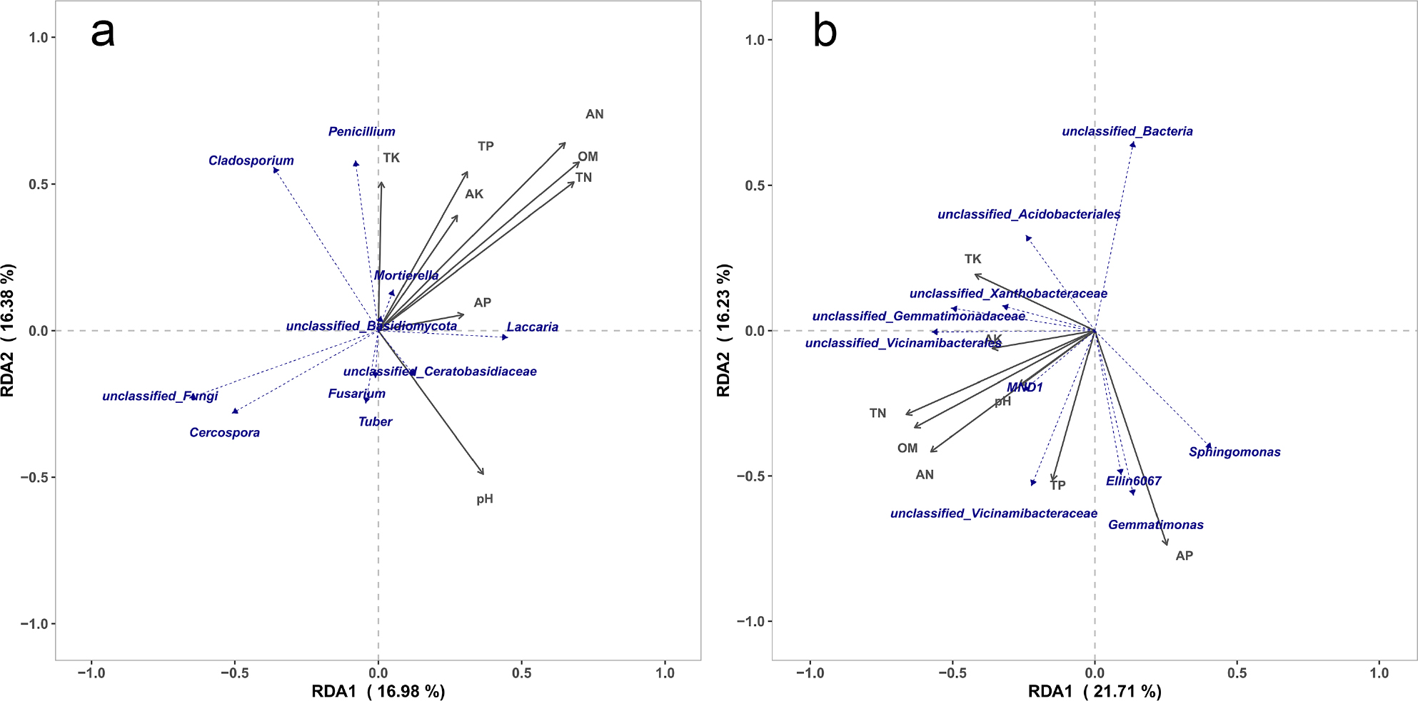
Fig. 6.
Redundancy analysis (RDA) of fungi (a) and bacteria (b) community structure and environmental factors.
Table I
Symbiotic network topological parameters.
| Network Parameters | Fungal | Bacterial |
|---|---|---|
| Nodes | 46 | 42 |
| Edges | 78 | 100 |
| Clustering coefficient | 0.364 | 0.607 |
| Graph density | 0.075 | 0.116 |
| Graph diameter | 18.615 | 24.27 |
| Average path length | 3.565 | 4.663 |
| Average degree | 3.391 | 4.762 |
| Betweenness centralization | 0.170 | 0.322 |
| Degree centralization | 0.146 | 0.177 |
| Modularity | 0.527 | 0.463 |
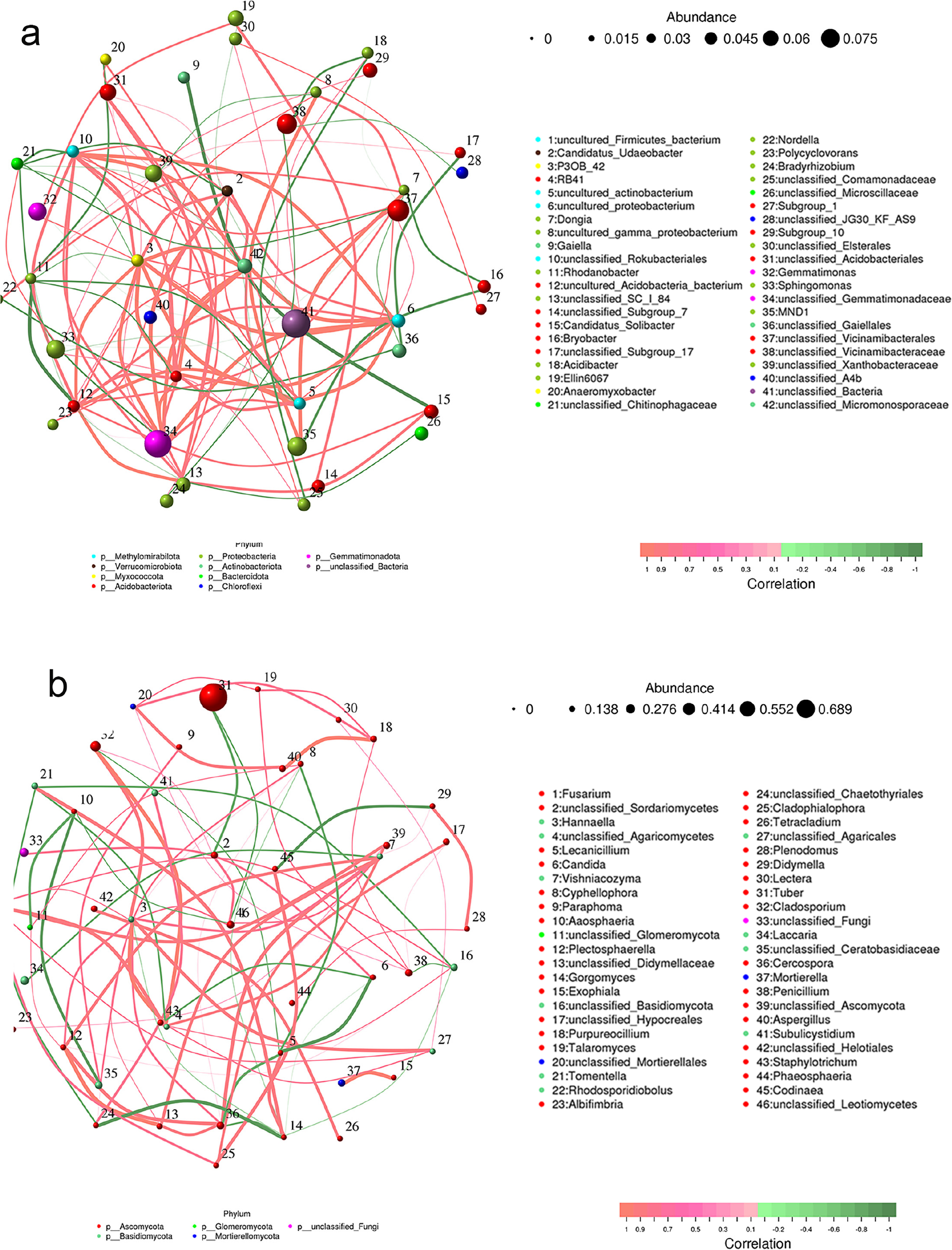
Fig. 7.
Topological profiling of bacterial (a) and fungal (b) symbiont networks.
The circle represents the species, and the size of the circle represents the average abundance of the species; line represents the correlation between the two species, the thickness of the line represents the strength of the correlation, the color of the line: red represents a positive correlation, green represents a negative correlation.
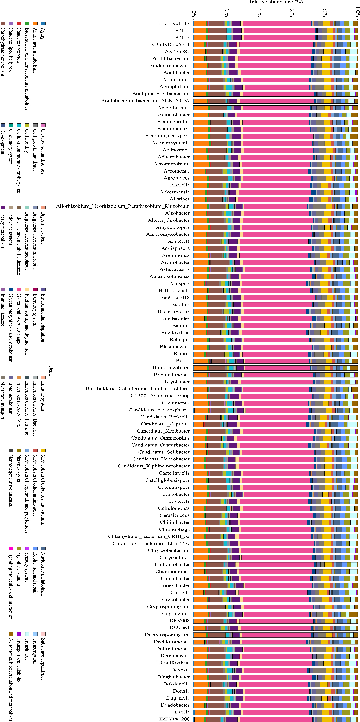
Fig. 8.
KEGG metabolic pathway histogram.
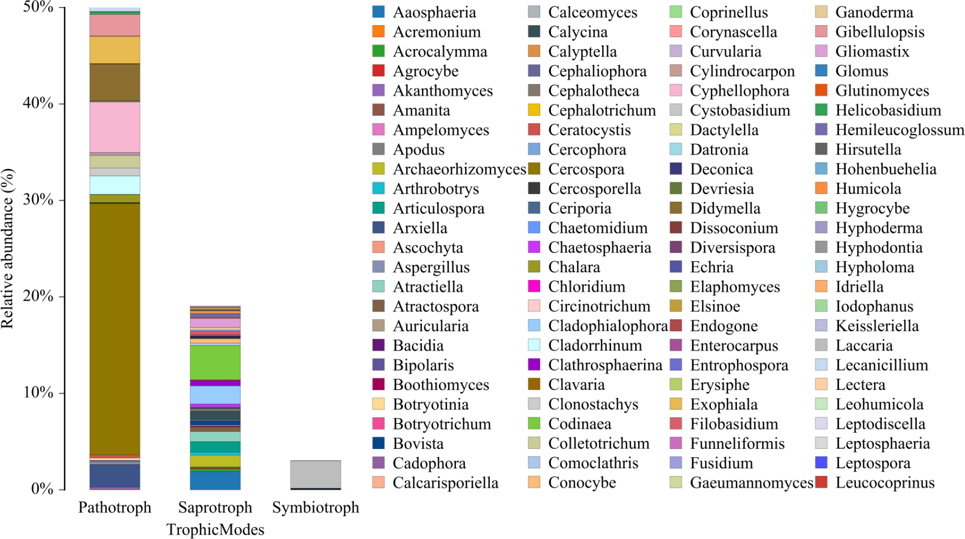
Fig. 9.
Funguild taxonomic composition bar plot.