Fig. 1.

Fig. 2.
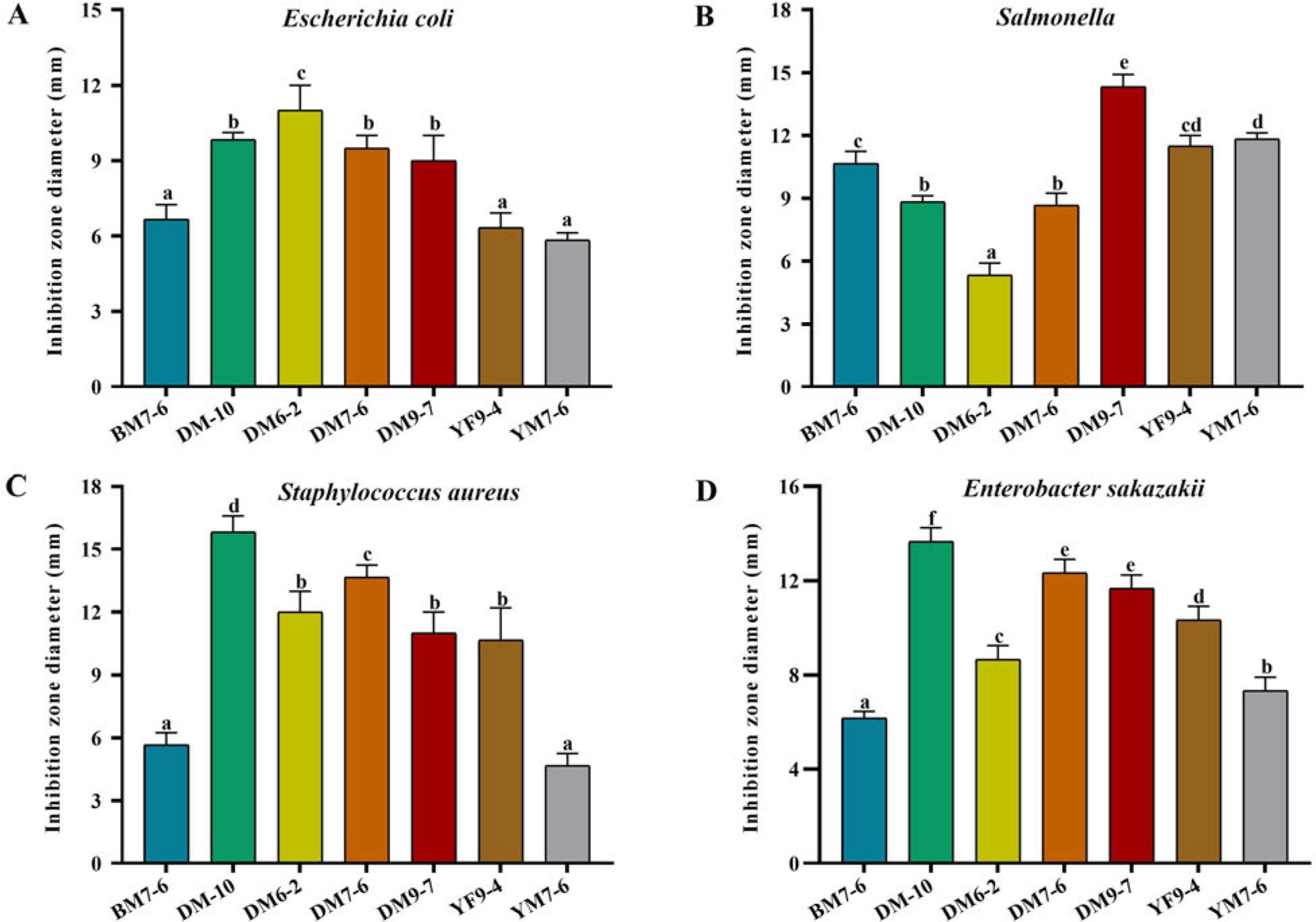
Fig. 3.

Fig. 4.
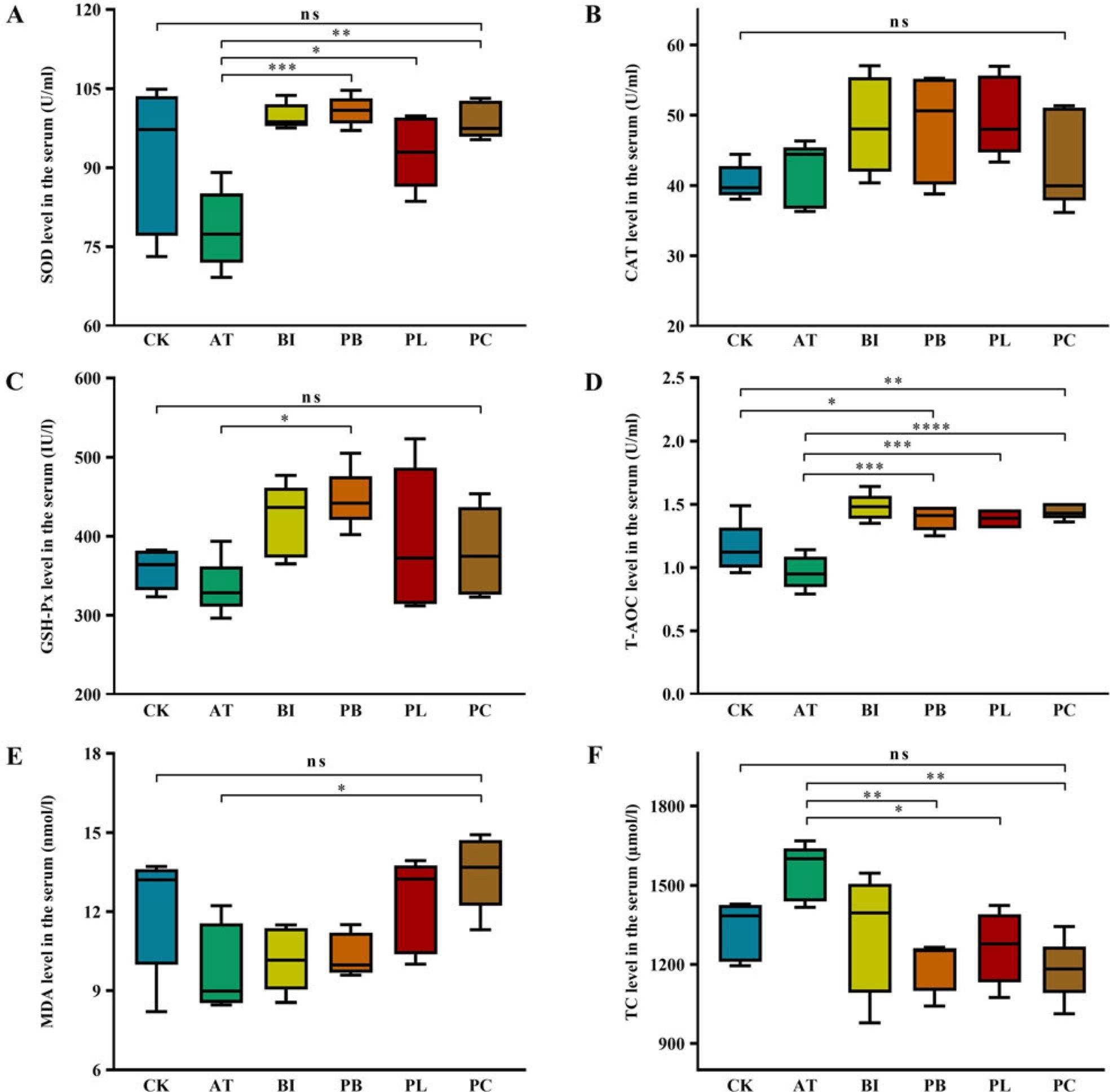
Fig. 5.
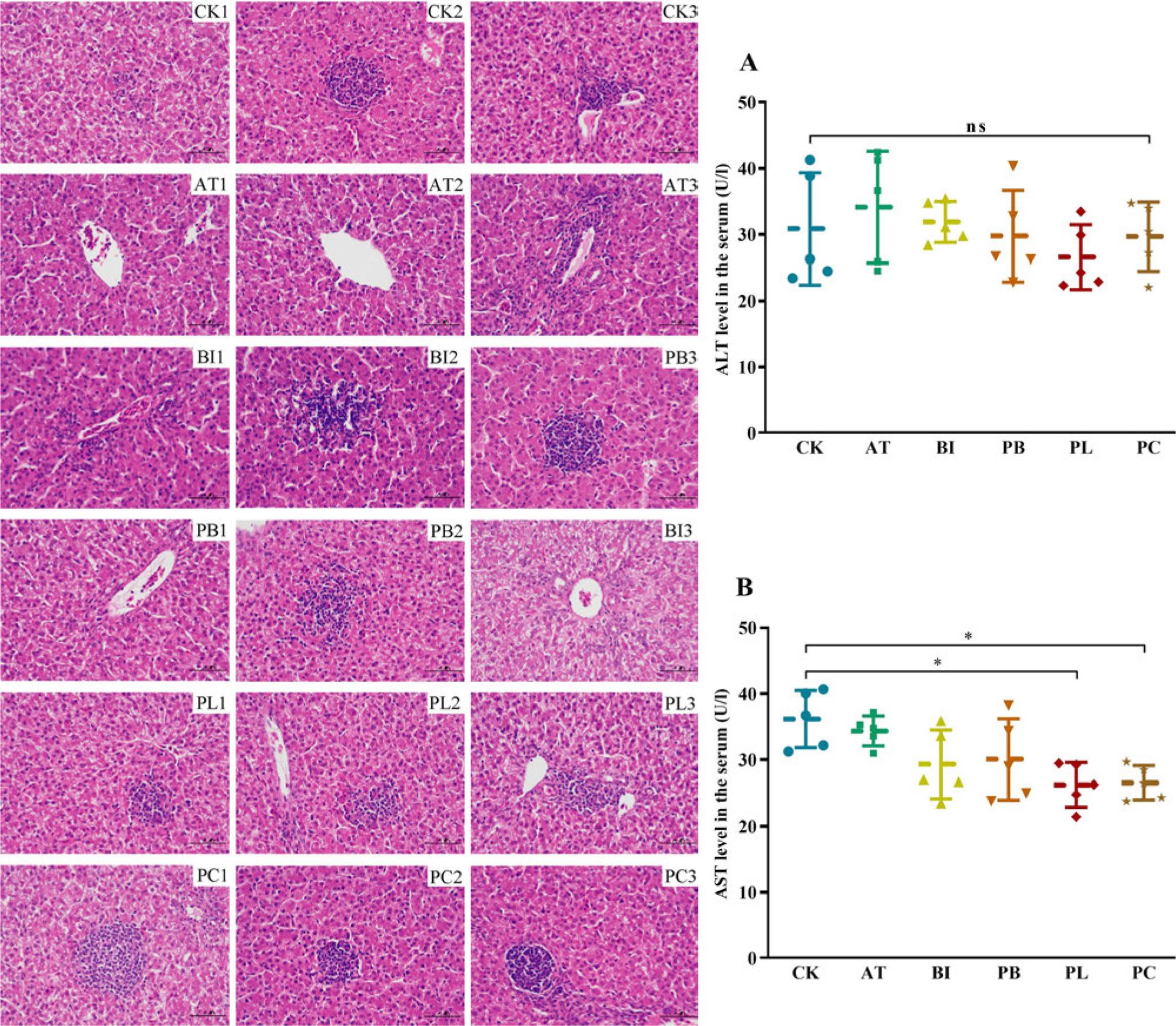
Fig. 6.

Fig. 7.

Fig. 8.

Fig. 9.

The susceptibility of candidate lactic acid bacteria (LAB) strains to different antibiotics_
| Antibiotics | Dose (μg/disc) | Susceptibility | ||||||
|---|---|---|---|---|---|---|---|---|
| BM7-6 | DM-10 | DM6-2 | DM7-6 | DM9-7 | YF9-4 | YM7-6 | ||
| Erythromycin | 15 | S | R | I | S | I | R | S |
| Amikacin | 30 | S | N | S | N | N | R | R |
| Ampicillin | 10 | I | I | I | S | S | S | I |
| Penicillin | 10 | S | N | S | S | N | S | N |
| Chloramphenicol | 30 | S | S | S | S | S | I | S |
| Ceftazidime | 30 | S | R | R | I | I | S | S |
| Tetracycline | 30 | S | N | I | S | N | S | R |
| Ciprofloxacin | 5 | S | N | N | N | N | I | S |
| Clindamycin | 2 | S | R | R | I | N | S | R |
| Azithromycin | 10 | S | S | S | S | S | I | S |
The growth of probiotics in artificial gastric juice and intestinal juice_
| Gastric juice | Intestinal juice | ||||||
|---|---|---|---|---|---|---|---|
| 0 h (CFU/ml) | 3 h (CFU/ml) | Survival rate (%) | 7 h (CFU/ml) | Survival rate (%) | 11 h (CFU/ml) | Survival rate (%) | |
| BM7-6 | 3.0 ± 0.5 × 10⁸ | 7.8 ± 0.8 × 10⁷ | 26.11 | 9.6 ± 1.0 × 10⁶ | 3.2 | 8.7 ± 0.7 × 10⁶ | 2.9 |
| DM-10 | 2.8 ± 0.4 × 10⁸ | 1.8 ± 0.3 × 10⁷ | 6.55 | < 105 | < 1 | < 105 | < 1 |
| DM6-2 | 1.7 ± 0.2 × 10⁹ | 1.4 ± 0.4 × 10⁸ | 7.92 | < 105 | < 1 | < 105 | < 1 |
| DM7-6 | 3.1 ± 0.4 × 10⁹ | 2.5 ± 0.6 × 10⁹ | 79.79 | 1.5 ± 0.3 × 10⁹ | 48.39 | 1.9 ± 0.7 × 10⁹ | 61.29 |
| DM9-7 | 5.8 ± 0.9 × 10⁹ | 3.7 ± 0.8 × 10⁹ | 63.22 | 2.5 ± 0.6 × 10⁹ | 43.10 | 2.6 ± 0.5 × 10⁹ | 44.82 |
| YF9-4 | 7.6 ± 1.0 × 10⁹ | 5.4 ± 0.7 × 10⁹ | 71.05 | 4.2 ± 0.5 × 10⁹ | 55.26 | 4.2 ± 1.4 × 10⁹ | 55.26 |
| YM7-6 | 2.3 ± 0.5 × 10⁸ | 8.7 ± 0.8 × 10⁷ | 37.14 | 2.7 ± 0.3 × 10⁷ | 11.74 | 2.3 ± 0.6 × 10⁷ | 10.00 |